
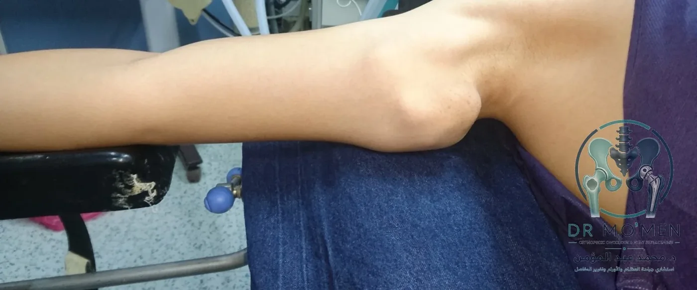
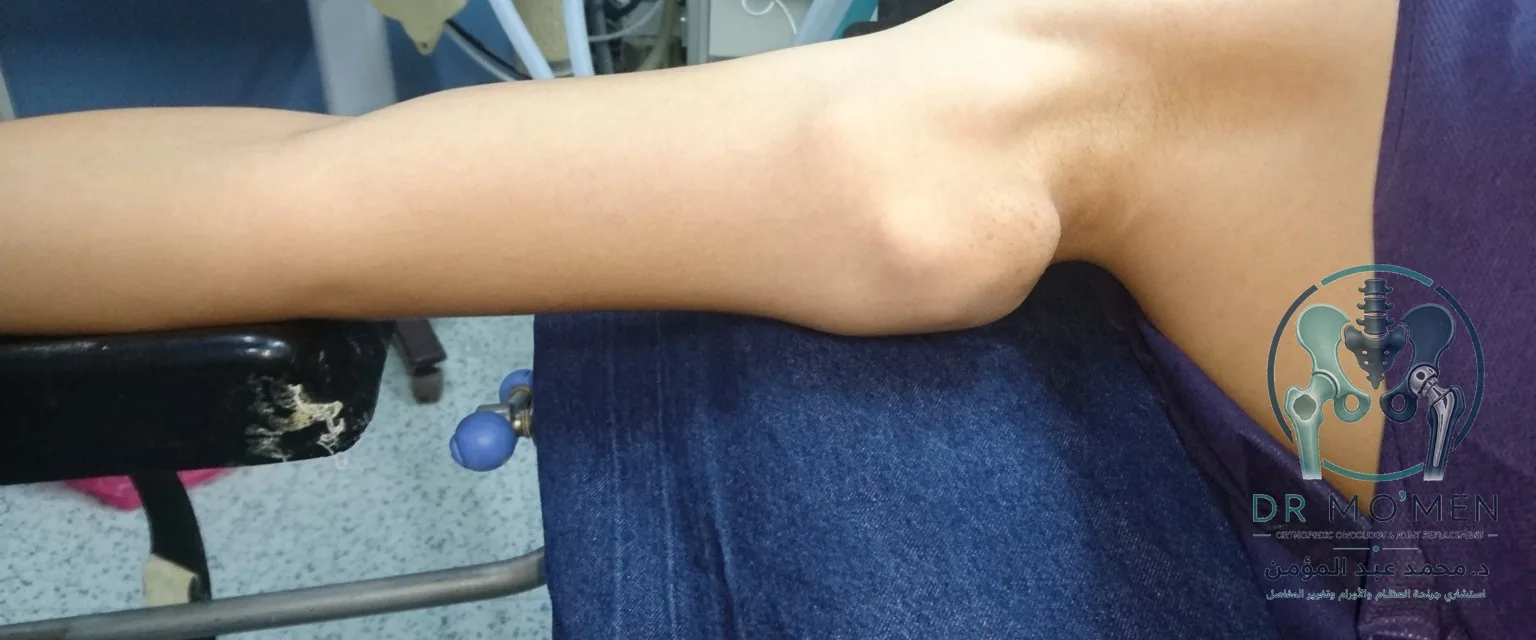
This case demonstrates a proximal humerus osteochondroma in a 12-year-old male patient presenting with visible medial arm swelling. Surgical planning was important because the lesion was large and close to major neurovascular structures in the medial arm.
The case in one minute
This case presents a proximal-humerus osteochondroma in a child. Osteochondroma is usually benign, but size, growth, pain, and its relationship to nerves and vessels determine surveillance or surgery.
- Clinical category
- Benign osteochondroma in a child
- Anatomical site
- Proximal humerus
- Decision illustrated
- Specialist imaging assessment of critical tissue relationships and treatment need
- Published reporting scope
- The page presents assessment and images; detailed histology and long-term outcome are not published.
Related pages
Case summary
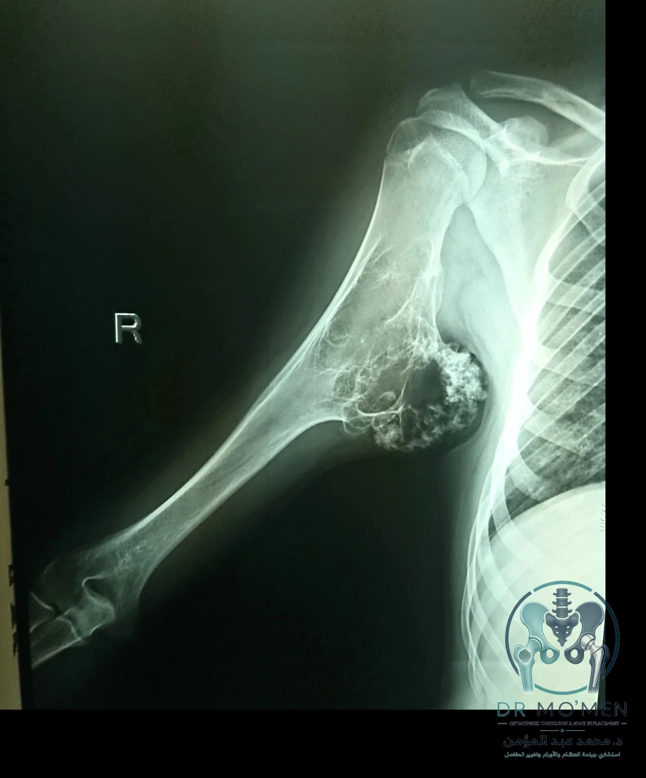
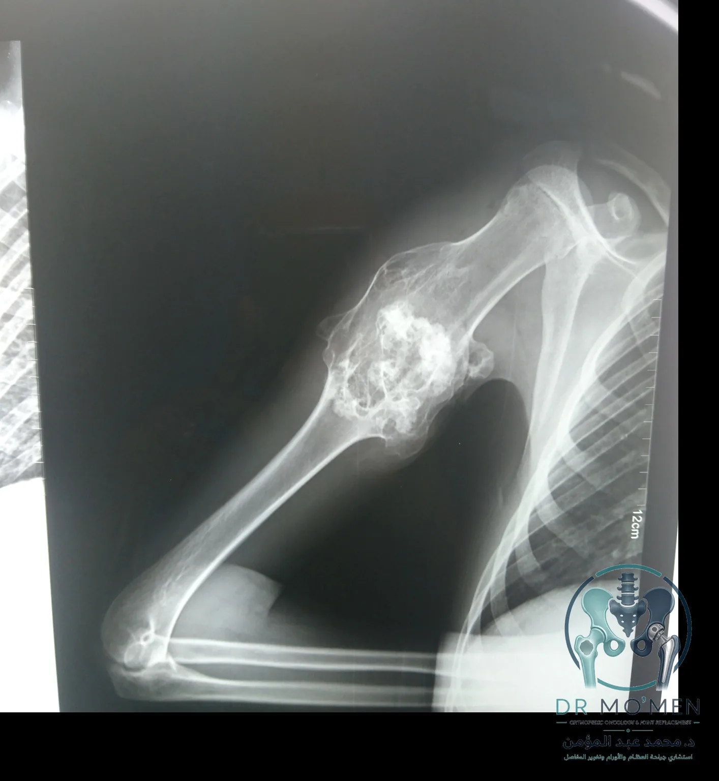
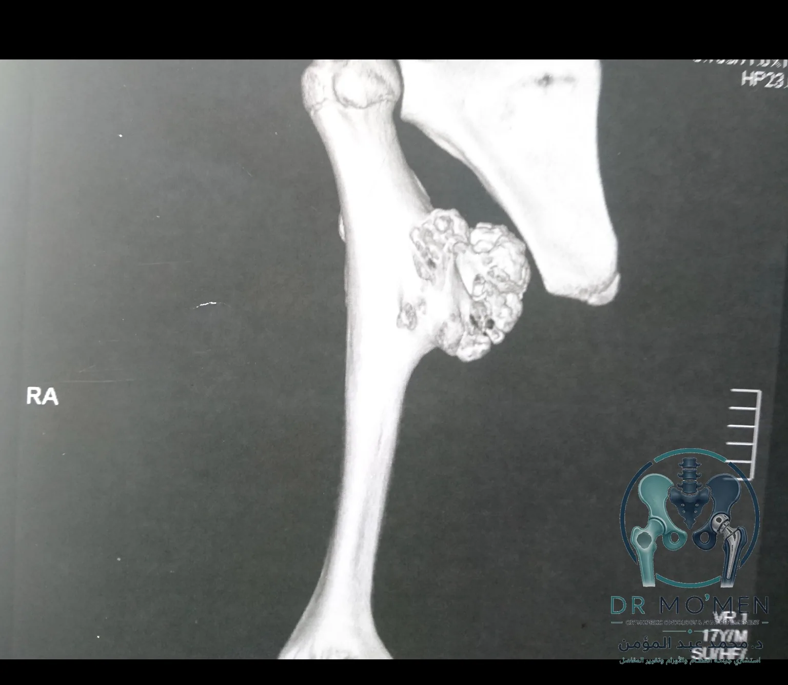
Clinical assessment and imaging showed an exophytic bony lesion arising from the medial aspect of the proximal right humerus. Plain radiographs and 3D CT reconstruction demonstrated a large osteochondroma projecting toward the medial arm.
The final diagnosis was osteochondroma. Because of the lesion size and anatomical location, surgical management required careful dissection and protection of the surrounding neurovascular structures.
Why specialist assessment matters
Many osteochondromas are benign lesions, but symptoms, size, growth behavior, pain, neurovascular proximity, and imaging features determine whether observation or surgery is more appropriate. In children, evaluation should also consider skeletal maturity and the relationship of the lesion to nearby growth regions and soft tissues.
Clinical and radiographic documentation
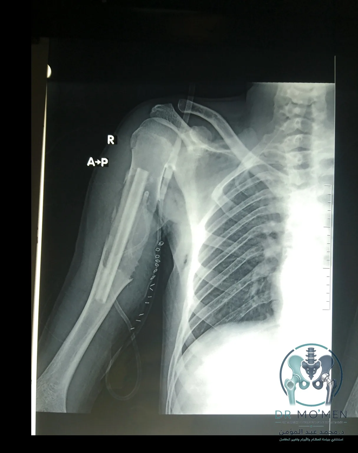
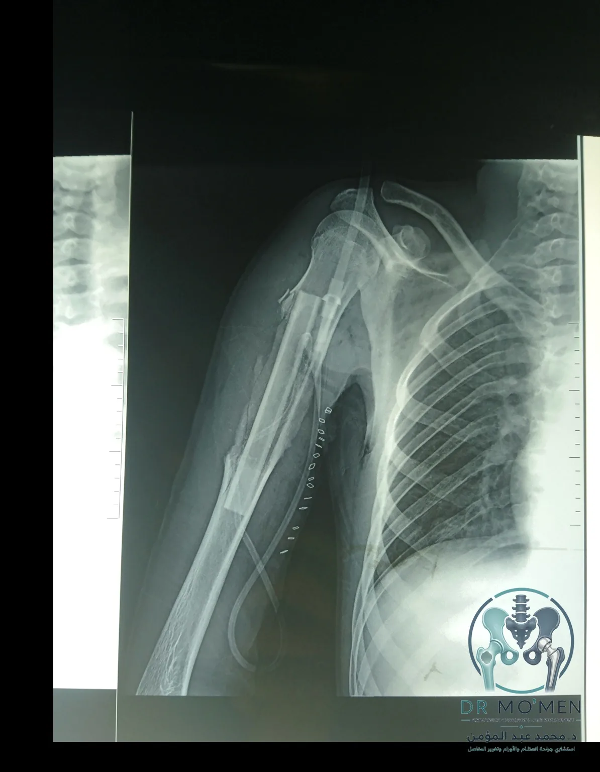
The following de-identified, logo-watermarked images show the clinical swelling, preoperative X-rays, 3D CT planning images, and postoperative radiographs.
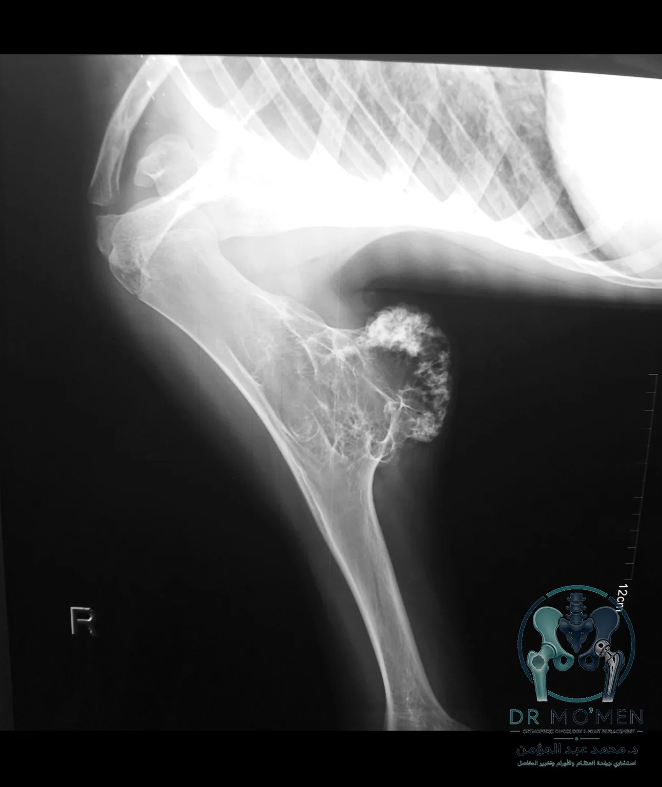
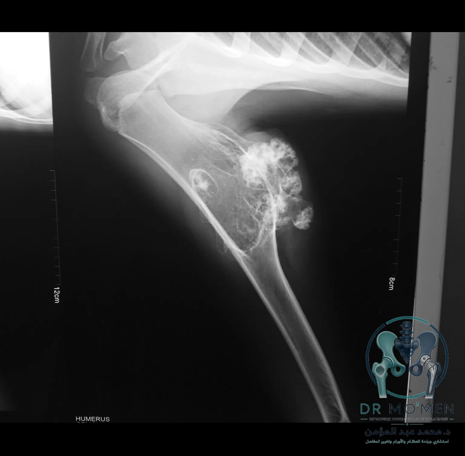
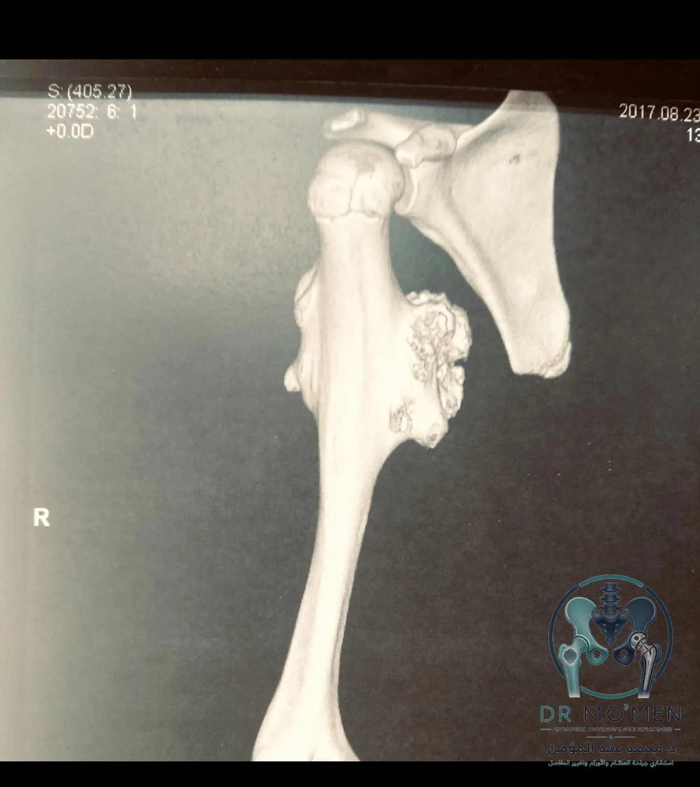
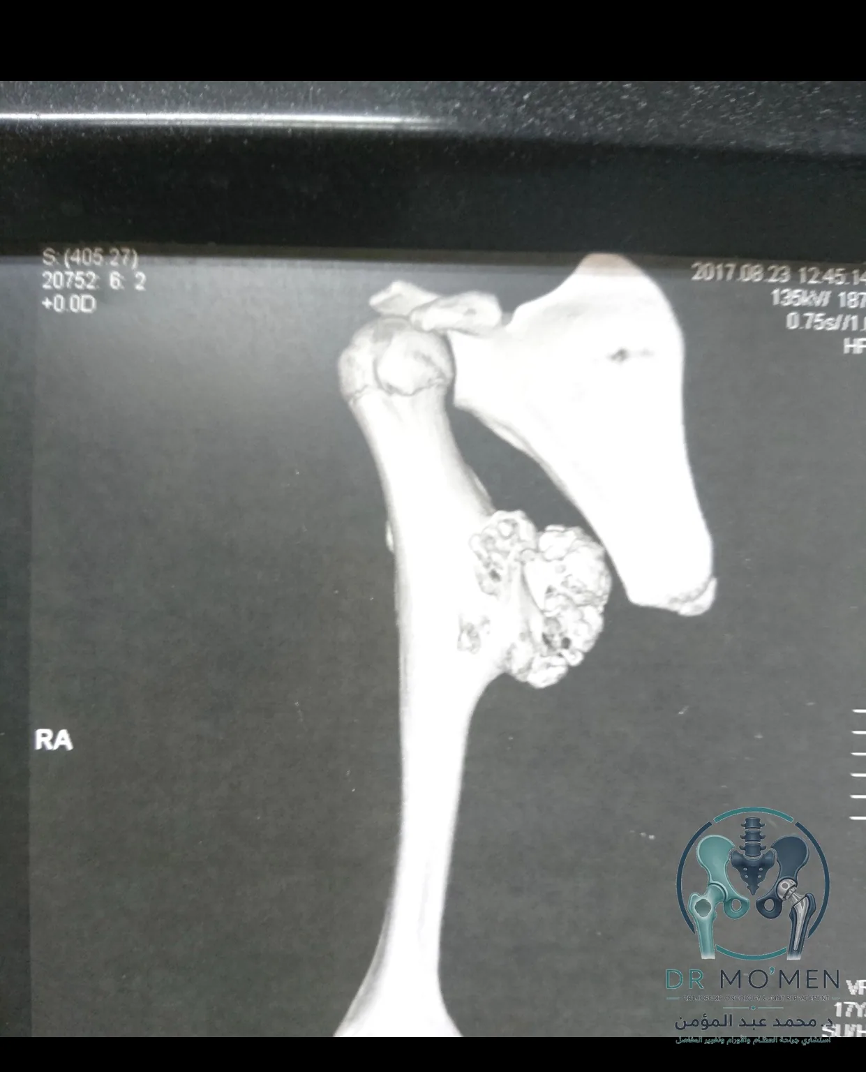
Clinical learning points
- The final diagnosis in this case was osteochondroma.
- No malignant diagnosis is stated in this website case summary.
- Medial proximal humerus lesions require careful planning because of nearby vessels and nerves.
- Treatment decisions depend on clinical examination, complete imaging, lesion behavior, and operative findings.
FAQ
Is osteochondroma usually benign?
Osteochondroma is commonly benign, but each case should be assessed according to symptoms, growth behavior, imaging appearance, and anatomical risk.
Why was CT useful in this case?
3D CT helped clarify the shape, size, and direction of the lesion and its relationship to the proximal humerus, which supports operative planning.
Can an osteochondroma be close to nerves and vessels?
Yes. Depending on location and projection, an osteochondroma can be close to important neurovascular structures, especially in the medial arm.
Does every osteochondroma need surgery?
No. Management is individualized. Observation may be suitable in selected cases, while surgery may be considered for symptoms, size, risk to nearby structures, or concerning clinical features.
Child with a bony swelling or bone tumor report?
Send X-rays, CT/MRI images, and reports through WhatsApp for guidance on whether clinic assessment or online review is appropriate.
Send scans and reportsTerms used in this case
- Osteochondroma
- A cartilage-capped bony projection that is usually benign and commonly appears during growth.
- Cartilage cap
- The cartilage layer on the lesion surface; its thickness and behavior are assessed on imaging.
- Neurovascular bundle
- Nearby nerves and blood vessels whose relationship to the lesion must be understood before surgery.