
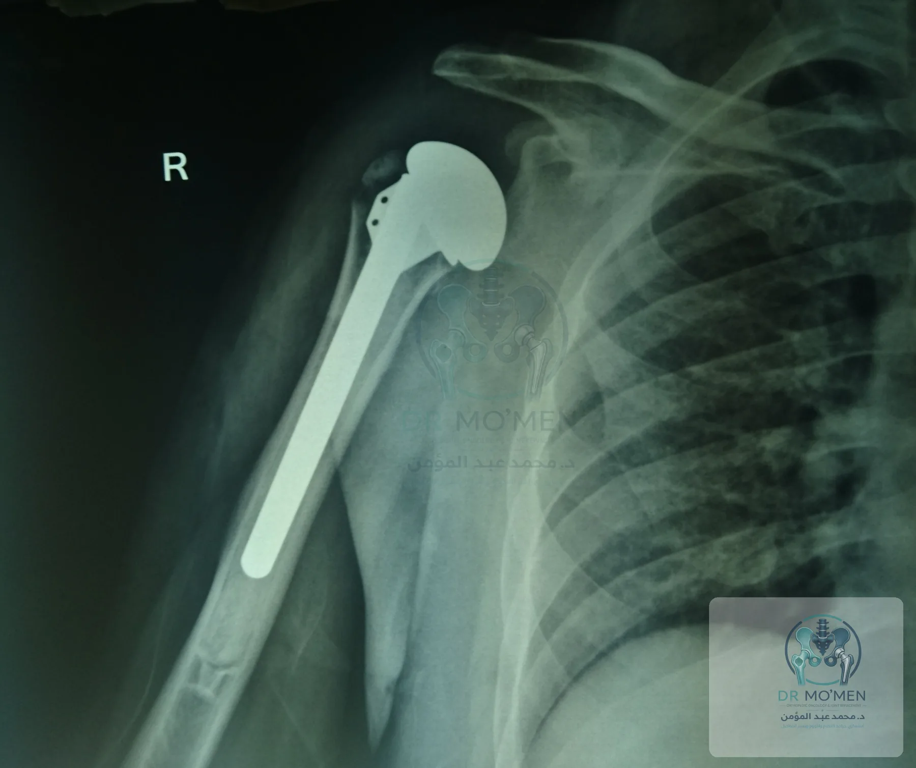
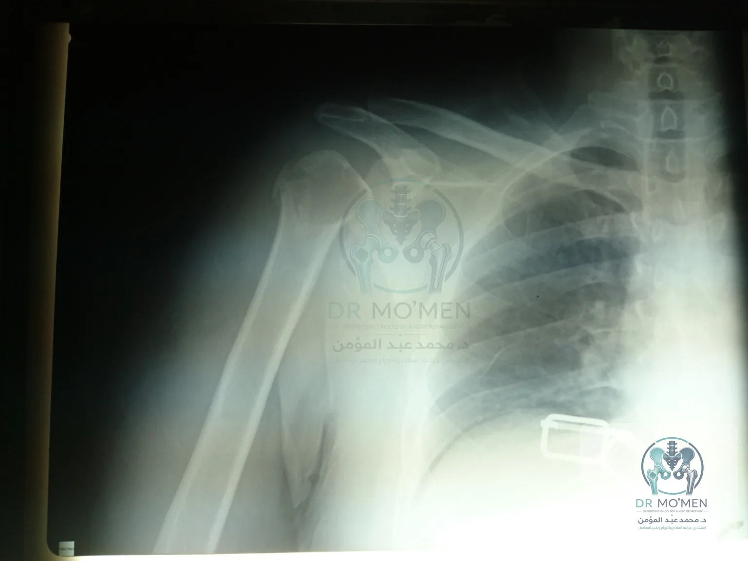
This case shows a severe right proximal humerus fracture-dislocation in a 38-year-old male after motorcycle trauma. Imaging demonstrated a displaced surgical-neck fracture with humeral head dislocation, requiring CT-based assessment and individualized operative planning.
The case in one minute
This case presents a complex proximal-humerus fracture-dislocation treated with shoulder hemiarthroplasty. Selection among fixation, hemiarthroplasty, and reverse replacement depends on age, fracture pattern, bone quality, and rotator-cuff status.
- Clinical category
- Complex proximal-humerus fracture-dislocation
- Anatomical site
- Shoulder joint
- Decision illustrated
- Shoulder hemiarthroplasty when preservation of the humeral head was not appropriate in this case
- Published reporting scope
- The page presents imaging and operative documentation; detailed functional outcome and follow-up duration are not published.
Related pages
Why this injury is complex
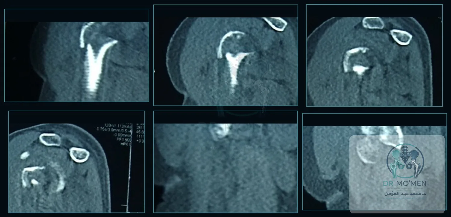
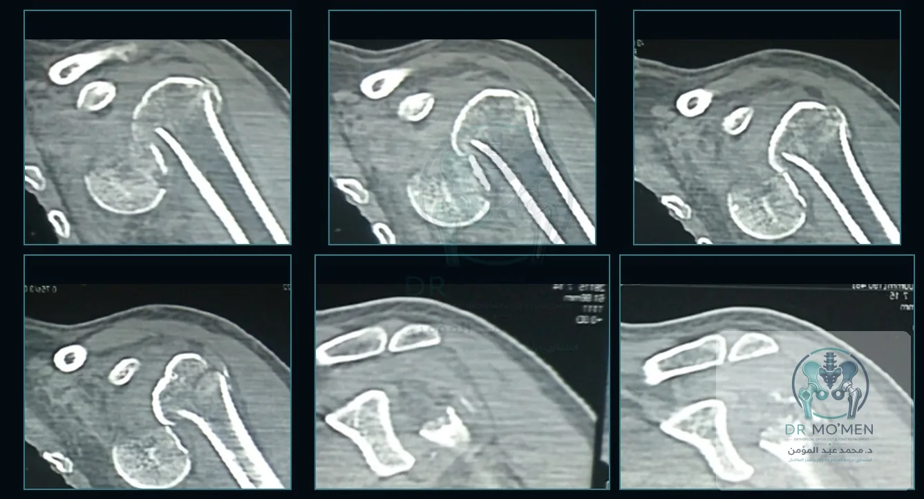
A proximal humerus fracture-dislocation is different from a simple shoulder fracture. The fracture pattern can affect joint congruity, humeral head viability, tuberosity healing, and soft-tissue repair. CT imaging is often required to understand the fragments and the position of the humeral head before deciding on treatment.
Why hemiarthroplasty was selected
In young adults, preservation of the native joint is considered whenever feasible. In selected complex fracture-dislocations, however, hemiarthroplasty may be chosen when fixation of the injured humeral head is not considered the most reliable option after imaging and intra-operative assessment.
In shoulder hemiarthroplasty, the damaged humeral head is replaced while the glenoid side of the joint is preserved. The objective is to restore a stable relationship between the prosthetic humeral head and the glenoid and allow structured rehabilitation.
Radiographic and operative documentation
The following de-identified, logo-watermarked images are presented for patient education and case-library documentation.
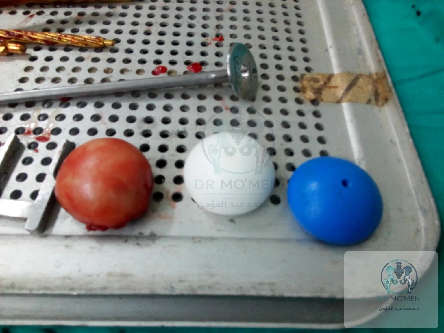
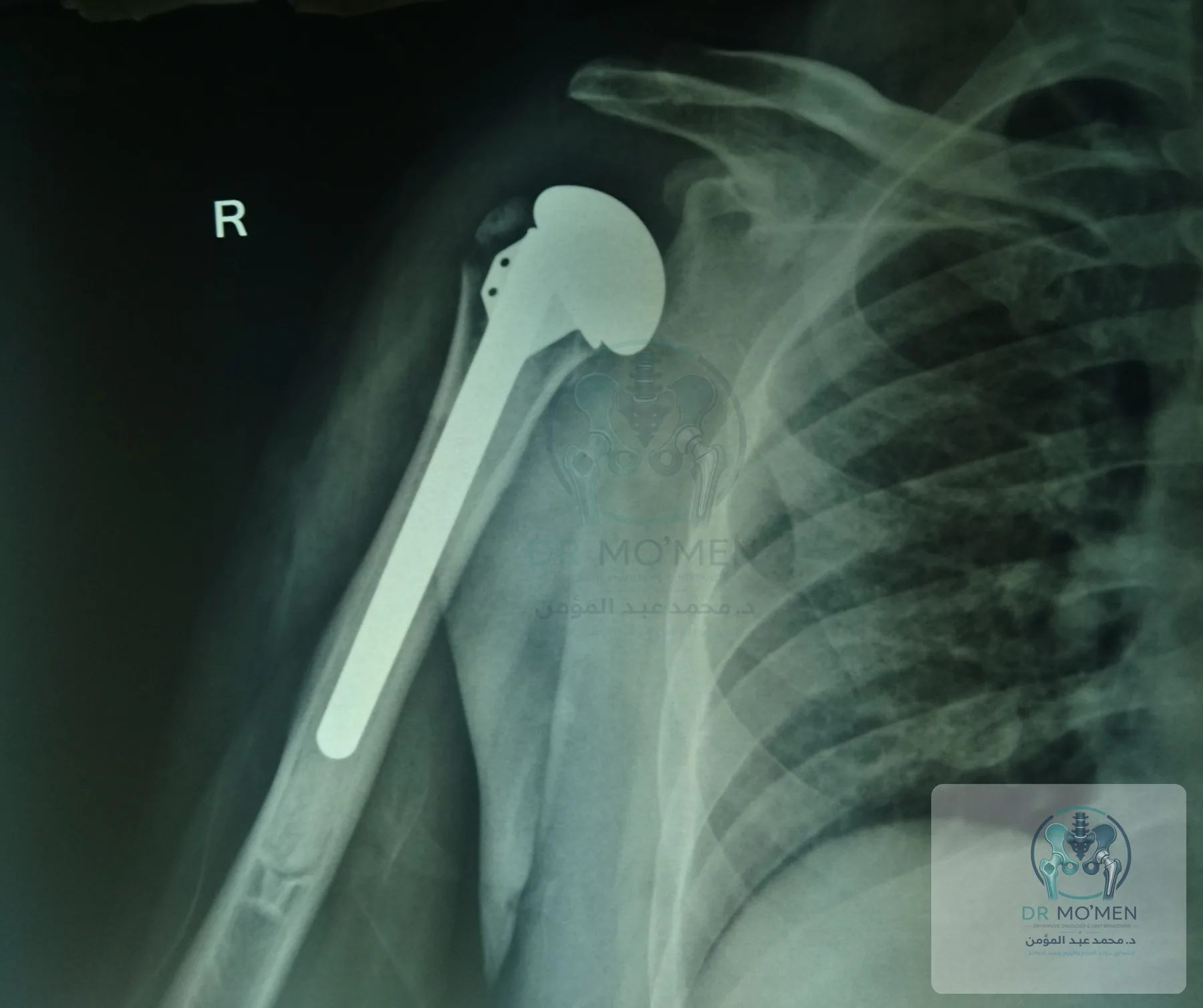
Clinical learning points
- Fracture-dislocation around the shoulder often needs CT imaging, not plain X-ray alone.
- The choice between fixation and arthroplasty depends on fracture morphology, bone quality, humeral head viability, tuberosity status, patient demand, and intra-operative findings.
- Post-operative rehabilitation is a core part of recovery and should protect soft-tissue repair and tuberosity healing when relevant.
- This case does not mean every proximal humerus fracture needs arthroplasty.
FAQ
Does every proximal humerus fracture-dislocation need hemiarthroplasty?
No. Some injuries can be treated with fixation or other reconstructive strategies. The decision depends on imaging, patient factors, and intra-operative assessment.
Why is CT important in this injury?
CT helps define the number and position of fracture fragments and the relationship of the humeral head to the glenoid, which supports safer surgical planning.
What is shoulder hemiarthroplasty?
It is replacement of the humeral head component of the shoulder joint while preserving the glenoid side.
Is rehabilitation required after surgery?
Yes. A structured rehabilitation plan is essential, but timing and restrictions must be individualized by the treating surgeon and physiotherapy team.
Need review of a complex shoulder fracture?
Send X-rays, CT images, and reports through WhatsApp to determine whether clinic assessment or online review is appropriate.
Send scans and reports