

Noticing that the front of your foot no longer lifts normally can be alarming. Your toes may catch the floor, the foot may slap down when you walk, or you may lift the knee higher than usual to clear the ground.
This is commonly called foot drop. Foot drop is not a diagnosis by itself; it is a clinical sign caused by weakness of the muscles responsible for lifting the ankle and toes. The problem can occur at different points along the nervous system, from the brain or spinal cord to the lumbar nerve roots, sciatic nerve, common peroneal nerve or the muscles themselves. Identifying the anatomical level and cause is therefore essential before choosing treatment.
What Is Foot Drop?
Foot drop describes difficulty lifting the front of the foot upward, known medically as ankle dorsiflexion.
- Toes catching on the floor
- Difficulty walking on the heel
- The foot slapping the ground
- Repeated stumbling
- Weakness lifting the ankle or toes
- Numbness over parts of the leg or top of the foot
- A high-stepping compensation known as steppage gait
Because foot drop can increase the risk of tripping and falling, the functional consequences may be important even before the underlying diagnosis is established.
What Causes Foot Drop?
A useful approach is anatomical: central nervous system → lumbar nerve roots → lumbosacral plexus → sciatic nerve → common peroneal nerve → deep peroneal nerve → dorsiflexor muscles. A lesion at more than one of these levels can produce a similar outward symptom.
1. Common Peroneal Nerve Compression
The common peroneal nerve travels around the fibular head on the outer side of the knee. Its superficial position makes it susceptible to compression and injury.
- Prolonged external pressure around the fibular head
- Prolonged leg crossing or sustained positions
- Substantial weight loss in some patients
- A tight cast or brace
- Trauma, fracture or dislocation around the knee
- Certain postoperative or positioning-related injuries
- Cysts, tumors or other masses affecting the nerve
A peripheral nerve problem at the knee can sometimes resemble a nerve-root problem originating from the lumbar spine.
2. L5 Radiculopathy
L5 nerve-root dysfunction is another major differential diagnosis. Possible causes include lumbar disc herniation, foraminal narrowing, degenerative spinal disease, spinal stenosis or other lesions affecting the root. Some patients have lower-back pain, radiating leg pain, tingling or weakness involving additional muscle groups, but the absence of back pain does not by itself exclude a spinal cause.
3. Sciatic Nerve and Lumbosacral Plexus Disorders
A sciatic nerve lesion can affect the peroneal division and produce dorsiflexion weakness. Lumbosacral plexus disorders can also cause foot drop, usually with a broader motor or sensory pattern than an isolated peroneal neuropathy.
4. Peripheral Neuropathy and Central Neurological Causes
More generalized peripheral neuropathies can cause unilateral or bilateral weakness. Less commonly, brain or spinal-cord disorders can present with foot drop. Additional weakness, altered reflexes, spasticity or other central neurological signs should redirect the diagnostic pathway.
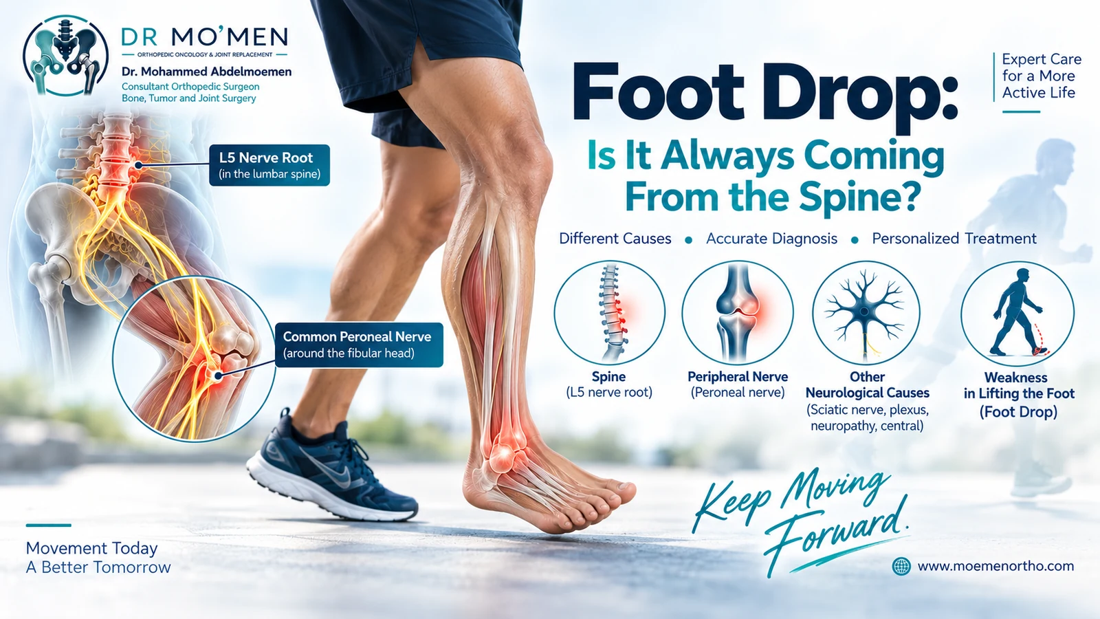
L5 Radiculopathy vs Common Peroneal Neuropathy
This distinction is clinically important because both conditions can weaken ankle dorsiflexion but represent different anatomical problems and may require different treatment.
- Peroneal neuropathy may cause weak dorsiflexion, toe extension and eversion, with sensory disturbance in a compatible peroneal distribution.
- L5 radiculopathy can affect muscles supplied through different peripheral nerves. Examination may therefore include inversion, eversion, great-toe extension, proximal muscle groups, sensation and reflexes.
- No single movement or MRI finding should be interpreted in isolation.

A Common Diagnostic Trap: Treating the MRI Instead of the Patient
A lumbar MRI may report an L4-L5 disc protrusion, but that finding should not automatically be assumed to explain foot drop. Imaging must be correlated with the side of weakness, muscles affected, sensory findings, symptom onset and the neurological examination. Electrodiagnostic testing may be helpful when localization remains uncertain.
Key principle: the imaging abnormality must fit the clinical localization.
When Is Foot Drop Urgent?
New foot drop deserves timely medical assessment, particularly if weakness is significant or progressing. Seek urgent or emergency evaluation when it occurs with:
- Sudden facial or arm weakness or speech difficulty
- New loss of bladder or bowel control
- Saddle-region numbness
- Rapidly progressive weakness in one or both legs
- Severe trauma around the knee or leg
- A cold, pale or poorly perfused foot after trauma
- Rapidly worsening neurological symptoms
How Is Foot Drop Diagnosed?
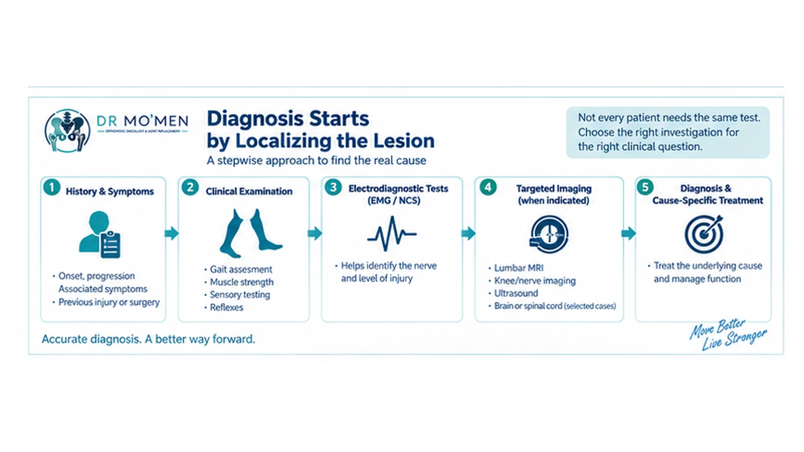
The preferred sequence is: History → Clinical examination → Anatomical localization → Targeted investigations → Cause-specific treatment.
Clinical and Neurological Examination
- Observation of gait
- Ankle dorsiflexion and great-toe extension
- Inversion and eversion
- Strength of other lower-limb muscle groups
- Sensory testing and reflexes
- Lumbar assessment
- Examination around the fibular head
- Assessment for central neurological signs
What Is the Role of EMG and Nerve-Conduction Studies?
EMG and nerve-conduction studies can be useful when the site or nature of the neurological lesion is uncertain. They may help distinguish common peroneal neuropathy, L5 radiculopathy, sciatic neuropathy, plexopathy or a more generalized peripheral neuropathy. They complement rather than replace clinical examination, and timing matters because electrodiagnostic abnormalities evolve after nerve injury.
Does Every Patient Need a Lumbar MRI?
No. Lumbar MRI is valuable when the history and examination suggest a lumbar nerve-root lesion or another spinal cause. If examination suggests a lesion around the common peroneal nerve, targeted ultrasound, MRI or other imaging may be more appropriate. Central imaging is reserved for patients whose neurological findings suggest brain or spinal-cord pathology.
Can Foot Drop Recover?
Sometimes, but recovery cannot be predicted from the label 'foot drop' alone. Prognosis depends on the underlying cause, severity and duration of nerve injury, whether compression is ongoing, nerve continuity and the patient's overall neurological condition. No universal recovery timeline or guarantee is appropriate.
How Is Foot Drop Treated?
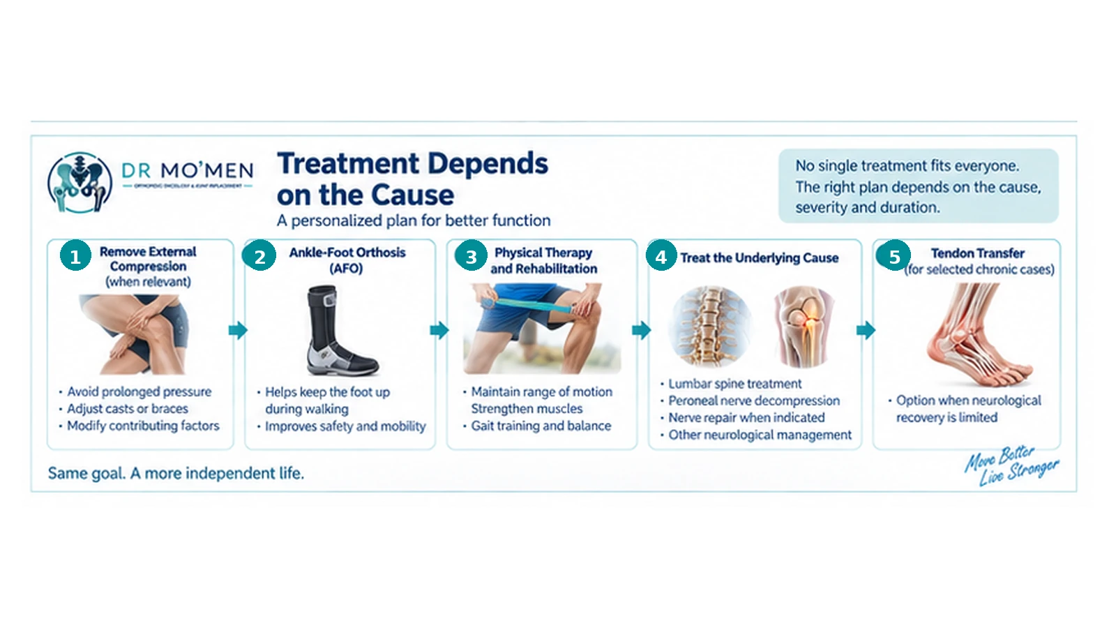
There is no universal treatment. Management should address both the underlying cause and the functional consequences of weak dorsiflexion.
1. Remove External Compression When Relevant
When an external factor contributes to common peroneal nerve compression, management may include modifying sustained pressure or reviewing restrictive casts or braces.
2. Ankle-Foot Orthosis (AFO)
An AFO can support the foot, improve toe clearance and reduce tripping. It is a functional aid; it does not establish the diagnosis or necessarily correct the underlying neurological lesion.
3. Physical Therapy and Rehabilitation
Rehabilitation may focus on maintaining ankle motion, reducing contracture risk, strengthening useful muscle function, gait training, balance and fall prevention.
4. Treat a Confirmed Lumbar Cause
When examination and investigations demonstrate clinically relevant nerve-root compression, treatment is individualized according to severity, progression, duration, pain, neurological findings, imaging and the underlying pathology. A disc abnormality on MRI does not automatically mean surgery is required.
5. Selected Peripheral Nerve Surgery
Nerve decompression or reconstructive procedures may be considered in selected cases involving persistent structural compression, traumatic injury or other clearly defined pathology. There is no single operation appropriate for every peroneal palsy.
6. Tendon Transfer for Selected Chronic Cases
When useful neurological recovery is not expected and foot drop is established, tendon transfer may be considered in selected patients to improve active dorsiflexion and gait. Patient selection and functional goals are central to the decision.
Why Early Diagnosis Matters
The reason for timely assessment is not that every patient needs urgent surgery. Most do not. The reason is that lumbar root compression, peroneal nerve compression, traumatic nerve injury and central neurological disease are fundamentally different problems. The management pathway depends on correct localization.
When Should You Seek Specialist Assessment?
- Your toes repeatedly catch the floor
- You cannot lift the front of the foot normally or walk on the heel
- Weakness developed after trauma or surgery
- There is numbness over the dorsum of the foot
- Back or leg pain accompanies weakness
- MRI findings do not seem to match the examination
- Weakness persists or progresses despite initial treatment
- Surgery has been proposed but the anatomical cause remains uncertain
- Chronic foot drop is interfering with walking and reconstructive options are being considered
When Is a Second Opinion Useful?
A second opinion can be useful when spine surgery has been proposed primarily because an MRI shows a disc abnormality, when peroneal nerve surgery is being considered, when EMG/NCS and imaging appear discordant, or when the anatomical source of persistent weakness remains uncertain. The purpose is to ensure that the diagnosis, localization and proposed treatment correspond to each other.
Foot Drop Assessment in Heliopolis, Cairo
Patients who need an in-person examination can attend the main MoemenOrtho clinic at 50 El Hegaz Street, Heliopolis, Cairo, beside Heliopolis Metro Station. Foot-drop assessment may include gait and motor examination, sensory testing, examination around the fibular head and knee, review of lumbar or lower-limb imaging, and interpretation of EMG/NCS when available.
Bring the original MRI, X-ray or ultrasound images when possible—not only the written report—plus any EMG/NCS results, previous operation notes and the exact timeline of weakness. See the Heliopolis orthopedic clinic page for directions, clinic hours and visit preparation.
The Role of Dr. Mo’men in Foot Drop Assessment
Dr. Mohammed Abdelmoemen Abuelhadid evaluates complex orthopedic and lower-limb conditions with attention to correlating symptoms and examination with imaging and investigations. Foot-drop assessment may include gait and motor examination, sensory findings, the knee and fibular-head region, lumbar imaging, EMG/NCS, previous trauma or surgery, rehabilitation and bracing requirements, and whether additional neurological, spine or peripheral-nerve assessment is appropriate.
For patients outside Egypt, the orthopedic second-opinion pathway can review existing reports, imaging and electrodiagnostic studies before deciding whether travel for in-person assessment is appropriate. The International Patients pathway explains record review and pre-travel planning. Remote review cannot replace a direct neurological examination when localization remains uncertain or emergency signs are present.
Selected Evidence Sources
- Dong K, et al. Foot Drop in Orthopaedic Surgery. JBJS Reviews. 2025.
- Peroneal nerve-conduction studies for differentiating L5 radiculopathy, peroneal neuropathy and sciatic neuropathy.
- Mayo Clinic: Foot drop — diagnosis and treatment.
- Tendon transfer in foot drop: systematic review.
Medical Disclaimer
This article is for patient education and does not replace a medical examination, review of imaging or appropriate investigations. New or progressive weakness, major trauma, or associated emergency neurological symptoms require appropriate medical assessment.