

Heel pain during the first few steps after getting out of bed is a common orthopedic complaint. Many patients are told they have a heel spur, particularly after an X-ray shows a small bony projection from the calcaneus. However, the presence of a heel spur does not automatically mean that the spur is causing the pain.
A common source of characteristic morning heel pain is the plantar fascia, the strong band of tissue running along the bottom of the foot.
The useful clinical question is not simply, “Is there a heel spur?” It is: What structure is actually causing the pain?
What Is Plantar Fascia Pain?
The plantar fascia extends from the heel bone toward the front of the foot and helps support the arch during standing and walking.

Repeated mechanical loading can produce pain near its attachment to the calcaneus. The term plantar fasciitis is widely used, although persistent cases may involve chronic tissue changes rather than simple acute inflammation alone.
Typical symptoms
- Pain underneath or toward the inner side of the heel.
- Marked discomfort with the first steps in the morning.
- Pain when standing after prolonged sitting.
- Partial improvement after walking for a short period.
- Recurrence after prolonged standing or walking.
- Symptoms following a sudden increase in exercise, running, or walking.
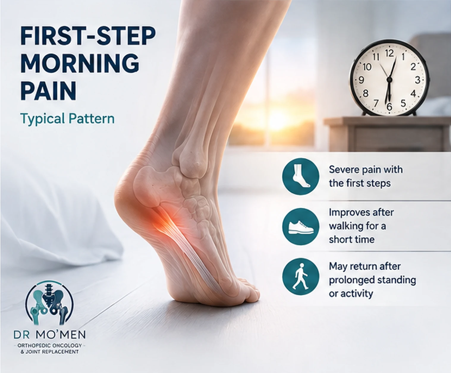
Is the Heel Spur Causing the Pain?
Not necessarily.
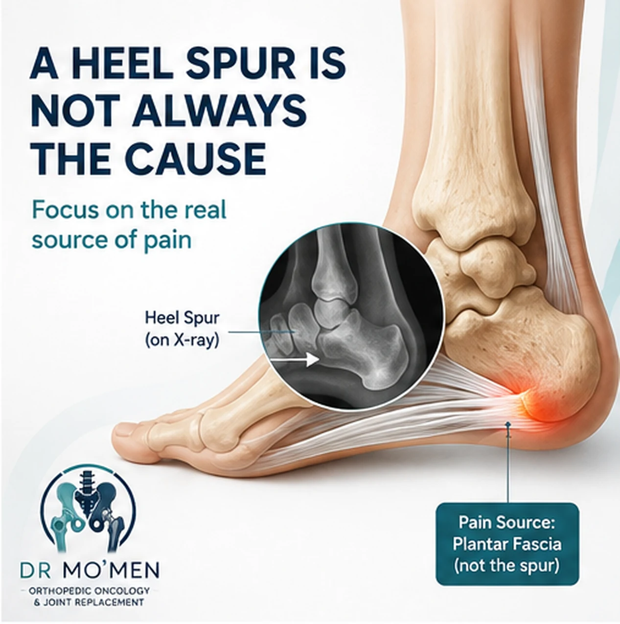
A heel spur may appear on an X-ray in someone with heel pain, but bony spurs can also be present in people without symptoms. Treatment therefore should target the actual pain generator, rather than an imaging finding alone.
A person can have a prominent spur without pain, while another may have typical plantar fascia pain with little or no visible spur.
Causes and Risk Factors
Factors that may increase mechanical load on the plantar fascia include:
- Prolonged standing or walking.
- A sudden increase in running or exercise.
- Repetitive activity on hard surfaces.
- Increased mechanical load on the foot.
- Tight calf muscles or Achilles tendon.
- Poorly supportive footwear.
- Some flat-foot or high-arch patterns.
- Returning too quickly to activity after a period of reduced exercise.
These factors may increase risk, but they do not prove the diagnosis.
Is All Heel Pain Plantar Fasciitis?
No.
Pain at the back of the heel
This may originate from the Achilles tendon, its insertion, or surrounding structures.
Pain after trauma
A calcaneal fracture, tendon injury, or ankle injury may need to be excluded. For traumatic heel pain or suspected fracture, see the Fractures service.
If symptoms followed an ankle sprain, related guidance is available here:
Cast or Physiotherapy After an Ankle Sprain?
Cast or Physiotherapy After an Ankle Sprain?
Burning pain or numbness
Neurological causes may need consideration.
Heel pain in children
The differential diagnosis differs from adults and may include disorders related to the calcaneal growth region.
Red Flags: When Does Heel Pain Need Faster Assessment?
Seek medical assessment sooner when heel pain is associated with:
- Inability to bear weight.
- Significant trauma or a fall.
- Marked swelling, redness, or warmth.
- Fever or systemic illness.
- Persistent numbness or weakness.
- A palpable mass.
- Unusual constant rest or night pain.
- Unexplained weight loss.
- Progressive symptoms that do not follow the typical plantar fascia pattern.
These findings do not automatically mean a serious diagnosis, but they should not simply be assumed to represent a routine heel spur.
Diagnosis and Investigations
In a typical presentation, diagnosis is mainly based on:
history + pain location + physical examination.
Assessment may include:
- The exact site of maximal tenderness.
- Ankle range of motion.
- Calf and Achilles tightness.
- Foot alignment and loading.
- Tenderness elsewhere around the calcaneus.
- Tendon and neurological examination when indicated.
Do I Need an X-Ray or MRI?
Not every patient requires advanced imaging.
Imaging becomes more relevant after trauma, with atypical or persistent symptoms, when a stress fracture is suspected, or when examination suggests another diagnosis.
If an X-ray identifies an unusual bone lesion rather than a conventional heel spur, it should be evaluated on its own merits before treatment is planned.
Treatment Options
Most patients start with nonsurgical treatment.

1) Activity and load modification
The aim is usually not complete inactivity. Activities that repeatedly aggravate symptoms may need temporary reduction followed by gradual progression.
2) Stretching
Calf, Achilles, and plantar fascia stretching may be useful as part of an appropriate rehabilitation program.
3) Supportive footwear
Shoes with appropriate cushioning and support can reduce mechanical stress. Heel cups or inserts may help selected patients as part of the overall plan.
4) Physiotherapy
Structured rehabilitation can address calf tightness, reduced ankle mobility, weakness, loading mechanics, and graded return to activity. For sports-related foot and ankle symptoms, see the Sports Injuries service.
5) Medication
Short courses of appropriate analgesic or anti-inflammatory medication may be considered for selected patients, depending on medical history and other medication.
6) Night splints
Night splints may be considered in selected patients with persistent first-step morning pain.
Do I Need a Heel Injection?
Not every patient does.
An injection should not automatically be offered simply because an X-ray shows a heel spur. It may be discussed in selected persistent cases after reviewing the diagnosis, previous conservative treatment, potential benefit, and risks.
The better question is: Why does my heel hurt, and is an injection likely to add meaningful benefit?
What About Shock-Wave Therapy?
Extracorporeal shock-wave therapy (ESWT) may be considered for selected patients with persistent plantar heel pain that has not improved sufficiently with appropriate conservative management. Response varies, and it is not mandatory for every case.
Does Plantar Fasciitis Require Surgery?
Surgery is uncommon and is generally reserved for carefully selected patients with persistent symptoms despite a structured nonsurgical program, after confirming that the diagnosis and pain generator are correct.
A heel spur on an X-ray alone is not an indication for surgery.
When Should You See an Orthopedic Specialist?
Assessment is reasonable when:
- Pain continues despite appropriate initial treatment.
- Walking or work becomes difficult.
- Symptoms repeatedly return.
- The diagnosis remains uncertain.
- Imaging and symptoms do not match.
- Repeated injections or surgery are being considered.
- There is a mass, unusual night pain, or an unexplained bone abnormality.
Role of Dr. Mo’men in Unclear or Complex Cases
Most typical plantar heel pain can be managed conservatively. Specialist assessment becomes more important when symptoms do not fit the expected pattern, repeated treatment has failed, or imaging shows findings that need interpretation.
Assessment may include review of previous imaging, differentiation between plantar fascia pain and tendon, fracture, or neurological causes, and determining whether additional imaging, injection, or surgery is actually necessary.
For appointments or imaging review: 01021690693
Patients outside Egypt can also use the second-opinion pathway to have existing reports and imaging reviewed before arranging travel: