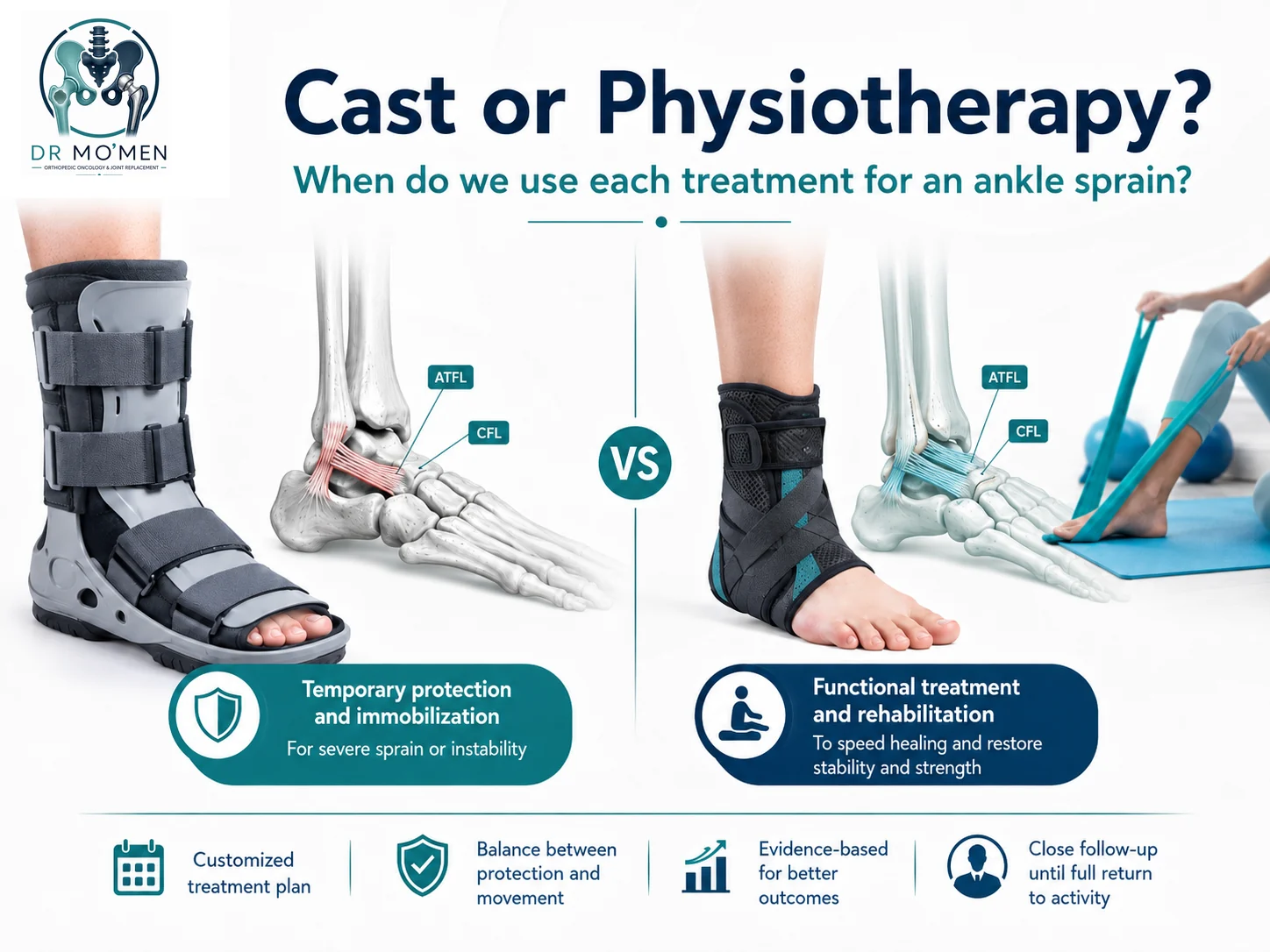
An ankle sprain is one of the most common lower-limb injuries. It may occur after stepping on an uneven surface, missing a stair, landing awkwardly, or changing direction during sport. The immediate question is often: “Does this injury need a cast, or is physiotherapy enough?”
The answer depends on the severity and stability of the injury. Most mild and moderate sprains are treated functionally with an ankle brace, progressive weight bearing, and rehabilitation exercises. Severe sprains may require a short period of rigid protection or immobilization, followed by early controlled movement and physiotherapy. Prolonged casting without a clear indication can contribute to stiffness, muscle weakness, and delayed return to activity.
What Happens in an Ankle Sprain?
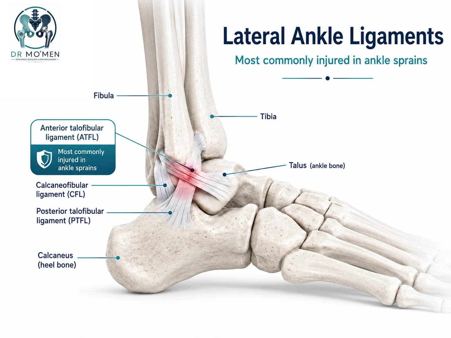
An ankle sprain occurs when the joint moves beyond its normal range and one or more stabilizing ligaments are stretched or torn. The most common mechanism is inversion, in which the sole turns inward and the outer ankle ligaments are injured. The anterior talofibular ligament (ATFL) is most frequently affected; a stronger injury may also involve the calcaneofibular ligament (CFL).
Grades of ankle sprain
- Grade I: Mild stretching or microscopic fiber injury, usually with limited swelling and the ability to walk, although walking may be painful.
- Grade II: Partial ligament tear, with more obvious swelling, bruising, tenderness, and difficulty walking.
- Grade III: Complete or near-complete ligament disruption, often with marked swelling, inability to bear weight, and possible mechanical instability.
The degree of swelling alone does not define the grade. A fracture, cartilage injury, tendon tear, or high ankle sprain can look similar to a routine ligament sprain. Treatment decisions therefore depend on the mechanism of injury, examination findings, walking ability, and imaging when indicated.
Symptoms and Warning Signs
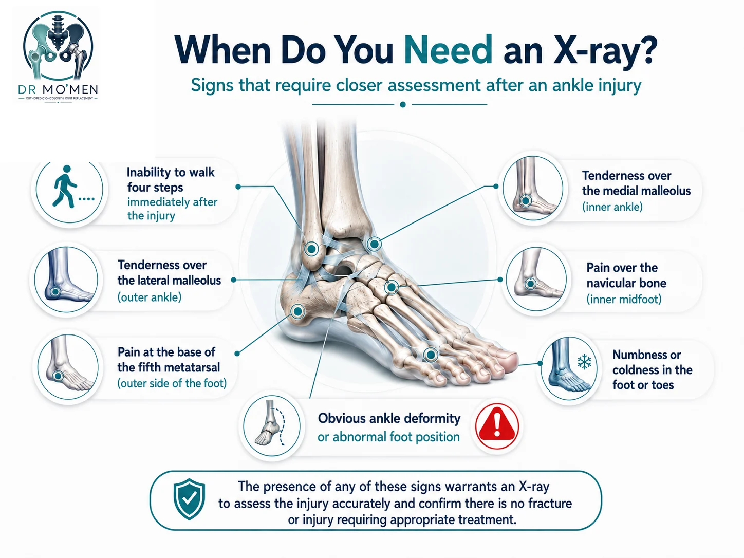
Typical symptoms may include lateral ankle pain, swelling, bruising, tenderness over the injured ligaments, and pain while standing or walking. Some findings require more urgent assessment because they may indicate a fracture, dislocation, neurovascular problem, or a more complex injury.
- Inability to take four steps immediately after the injury or during examination.
- Point tenderness directly over the medial or lateral malleolus.
- Pain at the base of the fifth metatarsal or over the navicular bone.
- Visible deformity or an abnormal position of the foot.
- Numbness, unusual coldness, marked color change, or weakness in the foot or toes.
- Pain above the ankle joint, which may suggest a syndesmotic or high ankle sprain.
Diagnosis and Imaging
Assessment starts with the mechanism of injury and a focused examination. The clinician checks the exact location of pain, bony tenderness, swelling, range of motion, ligament stability, walking ability, tendon function, circulation, and sensation. In a very painful acute injury, a repeat stability examination after several days may be more informative once swelling and guarding have decreased.
Does every ankle sprain need an X-ray?
No. Many uncomplicated sprains do not require routine radiographs. The Ottawa Ankle Rules help identify patients who are more likely to need an X-ray. Imaging is generally considered when the patient cannot take four steps or has focal bony tenderness at specified ankle or midfoot locations. Clinical judgment remains important, particularly in children, older adults, high-energy injuries, recurrent injuries, and patients with diabetes or impaired sensation.
MRI or CT may be considered when pain persists despite normal plain radiographs, or when there is concern for an osteochondral injury, tendon tear, occult fracture, syndesmotic injury, or another diagnosis that would change management.
When Are a Brace and Physiotherapy Preferred?
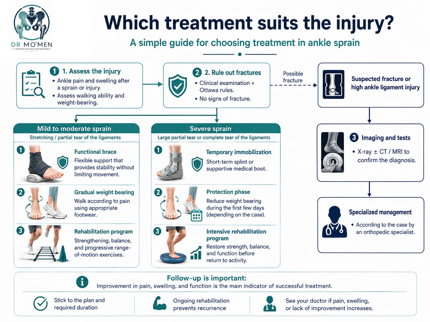
Functional treatment is generally preferred for most mild and moderate lateral ankle sprains once fracture and major instability have been excluded. Functional care aims to protect the healing tissues without unnecessarily preventing all movement.
- Use an appropriate external support, such as a semi-rigid brace, lace-up brace, or professionally applied supportive taping.
- Reduce painful activity during the first phase, but avoid complete inactivity when safe movement is possible.
- Use compression and elevation to help control swelling; ice may be used for short-term symptom relief when appropriate.
- Begin progressive weight bearing according to pain, often with crutches for a short period if needed.
- Start controlled range-of-motion exercises and advance gradually to strengthening, balance, and neuromuscular training.
- Use sport-specific rehabilitation before returning to running, jumping, or rapid changes of direction.
Can physiotherapy start immediately?
Some rehabilitation elements can begin early, but early treatment does not mean aggressive stretching, forceful manipulation, or painful exercise. During the first few days, the program may be limited to toe movement, gentle ankle motion within a comfortable range, swelling control, and protected walking. Resistance, balance drills, and impact activity are added progressively as pain, movement, and weight-bearing improve.
When Is a Cast or Temporary Immobilization Used?
Rigid protection may be appropriate when the injury is severe, very painful, unstable, or associated with an inability to bear weight. Immobilization may also be necessary when a fracture or dislocation is suspected, or when another associated injury requires stricter protection.
- A severe Grade III sprain with substantial pain, swelling, and functional instability.
- Pain that prevents safe weight bearing despite appropriate support.
- Suspected fracture, dislocation, osteochondral injury, or high ankle sprain.
- A patient who cannot reliably protect the injured ankle with a removable brace.
- An associated tendon, cartilage, or bone injury that requires a specific immobilization protocol.
In a severe isolated ligament sprain, immobilization is usually short rather than prolonged. Clinical guidelines allow a brief period of rigid support—often up to approximately 10 days in selected severe injuries—before transitioning to controlled movement, functional support, and rehabilitation.
Why Is Prolonged Casting Not Recommended for Every Sprain?
Unnecessary or prolonged immobilization can cause ankle stiffness, calf muscle weakness, altered walking mechanics, reduced proprioception, and delayed return to work or sport. Functional treatment allows protected movement and helps the patient regain motion and muscle control earlier. Evidence does not support one identical protocol for every injury, so severe sprains may still benefit from brief immobilization before rehabilitation.
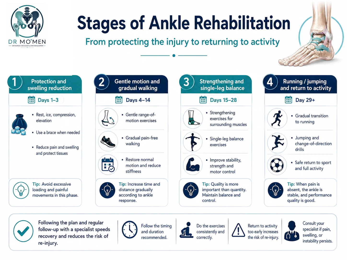
A Progressive Physiotherapy Plan
Phase 1: Protect the ankle and control symptoms
The early aim is to protect the injured tissues, limit excessive swelling, and maintain safe movement. A brace, compression, elevation, and temporary crutch use may be recommended. Weight bearing is adjusted to pain and clinical findings.
Phase 2: Restore motion and normal walking
Gentle ankle movement, calf stretching when appropriate, and gradual walking progression are introduced. The goal is to recover a comfortable range of motion and a normal gait without provoking increasing pain or swelling.
Phase 3: Strength, balance, and neuromuscular control
Exercises progress to resisted inversion, eversion, dorsiflexion, plantarflexion, calf raises, single-leg stance, and dynamic balance drills. This phase is important even when pain has improved because reduced proprioception and delayed muscular response can persist after swelling settles.
Phase 4: Return to running and sport
Return to sport should not be based only on the number of days since injury. The decision should consider pain, ankle range of motion, strength, confidence, balance, hopping, agility, change-of-direction ability, and the capacity to complete a full training session. The PAASS framework summarizes these domains: Pain, Ankle impairments, Athlete perception, Sensorimotor control, and Sport/functional performance.
When Should You Seek Specialist Assessment?
- You cannot stand or walk safely after the injury.
- Pain is directly over bone rather than mainly over the soft tissues.
- There is deformity, a suspected dislocation, or a clear cracking sensation with loss of function.
- The foot becomes numb, cold, pale, blue, or unusually weak.
- Pain extends above the ankle or along the lower leg.
- Swelling is severe, rapidly progressive, or associated with significant skin tension.
- Pain, limping, or swelling does not show a clear pattern of improvement.
- The ankle repeatedly gives way or sprains recur.
- You cannot return to work or sport despite an appropriate rehabilitation program.
The Role of Dr. Mo’men in Complex Ankle Injuries
An apparently simple sprain can sometimes conceal a small fracture, osteochondral lesion, tendon injury, syndesmotic disruption, or chronic ankle instability. Specialist assessment may help exclude fracture and associated injuries, determine injury stability, select a brace or short immobilization plan, develop staged rehabilitation, and investigate recurrent instability or persistent pain with targeted imaging when needed.
For appointments or review of X-rays and reports: 01021690693. Patients outside Egypt may request an initial online review of available imaging and reports to clarify the next steps before travel; a final treatment decision may still require an in-person clinical examination.

Medically reviewed by Dr. Mohammed Abdelmoemen Abuelhadid
Orthopedic oncology, joint replacement, sports injuries, and complex orthopedic care. About the doctor · ORCID profile
How this article was reviewed
This patient-education article was reviewed for clinical accuracy, patient safety, and decision-making clarity by Dr. Mohammed Abdelmoemen Abuelhadid. It is intended to support, not replace, clinical examination and review of the actual imaging and reports.
Authoritative medical sources
Request an appointment or imaging review
For a clinic appointment or preliminary review of available scans and reports, contact the clinic.