
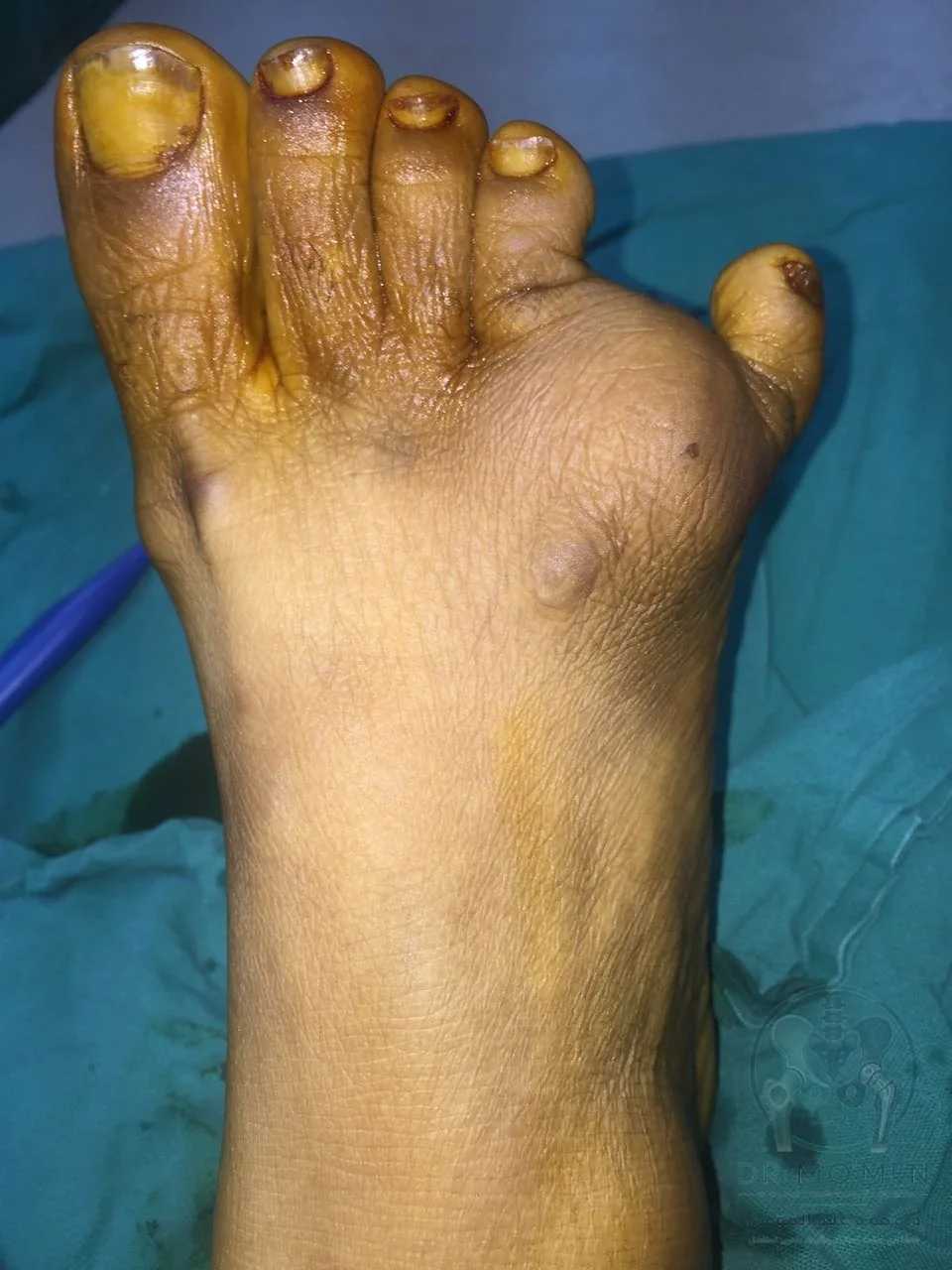
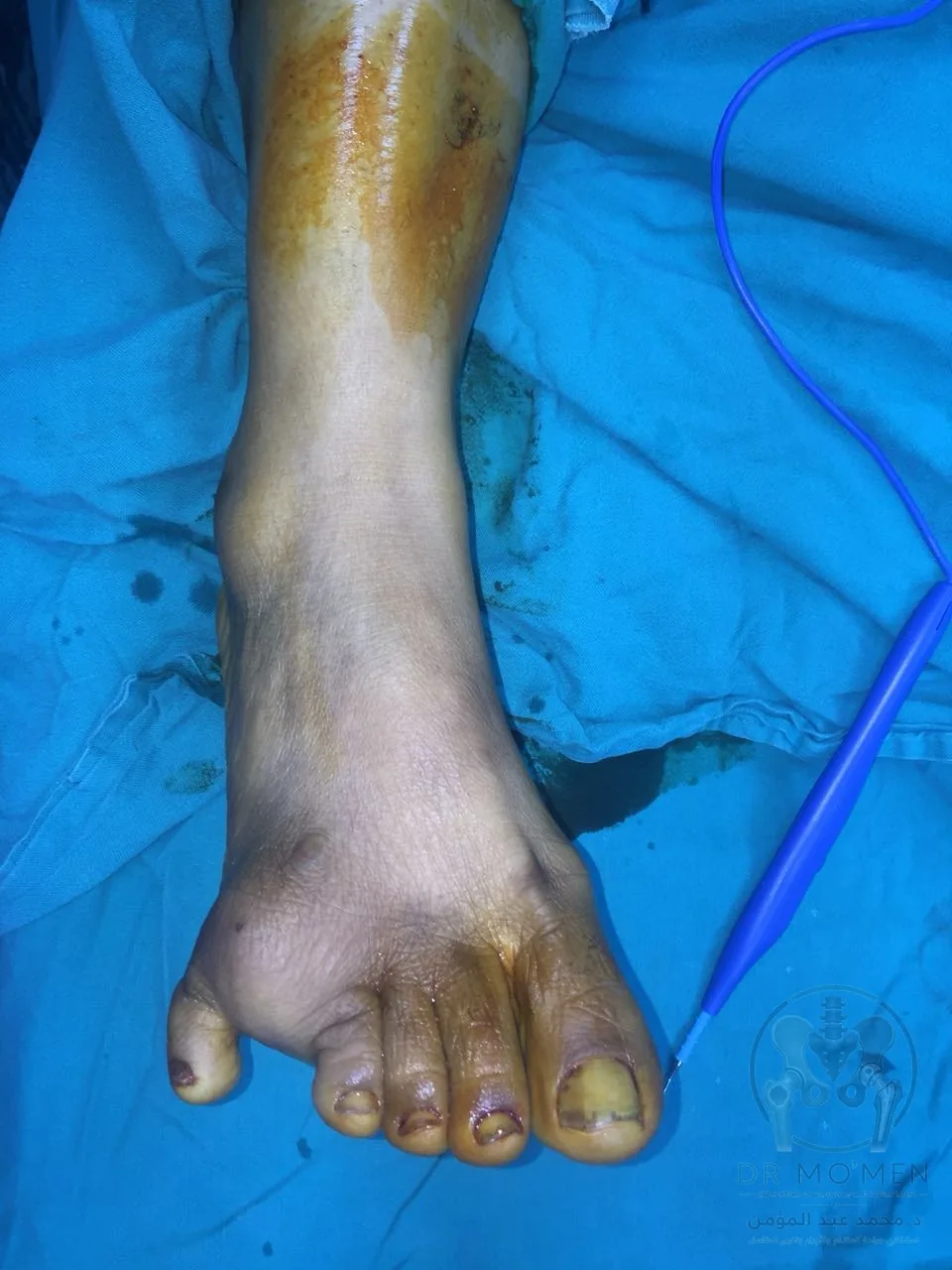
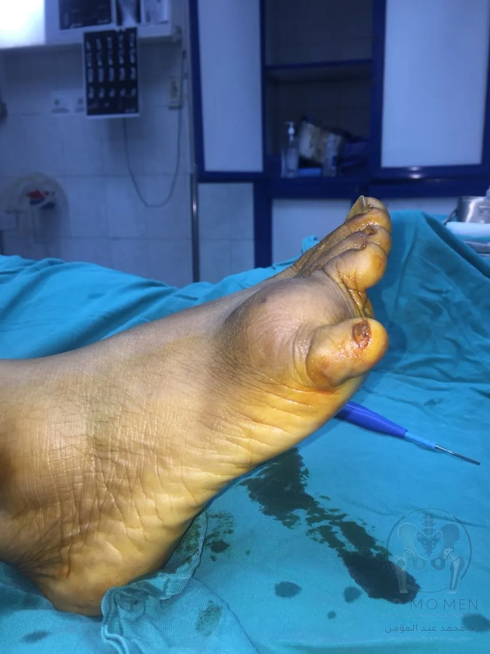
A 40-year-old patient presented with a foot mass that had been present for years, with a clear recent increase in size over the last three months. The mass was located at the lateral/anterior aspect of the foot near the fifth toe region and adjacent soft tissues.
The case in one minute
This case illustrates excision of a large foot mass with planning to preserve the small vessels supplying the toes. Safe treatment depends on the diagnosis, the mass relationship to nerves and vessels, and whether adequate excision can be achieved without unnecessary functional harm.
- Clinical category
- Foot soft-tissue tumor
- Anatomical site
- Foot and digital vascular structures
- Decision illustrated
- Planned excision with preservation of blood supply and critical tissues
- Published reporting scope
- The images document the procedure; detailed histology and follow-up duration are not published in this educational version.
Related pages
Case summary
- Age: 40 years.
- Presentation: foot mass present for years with rapid recent enlargement over three months.
- Location: lateral/anterior foot near the fifth toe and adjacent soft tissues.
- Imaging: X-ray showed a large soft-tissue mass with clear calcification/ossification. The exact diagnosis cannot be confirmed from images alone.
- Procedure: surgical excision of the mass while preserving the digital vascular supply as much as possible.
- Functional objective: remove the mass while maintaining toe perfusion and vital foot tissues whenever safely feasible.
Important medical note
This educational page is based on the clinical images and case information provided. It is not a final pathology report. The definitive diagnosis depends on histopathological examination of the excised specimen.
Key surgical planning points
- Planning the surgical approach to allow safe access to the mass without compromising digital perfusion.
- Gradual dissection around the tumor while protecting the small vessels supplying the toes.
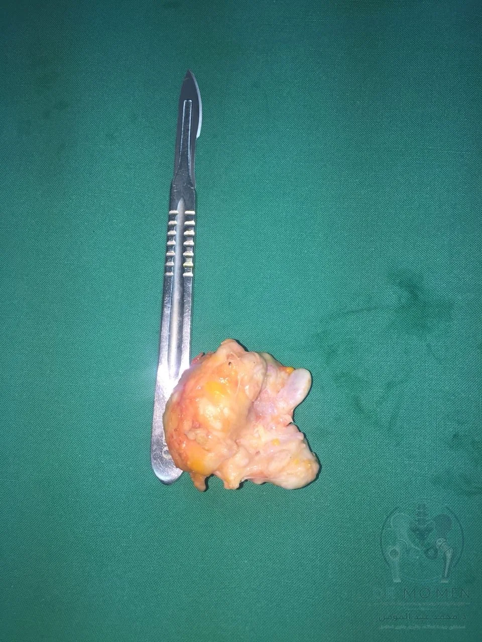
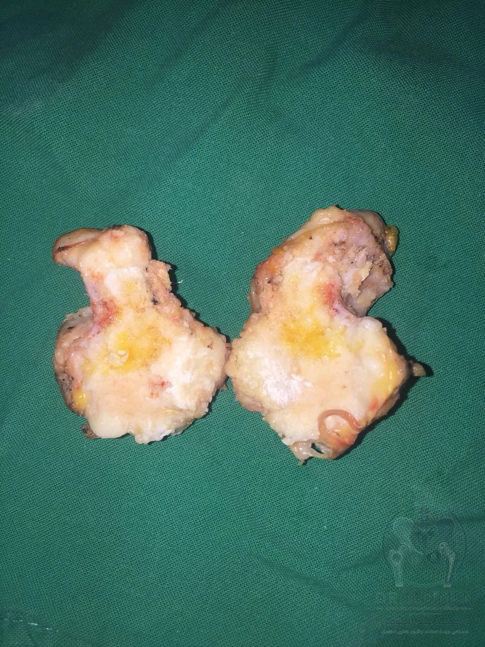
- Excision of the mass as completely as safely possible and sending the specimen for pathology.
- Inspection of the tumor bed after excision to confirm tissue viability and absence of visible vascular injury or bleeding.
Image sequence
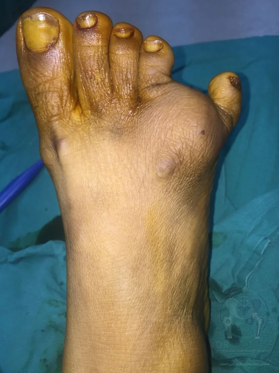
The following anonymized educational images show the preoperative appearance, X-ray findings, surgical dissection, tumor bed after excision, and the removed specimen.
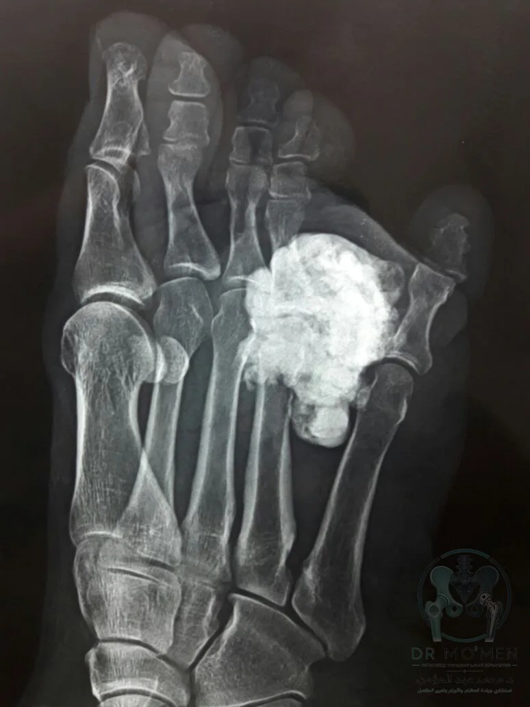
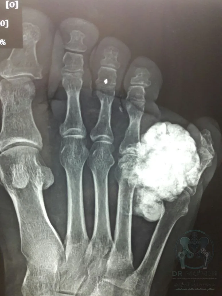
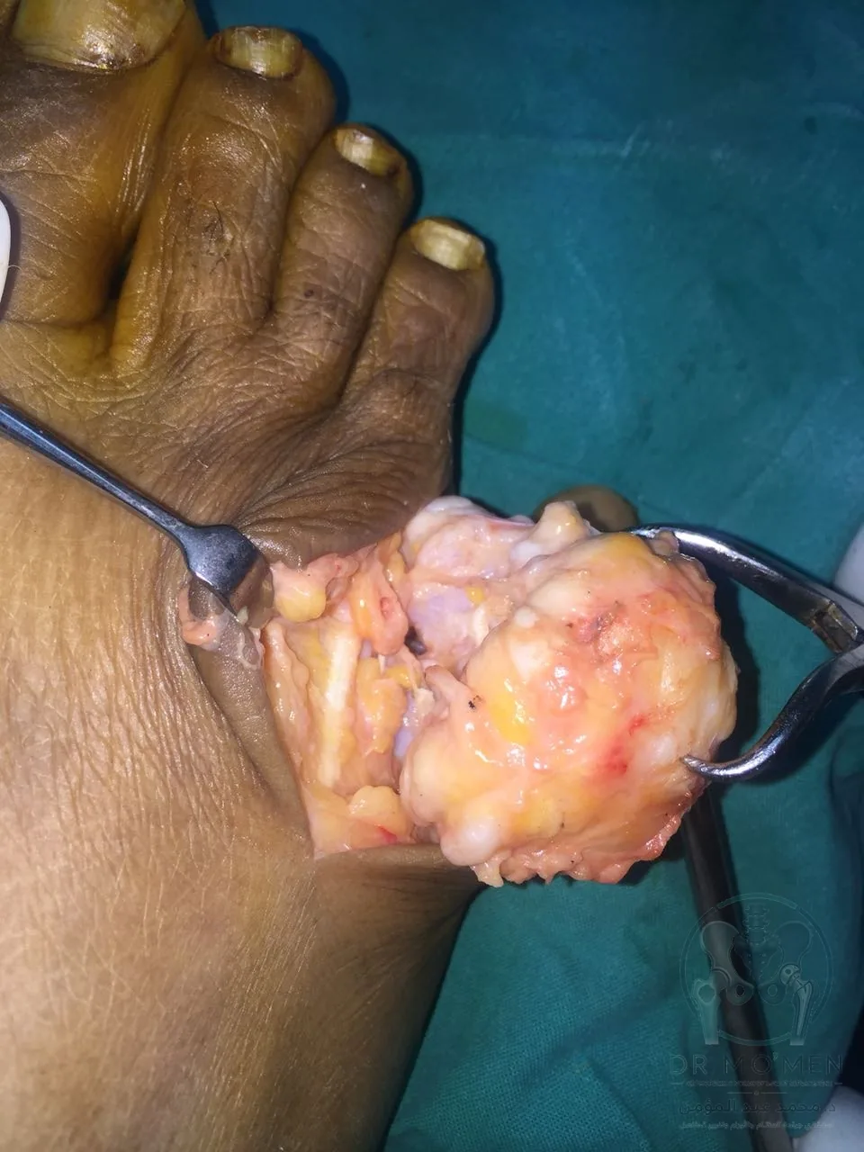
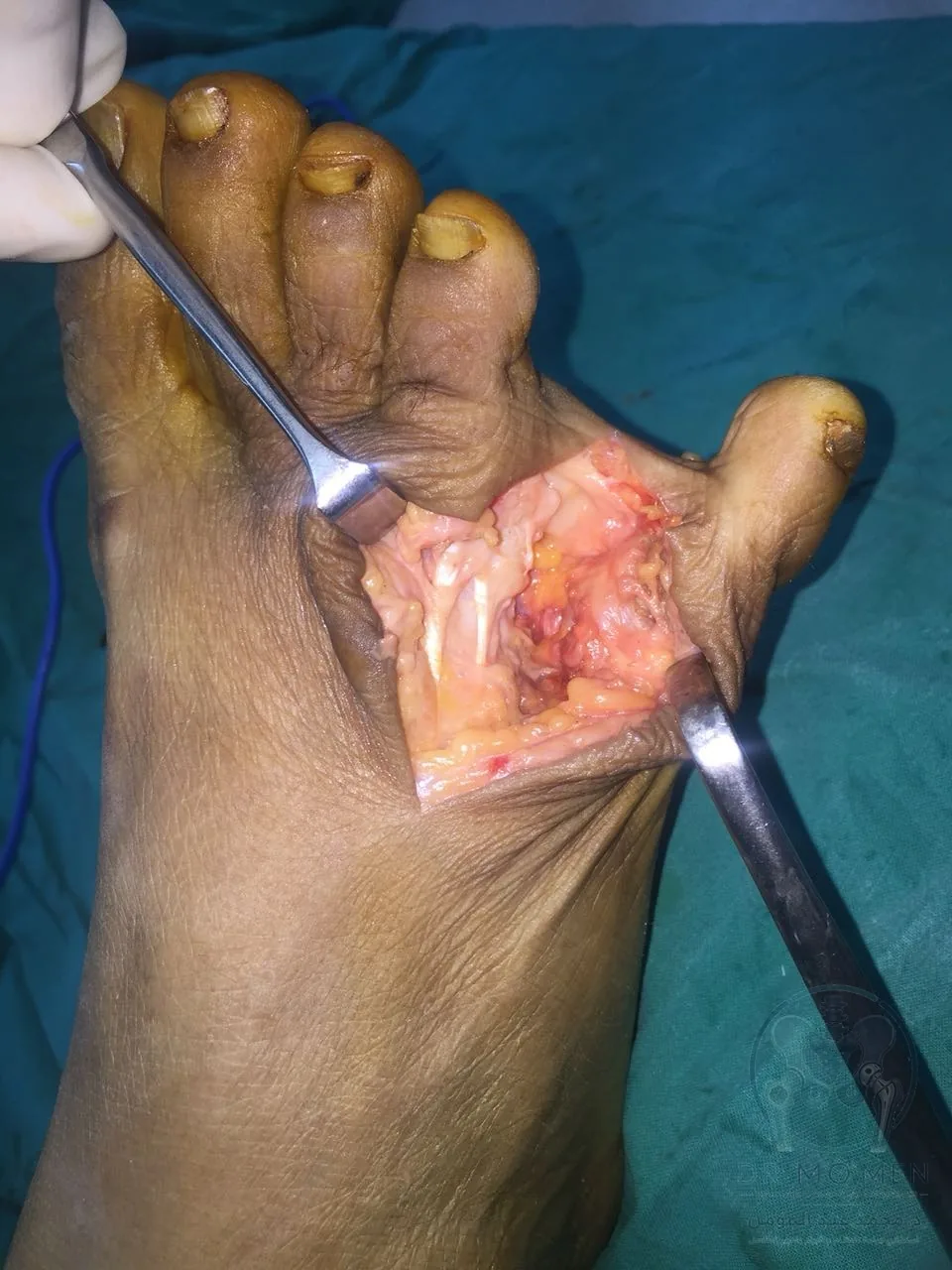
Learning points
- A long-standing foot mass with recent rapid enlargement needs careful clinical and radiological evaluation.
- Calcification or ossification inside a soft-tissue mass should not be treated as a final diagnosis by imaging alone.
- Foot tumor excision requires precise knowledge of local anatomy, especially digital vessels and surrounding soft tissues.
- Follow-up depends on pathology, wound healing, toe perfusion, and foot function.
Medical disclaimer
This case is shared for patient education. It does not replace direct clinical examination, imaging review, or pathology confirmation. Treatment plans vary according to each patient’s diagnosis, anatomy, pathology result, and functional requirements.
Terms used in this case
- Soft-tissue tumor
- A mass arising from muscle, fat, tendon, or surrounding tissues; it may be benign or malignant.
- Digital vessels
- The small arteries and veins supplying the toes.
- Surgical margin
- The tissue boundary around a mass that is assessed when judging adequacy of excision.