
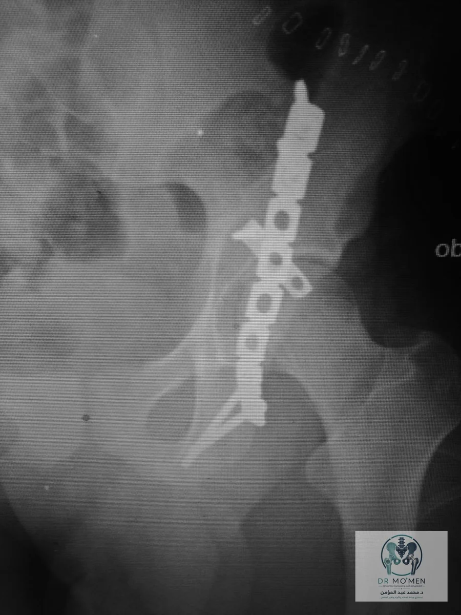
The patient presented to the emergency department with an acute traumatic hip dislocation. Urgent reduction was performed, followed by CT imaging to define the associated acetabular injury.
The case in one minute
This case presents a hip dislocation with a posterior-wall acetabular fracture. After urgent reduction and CT assessment, a spring plate supported a small fragment that was unsuitable for direct screw fixation.
- Clinical category
- Posterior-wall acetabular fracture with hip dislocation
- Anatomical site
- Hip and acetabulum
- Decision illustrated
- Urgent reduction followed by spring-plate fixation based on fragment pattern and hip stability
- Published reporting scope
- Post-fixation images are shown; long-term functional outcome and follow-up duration are not published.
Related pages
Initial Assessment and Emergency Management
The initial radiograph demonstrated hip dislocation. After urgent reduction in the emergency department, CT was obtained to assess associated acetabular injury that might not be fully characterized on plain radiographs. A different example of complex hip trauma can be seen in the left femoral neck fracture case treated with cementless total hip arthroplasty.
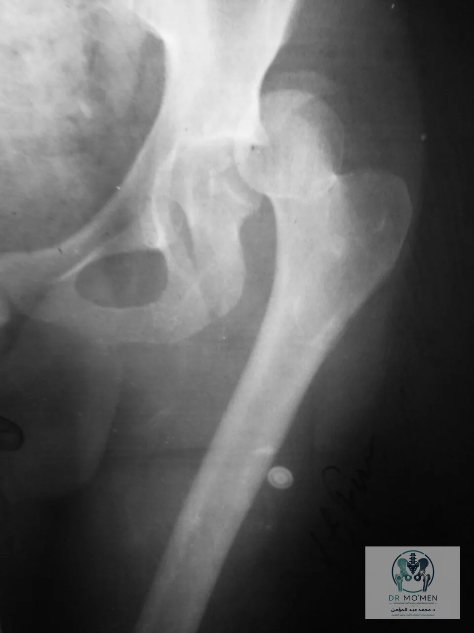
CT Diagnosis
Post-reduction CT demonstrated a posterior wall acetabular fracture with a very small posterior wall fragment. The fragment size made conventional direct screw fixation alone less suitable because there was limited bone available for secure screw purchase.
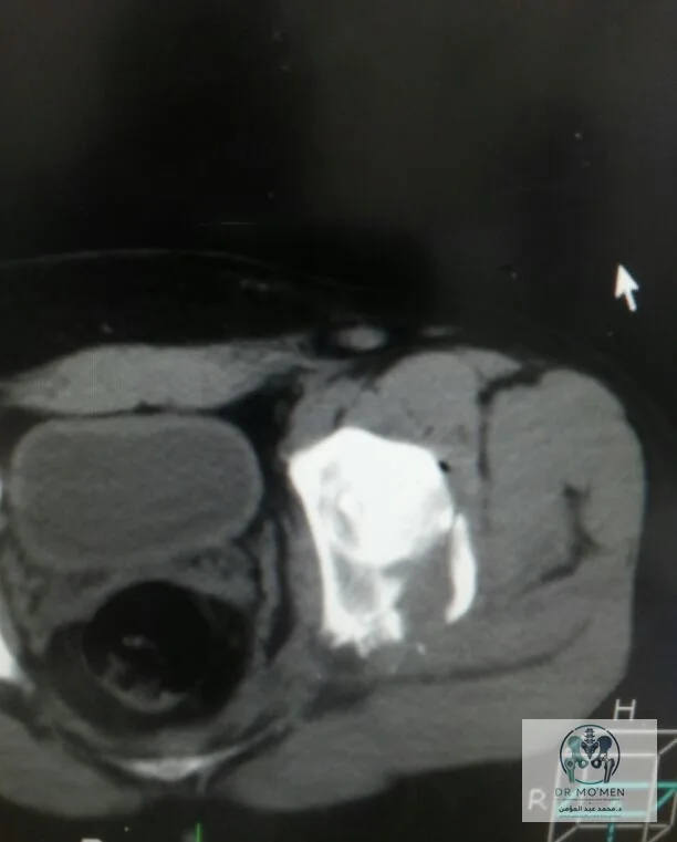
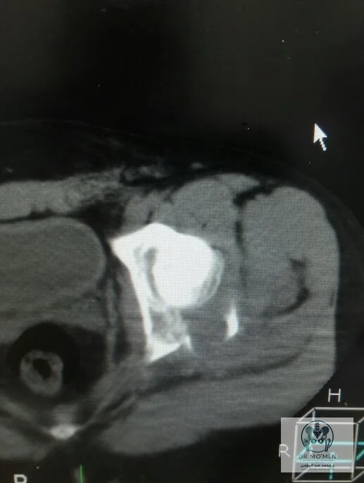
Why Use a Spring Plate?
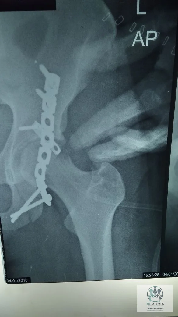
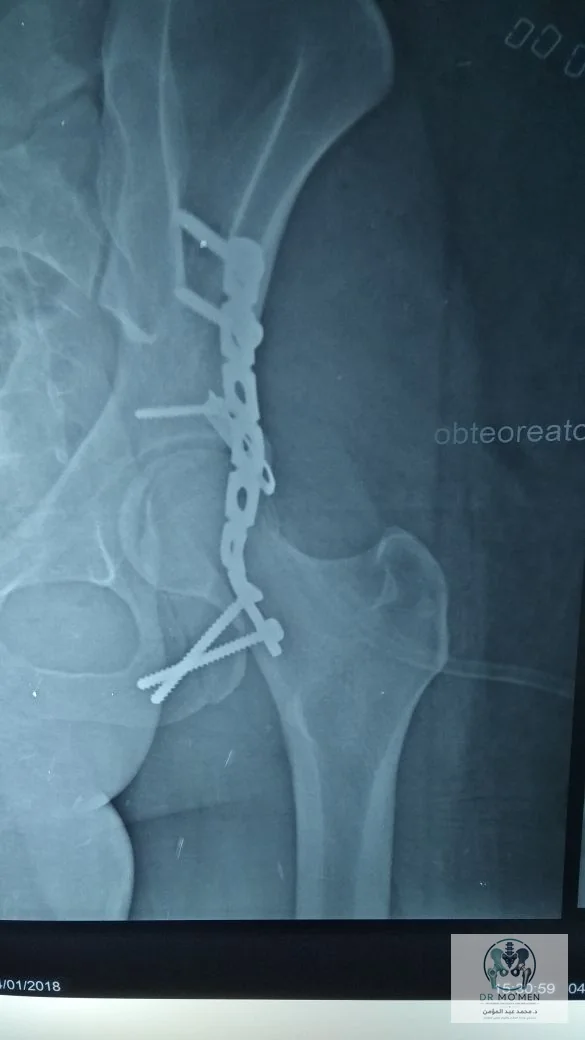
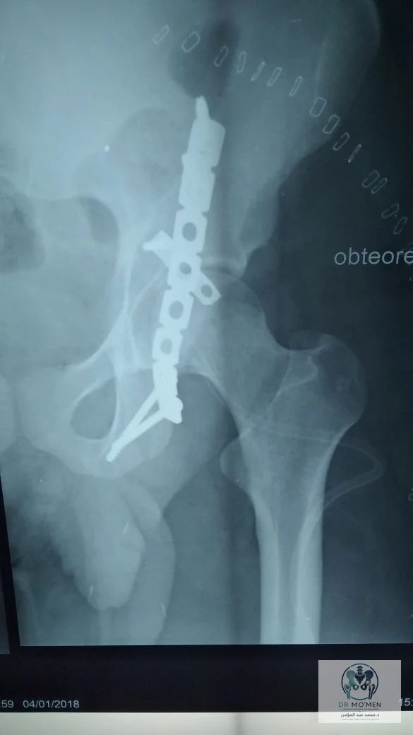
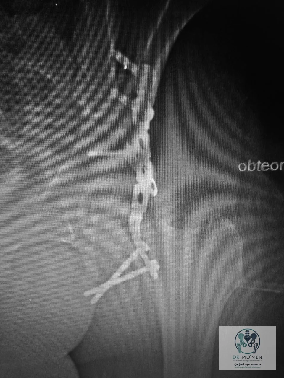
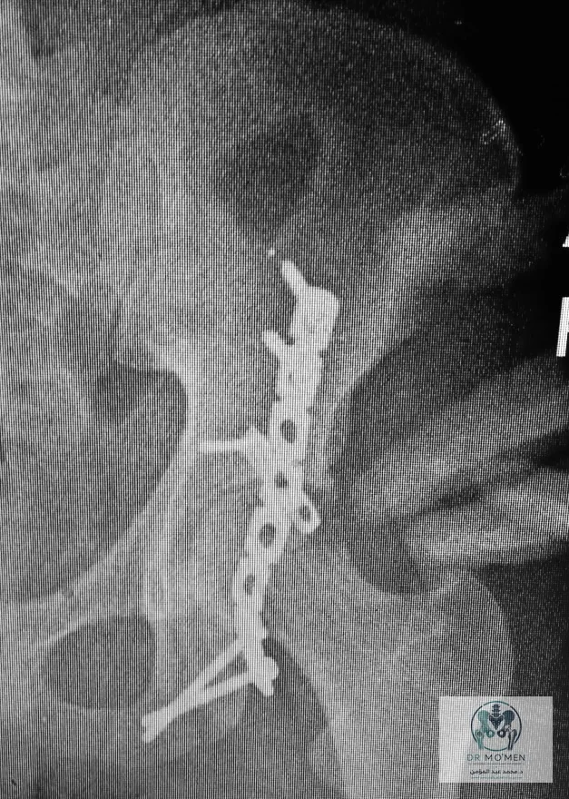
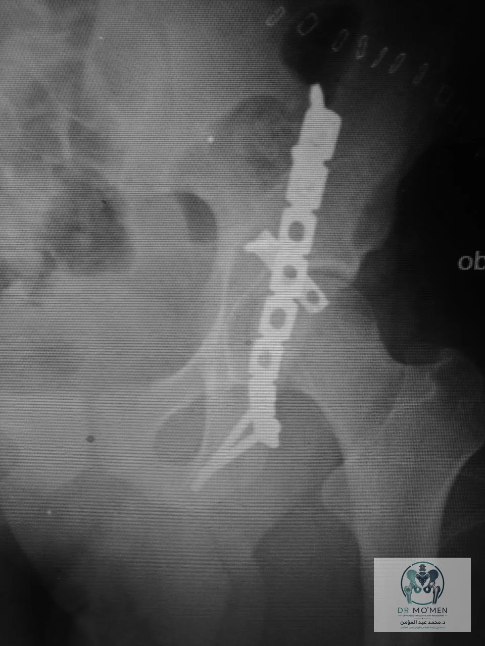
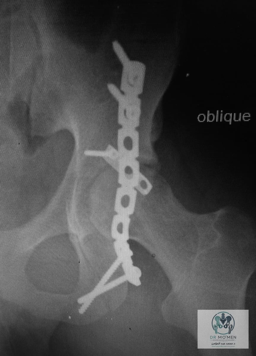
The objective was to support the very small posterior wall fragment without relying on a direct screw through limited bone. The spring plate acts as a fragment-supporting device and is reinforced by a buttress plate as part of the fixation construct.
Surgical Treatment
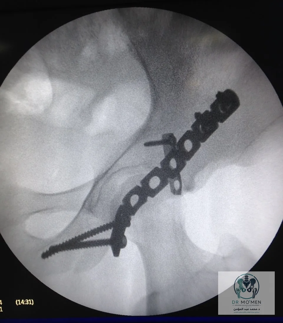
Open reduction and internal fixation of the posterior wall acetabular fracture was performed. Because the fragment was very small, a spring plate was used to support it, with a buttress plate reinforcing the construct.
- Reduction of the posterior wall fragment and restoration of posterior acetabular support.
- Use of a spring plate to capture and support the small osseous fragment.
- Reinforcement of the fixation with a buttress plate.
- Intraoperative imaging to assess implant position and the relationship of the fixation construct to the hip joint.
What Is the Spring Plate Technique?
The spring plate technique is useful in selected posterior wall acetabular fractures when the fragment is small or thin and cannot be safely secured with a conventional lag screw alone. The spring plate provides elastic fragment support, while the buttress plate adds stability to the overall construct.
Postoperative Imaging
Postoperative images demonstrate the reduced hip and the fixation construct from multiple projections. These views assess plate position, spring plate support, and the relationship of the implants to the joint.
Clinical Lesson
In a hip dislocation associated with a posterior wall acetabular fracture, management does not end with reduction of the dislocation. Post-reduction CT helps define fracture morphology, fragment size, and fixation strategy. Very small posterior wall fragments may require a specialized solution such as spring plate fixation rather than conventional screws alone.
Do you have an acetabular fracture or imaging that needs review?
Scans and reports can be sent by WhatsApp to determine whether an in-person assessment or an initial record review is more appropriate.
Book a clinic visit