
A 58-year-old woman presented with painful, progressively enlarging swelling around the distal femur and knee. Her symptoms were initially attributed to knee osteoarthritis, but the rapid progression of pain and swelling was atypical for uncomplicated degenerative disease.
The case in one minute
This case illustrates why progressive swelling and pain around the knee should not be assumed to be osteoarthritis when symptoms are rapidly worsening or disproportionate to the radiographic degenerative changes.
- Clinical category
- Orthopedic oncology
- Anatomical site
- Distal femur and knee region
- Final diagnosis
- Chondrosarcoma of the distal femur
- Treatment
- Wide distal femoral resection and modular distal femoral endoprosthesis
- Outcome
- Limb preserved with very good postoperative function
Case presentation
A 58-year-old woman presented with a painful, progressively enlarging swelling around the distal femur and knee. Her symptoms had initially been attributed to knee osteoarthritis and treated accordingly. The rapid progression of pain and swelling, however, was atypical for uncomplicated degenerative disease.
Imaging and diagnosis
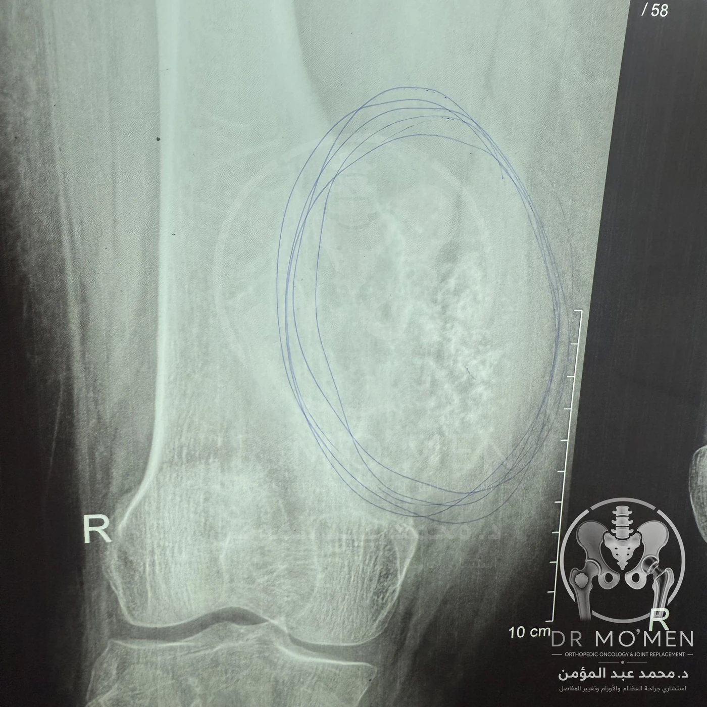
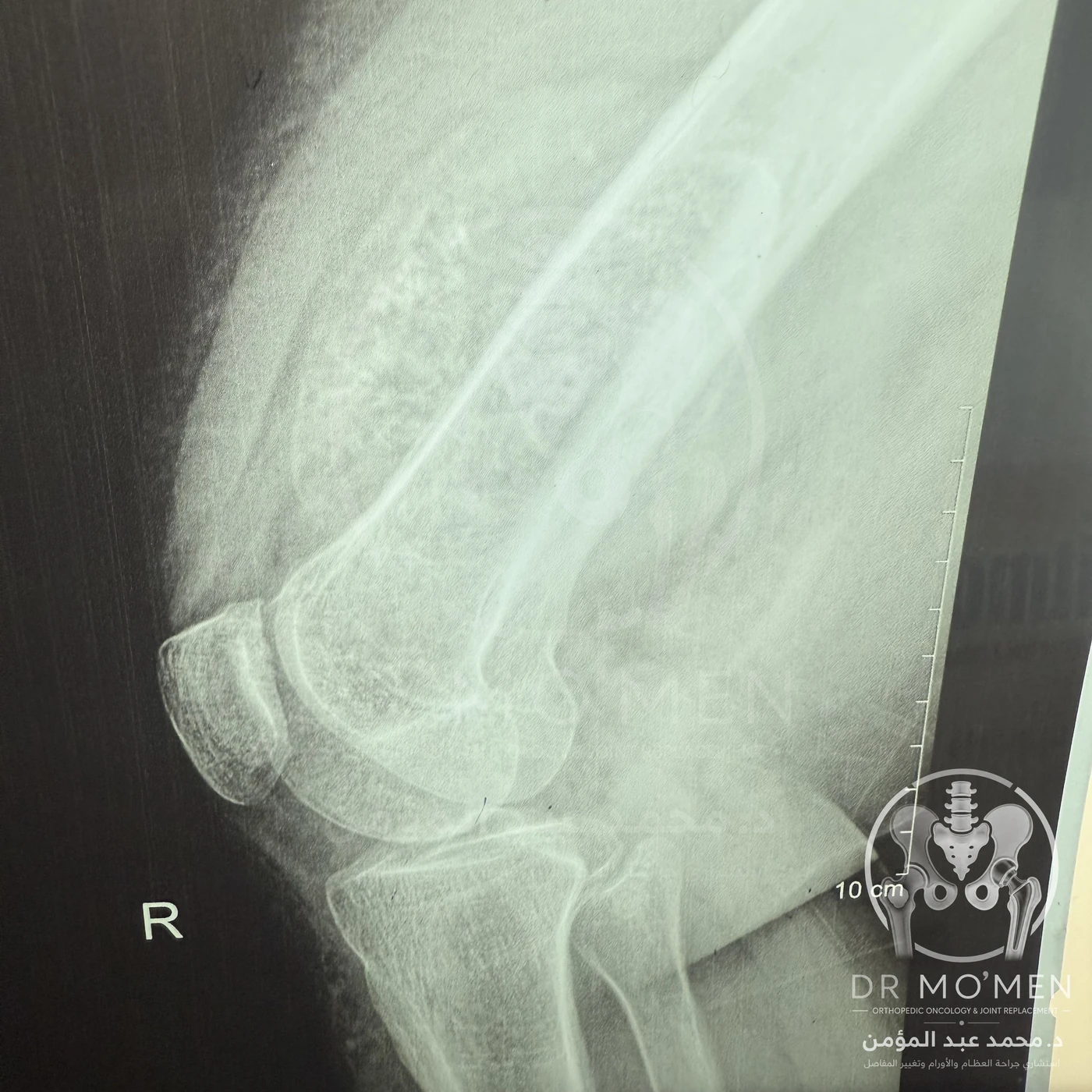
Plain radiographs demonstrated an aggressive osteolytic lesion of the distal femur with cortical destruction and internal mineralization, raising suspicion for a cartilage-forming malignant bone tumor. Further oncological imaging defined the local extent of the lesion. A planned biopsy confirmed chondrosarcoma, after which the patient was referred for specialized orthopedic oncology management.
For a related educational discussion, read about bone biopsy planning and orthopedic oncology care.
Definitive surgical management
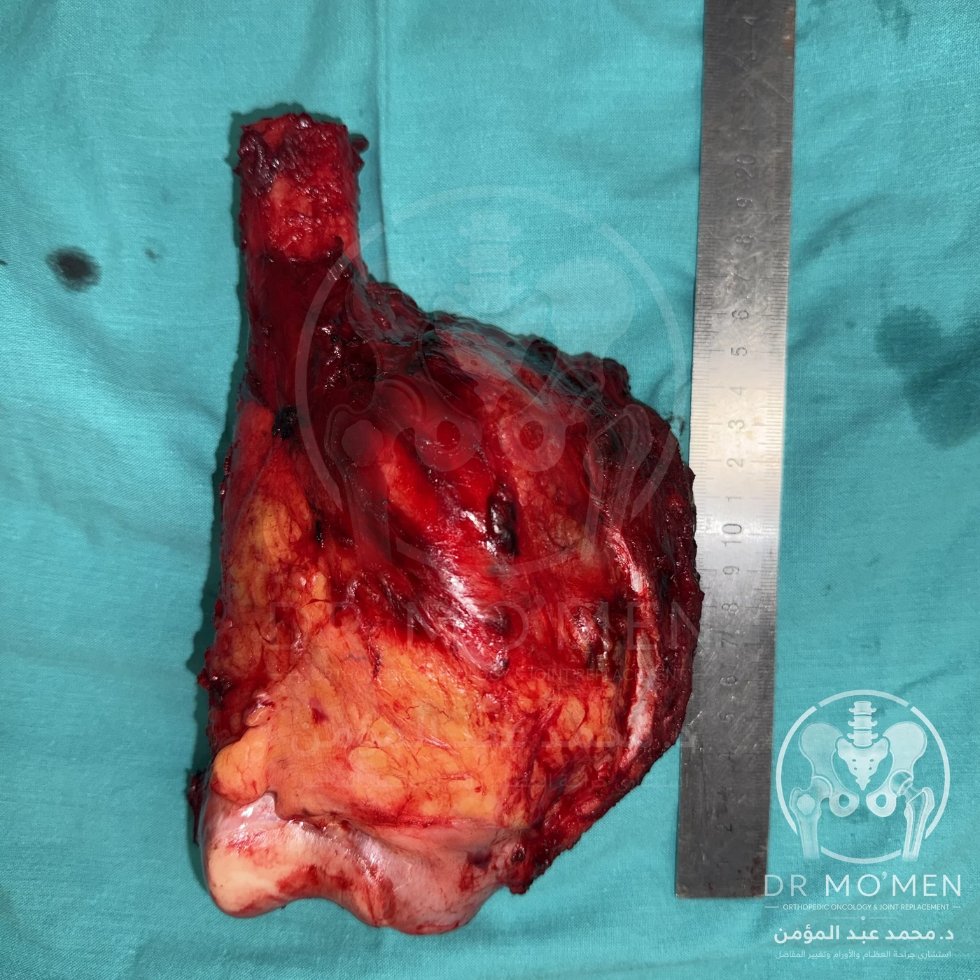
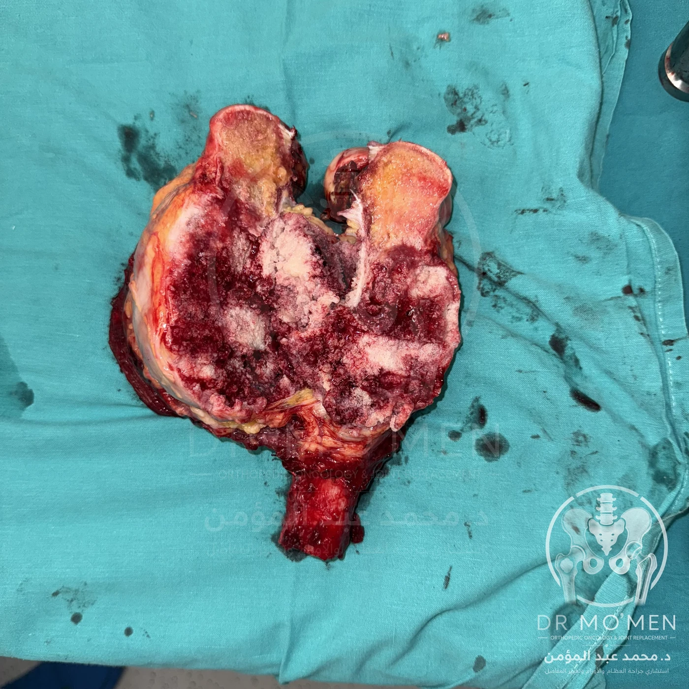
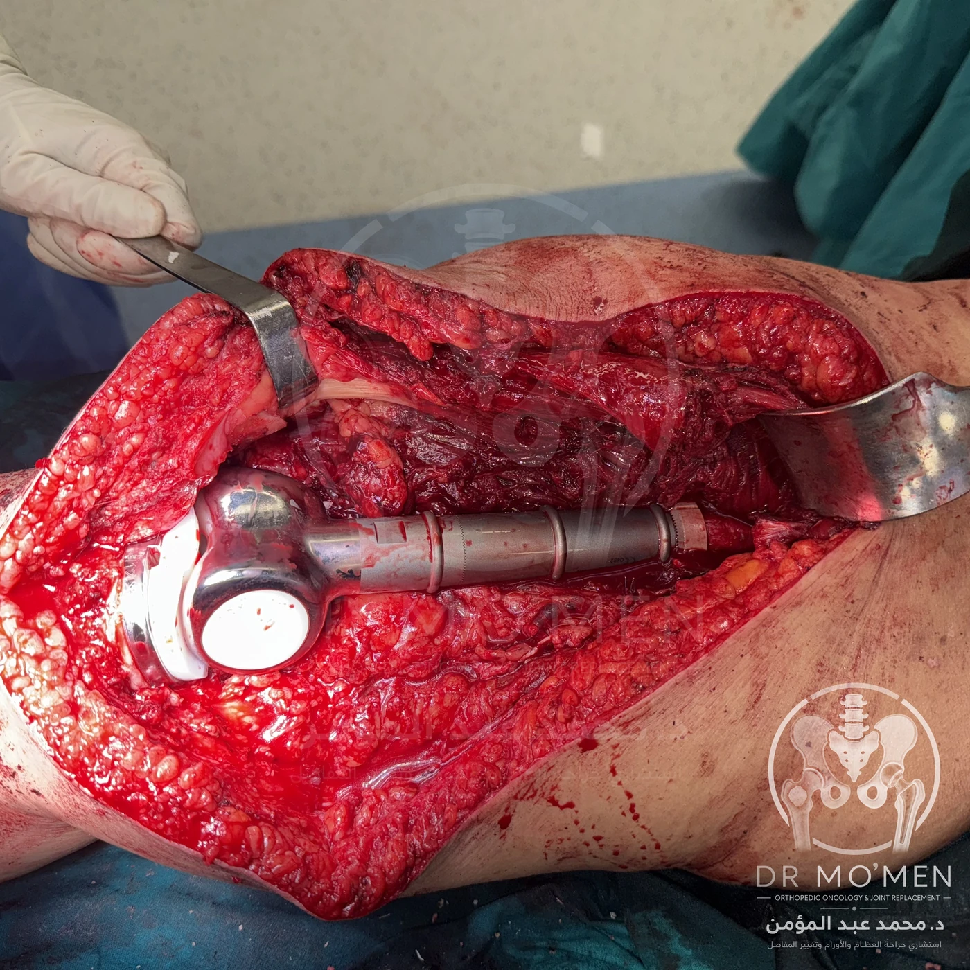
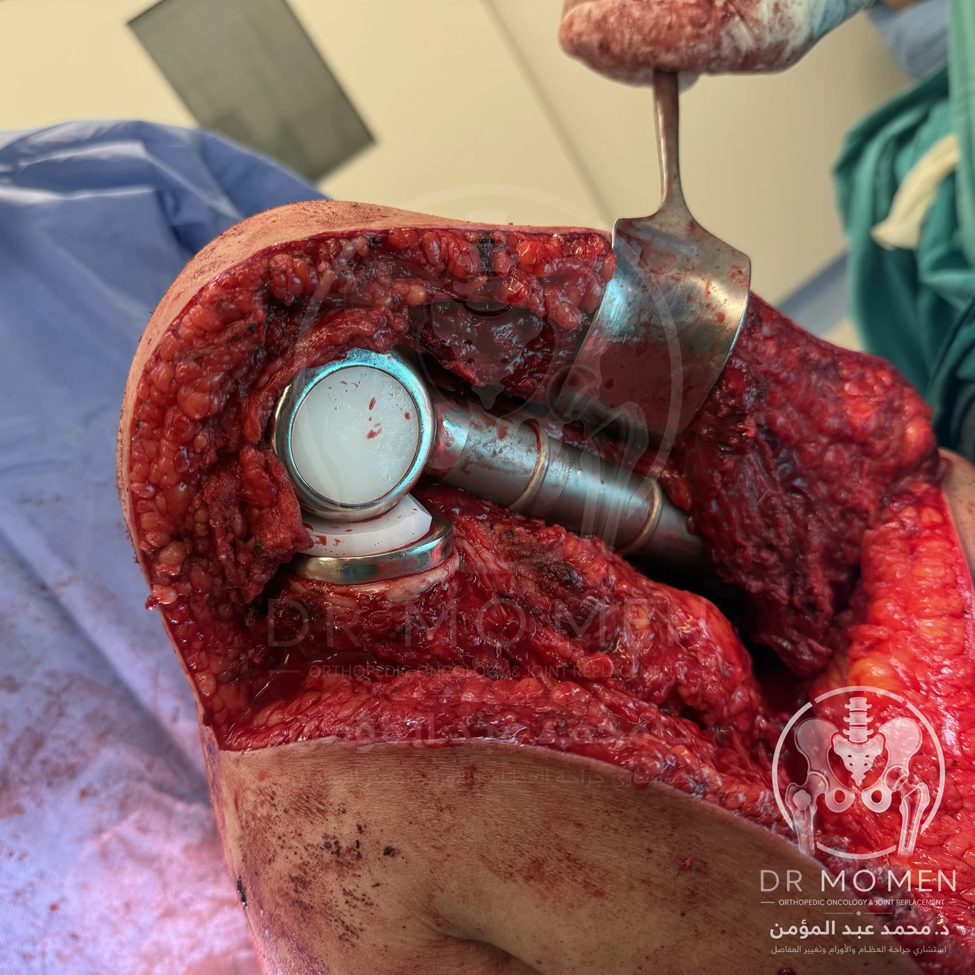
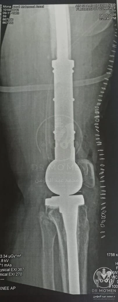
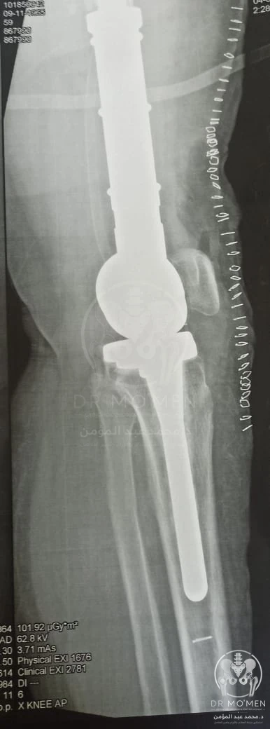
The patient underwent wide oncological resection of the involved distal femur with limb-salvage reconstruction using a modular distal femoral endoprosthesis. The objective was to achieve adequate oncological margins while preserving the limb and restoring knee function.
Systemic therapy and radiotherapy
For localized, resectable conventional chondrosarcoma, complete surgical excision with adequate margins is the mainstay of treatment. Conventional chondrosarcoma is relatively resistant to standard chemotherapy and radiotherapy; therefore, these modalities are not routinely required after adequate resection unless specific histological, anatomical, margin-related, or disease-stage considerations indicate otherwise.
Outcome
Following surgery and rehabilitation, the patient achieved a very good functional outcome with successful limb preservation. Long-term oncological and prosthetic surveillance remains essential.
Educational message
Progressive pain, a rapidly enlarging swelling, night or rest pain, or symptoms disproportionate to apparent osteoarthritis should prompt further investigation. Degenerative changes on imaging do not exclude an underlying bone tumor.
Clinical and operative image sequence
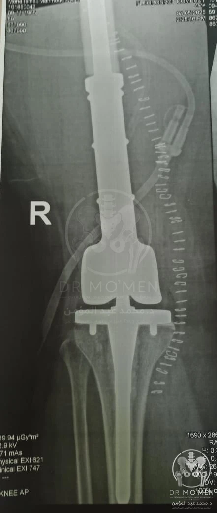
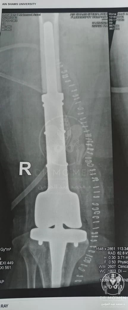
The images are arranged chronologically from preoperative radiographs, through wide resection and gross tumor specimen, to endoprosthetic reconstruction and postoperative radiographs.
Case learning points
Can knee osteoarthritis hide a bone tumor?
Degenerative changes may coexist with another pathology. Rapidly progressive swelling, rest pain, night pain, or symptoms disproportionate to typical osteoarthritis should prompt further investigation.
Why was wide resection required?
Chondrosarcoma is a malignant cartilage-forming bone tumor. When localized and resectable, treatment usually depends on complete surgical excision with appropriate margins.
Why use a distal femoral endoprosthesis?
After wide distal femoral resection, a modular tumor endoprosthesis can reconstruct the knee region and support limb preservation when oncologically appropriate.
Does chondrosarcoma always need chemotherapy or radiotherapy?
Conventional chondrosarcoma is relatively resistant to standard chemotherapy and radiotherapy. Management depends on histology, grade, margins, site and disease stage.