
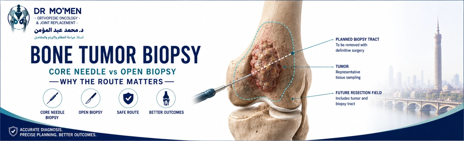
When an X-ray or MRI shows a suspicious bone tumor, patients often assume the next step is simply: “Take a biopsy and find out what it is.” The principle is correct—but in orthopedic oncology, how the biopsy is performed can matter almost as much as obtaining the tissue itself.
A biopsy is not an isolated diagnostic procedure. It can directly influence the definitive tumor operation. The biopsy route may need to be removed together with the tumor during later surgery. If a needle or incision crosses unnecessary muscles, joints, neurovascular structures or anatomical compartments, those tissues may potentially become part of the definitive oncological surgical field.
This is why biopsy planning for a suspected primary bone malignancy should ideally be coordinated with the team that would perform definitive tumor surgery.
The key question
Which biopsy can obtain sufficient representative tissue through the safest oncologically planned route without compromising definitive treatment?
Why Does a Bone Tumor Need Biopsy?
Imaging can provide important clues about a bone lesion, but different conditions can have overlapping appearances. A destructive lesion may represent:
- primary malignant bone tumor
- metastatic bone disease
- lymphoma
- myeloma
- locally aggressive benign tumor
- infection
- other less common conditions
These diagnoses can require completely different treatment. For example, osteosarcoma, Ewing sarcoma, chondrosarcoma, giant cell tumor and metastatic carcinoma may require very different combinations of surgery, chemotherapy, radiotherapy or systemic treatment. Biopsy provides tissue for pathological diagnosis before irreversible treatment decisions are made.
Does Every Bone Lesion Need a Biopsy?
No. Some bone lesions have sufficiently characteristic imaging and clinical features that biopsy may not be necessary. Others may be observed with follow-up imaging. Biopsy becomes particularly relevant when:
- the diagnosis remains uncertain
- imaging appears aggressive
- malignancy is suspected
- treatment would differ substantially depending on pathology
- major surgery is being considered
- systemic cancer treatment depends on tissue diagnosis
The decision should therefore be based on the complete clinical and radiological picture.
Imaging Should Usually Come Before Biopsy
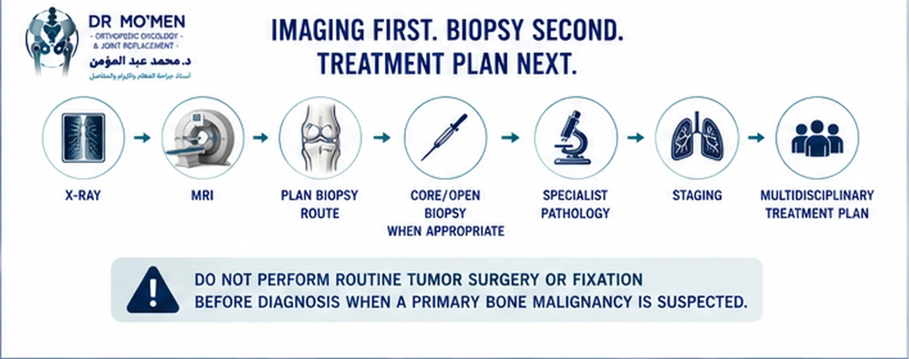
For a suspected primary bone tumor, the local imaging workup should ideally be completed before biopsy. MRI is particularly important because it can define:
- intramedullary tumor extent
- cortical destruction
- soft-tissue extension
- relation to the joint
- nearby muscles
- major nerves
- major blood vessels
- potential surgical resection levels
Biopsy can create bleeding, edema, hematoma, inflammatory change and tissue distortion. These changes may make subsequent imaging more difficult to interpret. When clinically feasible, obtaining appropriate imaging first provides the clearest map for biopsy and definitive treatment planning.
What Is a Core Needle Biopsy?
A core needle biopsy uses a relatively wide hollow needle to obtain cylinders or cores of tissue. It is different from a very fine needle used only to aspirate cells. Core biopsy aims to preserve enough tissue architecture for the pathologist to evaluate tumor morphology, cellular pattern, matrix production, immunohistochemistry, grading where possible, and molecular studies when needed.
Several cores may be obtained from representative regions of the lesion.
How Is a Core Needle Biopsy Performed?
Depending on tumor location, the biopsy may be guided using ultrasound, CT, fluoroscopy, or another imaging modality. Image guidance helps the team enter the correct part of the lesion, avoid major vessels and nerves, avoid necrotic areas when possible, limit unnecessary tissue contamination, and target viable representative tumor.
Is Core Needle Biopsy Usually Preferred?
For many musculoskeletal tumors, a properly planned image-guided core biopsy is commonly used as the initial tissue-sampling method when it can safely obtain adequate diagnostic tissue. Open biopsy remains valuable when core biopsy is technically inappropriate, unsafe, or nondiagnostic. The appropriate method depends on the lesion, anatomy and tissue requirements.
What Are the Advantages of Core Needle Biopsy?
- smaller skin opening
- less soft-tissue disruption
- lower procedural burden
- image-guided targeting
- ability to obtain multiple samples
- easier planning of a narrow biopsy tract
- shorter recovery than open biopsy in many cases
These advantages only apply when the biopsy is well planned and diagnostically adequate. A poorly placed needle biopsy can still compromise definitive surgery.
Can Core Needle Biopsy Give the Wrong Diagnosis?
No biopsy method is perfect. A core biopsy can occasionally be insufficient, nonrepresentative, predominantly necrotic, technically difficult to interpret, or unable to establish the exact subtype. This is particularly important in heterogeneous tumors, where different parts of the lesion may have different pathological appearances.
If you already have a biopsy result, see how to understand a bone tumor pathology report and what to do when pathology and imaging do not agree.
If imaging and pathology do not fit together, the case should be reassessed rather than forcing the diagnosis.
What Is an Open or Incisional Biopsy?
An incisional biopsy involves making a surgical incision and removing a portion of the tumor for diagnosis. The entire tumor is not removed. It can provide a larger tissue specimen than a needle biopsy, but it also creates a larger surgical field. The incision therefore needs careful oncological planning.
When Might Open Biopsy Be Needed?
- core biopsy is nondiagnostic
- adequate tissue cannot safely be obtained using a needle
- lesion location creates technical limitations
- substantial additional tissue is needed for diagnosis
- multidisciplinary review determines that open sampling is preferable
Open biopsy should not simply be performed because it appears easier to obtain a large sample.
Is Open Biopsy More Accurate Than Core Needle Biopsy?
Not necessarily. A larger specimen does not automatically mean a better biopsy. Diagnostic quality depends on correct lesion targeting, representative viable tissue, appropriate sample handling, experienced pathology, and clinical-radiological correlation. A carefully targeted core biopsy can be highly informative, while an open biopsy from the wrong location may still be misleading and may create unnecessary contamination.
Core Needle vs Open Biopsy: Which Is Better?
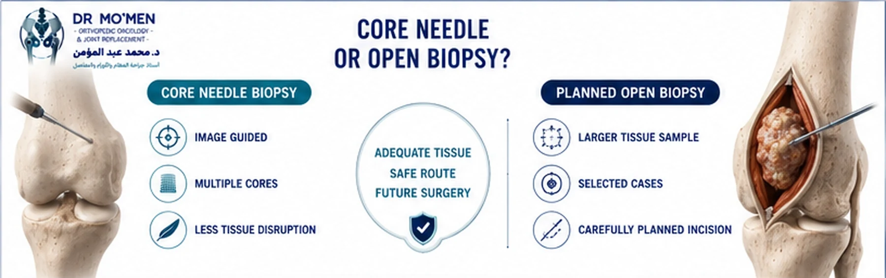
Core needle biopsy is often suitable when:
- there is a safe needle pathway
- representative tissue can be obtained
- adequate material can be collected
- image guidance allows accurate targeting
Open biopsy may be appropriate when:
- needle sampling remains nondiagnostic
- the lesion cannot be sampled safely by core needle
- tissue requirements cannot be adequately met
- tumor anatomy makes open sampling preferable
The decision should be individualized rather than based on a rigid rule.
Why the Biopsy Route Matters
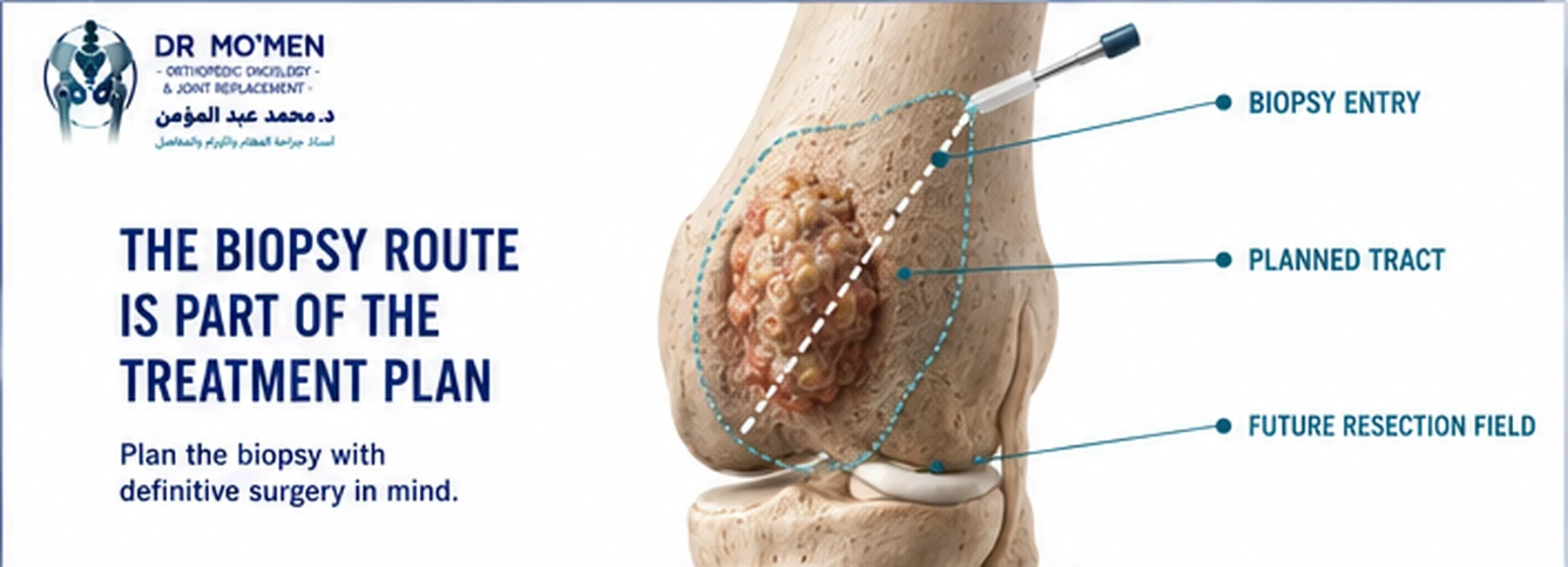
The route used for biopsy can influence definitive tumor surgery. The surgeon may need to remove the skin entry point, needle tract, open-biopsy scar and contaminated underlying tissues together with the tumor during definitive resection. Therefore, the biopsy route should ideally lie within the future planned surgical specimen.
The Shortest Route Is Not Always the Correct Route
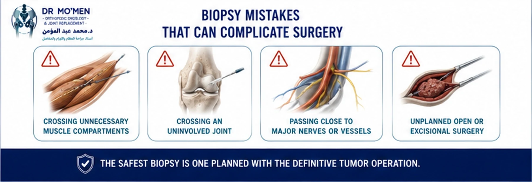
A radiologically simple straight-line route may not always be oncologically appropriate. The biopsy pathway should consider the future surgical incision, anatomical compartments, major muscles, neurovascular structures, joint capsule, planned reconstruction and the amount of tissue that may later need to be removed.
The goal is not simply to reach the lesion. The goal is to reach it without compromising future treatment.
Why Should Unnecessary Muscle Compartments Be Avoided?
If the biopsy crosses a muscle compartment that did not originally contain tumor, that additional pathway may potentially need to be considered during definitive surgery. This can affect muscle preservation, wound closure, reconstruction and postoperative function. Good biopsy planning therefore tries to limit contamination to the smallest reasonable surgical field.
Why Should a Biopsy Avoid Crossing the Joint?
Crossing an uninvolved joint can create a particularly difficult oncological problem. If the joint was initially free of tumor but the biopsy pathway unnecessarily enters it, definitive local treatment may become more complex. Biopsy planning should therefore respect joint anatomy whenever possible.
Why Are Major Nerves and Blood Vessels Important?
A biopsy tract placed close to or through major neurovascular structures can complicate definitive resection. The pathway should avoid unnecessary contact with arteries, veins and major nerves while still obtaining representative tissue safely.
Why the Biopsy Tract Should Be Simple
- direct
- short enough to be practical
- anatomically planned
- placed where it can be removed if required
- limited to the relevant surgical compartment
Why Incision Direction Matters
For extremity tumor surgery, the direction of an open biopsy incision may influence how easily that tract can later be included within definitive resection. Longitudinal planning often aligns better with future limb surgery than an unnecessarily broad transverse incision. The exact incision still depends on the individual anatomy and planned operation.
What About the Biopsy of a Soft-Tissue Component?
Some bone tumors extend through the cortex and create a soft-tissue mass. In selected situations, sampling the soft-tissue component can provide diagnostic tissue without weakening the bone further. This can be particularly relevant when fracture risk is significant, but the route still needs to be coordinated with the definitive surgical plan.
Why Pathological Fracture Risk Matters
A large hole through already weakened bone can increase mechanical risk. The biopsy team therefore needs to consider cortical destruction, weight-bearing location, lesion size, needle trajectory, existing fracture and impending-fracture risk. In selected high-risk lesions, temporary activity modification or protected weight-bearing may be appropriate while diagnosis is being established.
What Happens If the First Biopsy Is Nondiagnostic?
A nondiagnostic biopsy does not mean there is no tumor. Possible reasons include sampling necrotic tissue, missing the representative area, inadequate volume, difficult pathology, excessive decalcification, or lesion heterogeneity. Depending on the situation, the next step may include pathology review, imaging review, repeat image-guided core biopsy or a planned open biopsy.
Imaging and Pathology Must Agree
A fundamental tumor principle is radiological-pathological correlation. If imaging appears highly aggressive but pathology reports a completely innocuous process, the team should ask whether the tissue sample truly explains the imaging. If it does not, the case should be reconsidered before definitive treatment.
Why Specialist Musculoskeletal Pathology Matters
Primary bone tumors are rare. Accurate diagnosis may require assessment by a pathologist experienced in bone and soft-tissue tumors. Some diagnoses require immunohistochemistry, molecular testing, cytogenetic studies or special sample preparation. This can influence how tissue should be collected and preserved.
Why Sample Handling Matters
A technically perfect biopsy can still lose diagnostic value if tissue is handled incorrectly. Depending on the suspected diagnosis, samples may need to support conventional histology, immunohistochemistry, microbiology, molecular testing and cytogenetic studies. The pathology team should therefore be involved when unusual sample requirements are anticipated.
Why Excessive Decalcification Can Be a Problem
Bone samples often require processing before microscopic examination. Some decalcification methods can reduce the quality of nucleic acids and interfere with molecular testing. When molecular confirmation is likely to be important, tissue-handling requirements should be discussed in advance with pathology.
What About Fine-Needle Aspiration?
Fine-needle aspiration mainly retrieves individual cells rather than larger tissue cores. It can have useful roles in selected clinical contexts. However, many primary musculoskeletal tumors require architectural tissue, grading information, immunohistochemistry and sometimes molecular testing, so core biopsy is often more informative when sarcoma is suspected.
Should the Whole Small Tumor Simply Be Removed?
This is called an excisional biopsy. In carefully selected small superficial soft-tissue lesions, excisional biopsy may sometimes be appropriate. For a suspicious bone tumor or deep sarcoma, however, simply removing the lesion without oncological planning can violate tumor planes, contaminate surrounding tissue, produce inadequate margins and complicate reconstruction.
Important
“Take it out and send it to pathology” is not an appropriate default strategy for a suspicious musculoskeletal tumor.
What Is an Unplanned Excision?
An unplanned excision occurs when a mass is removed before sarcoma is recognized and appropriately staged or planned. If this happens, definitive management may require reassessment of pathology, preoperative imaging, the surgical scar, operative bed, drain tract and current MRI.
Related: Unplanned excision of a soft-tissue sarcoma — what should happen next?
What If a Bone Tumor Has Already Been Nailed or Plated?
This can create a difficult orthopedic oncology problem. If an undiagnosed primary bone malignancy is treated as a routine fracture, fixation may spread the surgical field through the medullary canal, screw tracks, plate exposure and surrounding tissues. Definitive surgery can become substantially more complex. This is one reason pathological fractures through suspicious lesions should be diagnosed before routine fixation whenever clinically feasible.
Should Biopsy Be Done at the Same Center as Definitive Surgery?
It is not always physically necessary for both procedures to happen in exactly the same hospital. What matters most is coordination. Ideally, the orthopedic oncology team responsible for definitive treatment should review the imaging, proposed biopsy route, target area and tissue requirements before biopsy. A radiologist, interventional radiologist, surgeon and pathologist can then work from the same treatment plan.
Is CT-Guided Biopsy Safe?
CT-guided core biopsy is widely used for appropriately selected deep bone lesions. Safety depends on lesion location, nearby vessels and nerves, fracture risk, patient factors, planned needle pathway and operator expertise. Potential complications can include bleeding, hematoma, pain, infection, pathological fracture in selected lesions and nondiagnostic sampling. The risk-benefit balance should be individualized.
Does the Biopsy Spread Cancer?
Patients frequently worry that inserting a biopsy needle will automatically cause cancer to spread through the body. That is not an accurate way to understand tumor biopsy. The relevant orthopedic oncology concern is local tract contamination, which is why the biopsy pathway is planned so it can be incorporated into definitive treatment when needed. This planning issue is not a reason to avoid a medically necessary biopsy.
What Happens After the Biopsy?
- Pathology review
- Correlation with imaging
- Tumor grading where appropriate
- Molecular testing when required
- Staging
- Multidisciplinary review
- Definitive treatment planning
The biopsy is therefore the beginning of the treatment pathway, not the end.
Why Multidisciplinary Planning Matters
A complex bone tumor case may involve orthopedic oncology, musculoskeletal radiology, interventional radiology, pathology, medical oncology, radiation oncology, reconstructive surgery and rehabilitation. The biopsy should fit within this larger plan.
Core Needle vs Open Biopsy — Practical Comparison
| Question | Core Needle Biopsy | Open / Incisional Biopsy |
|---|---|---|
| Tissue obtained | Multiple tissue cores | Larger tissue sample |
| Tissue disruption | Usually less | Greater |
| Image guidance | Commonly used | Not typically used in the same manner |
| Anaesthesia | Often local ± sedation depending on case | Often more procedural anaesthesia |
| Recovery | Usually shorter | Usually longer |
| Typical role | Frequently first-line when suitable | Selected cases |
| Potential limitation | May be nondiagnostic | Larger contaminated surgical field |
| Key principle | Correct tract and representative cores | Carefully positioned incision within future resection |
Planning principle
Planning the biopsy may preserve future surgical options.
The Most Important Rule: Do Not Rush the Biopsy
When a suspicious bone tumor is identified, there is usually value in spending a short period planning the biopsy properly rather than performing the fastest available procedure. Before biopsy, the team should know what the X-ray shows, what MRI shows, where the viable tumor is, which structures need to be avoided, where definitive surgery is likely to occur, and what tissue the pathologist requires.
International Patients: Send Imaging Before Biopsy When Possible
For patients outside Egypt who have been told that a biopsy is needed, preliminary specialist review before biopsy can be particularly useful. Useful records include:
- X-rays
- MRI
- CT
- PET/CT or other staging studies if already performed
- radiology reports
- clinical summary
- previous cancer history
- laboratory tests where relevant
A preliminary review may help determine whether imaging should be completed first, whether biopsy is actually required, how the biopsy route should be planned, whether core needle biopsy appears appropriate, whether specialist pathology preparation may be required, and whether the case should be evaluated by an orthopedic oncology team before intervention.
Remote review cannot determine every technical detail without examination and complete imaging, but it may help avoid an unnecessary or poorly planned initial procedure.
About Dr. Mohammed Abdelmoemen Abuelhadid
Dr. Mohammed Abdelmoemen Abuelhadid is an Orthopedic Oncology & Joint Replacement Surgeon and Lecturer of Orthopedic Surgery at Ain Shams University. His clinical work includes orthopedic oncology, bone and soft-tissue tumors, biopsy planning, limb-salvage surgery, wide tumor resection, megaprosthetic reconstruction, recurrent tumors, metastatic bone disease, complex orthopedic reconstruction and second opinions for complex musculoskeletal tumors.
Send imaging for preliminary review
If a biopsy has been recommended, available X-rays, MRI, CT and reports can be reviewed preliminarily before biopsy or travel.
For appointments or review of X-rays/MRI before biopsy: +20 102 1690693
Clinical principle
For a suspected primary bone tumor: plan the definitive treatment before choosing the biopsy pathway.
How this article was reviewed
This patient-education article was reviewed for clinical accuracy, patient safety and decision-making clarity by Dr. Mohammed Abdelmoemen Abuelhadid. It is intended to support, not replace, specialist imaging review, biopsy planning, pathology, staging or multidisciplinary tumor care. See the editorial policy.
Before Bone Tumor Biopsy: A Practical Checklist
Direct answer: before tissue sampling, the team should confirm that the lesion truly needs biopsy, complete the appropriate imaging when clinically feasible, choose a representative target, and plan a tract that will not compromise definitive tumor surgery. Pathology requirements and pathological-fracture risk should also be considered before the procedure.
- Review the radiographs and complete MRI or CT when indicated before choosing the biopsy target.
- Confirm that a classic benign-appearing lesion is not being biopsied unnecessarily.
- Coordinate suspected primary malignant bone tumors with the orthopedic oncology team responsible for definitive treatment.
- Plan a simple trajectory that avoids unnecessary uninvolved compartments, major neurovascular structures and joint contamination.
- Target viable, representative tissue rather than a necrotic area whenever imaging allows.
- Tell pathology the clinical and radiological differential and anticipate tissue needs for histology, immunohistochemistry and molecular testing when relevant.
- Consider pathological-fracture risk and avoid unplanned fixation of an undiagnosed destructive lesion when diagnosis can be established first.
Evidence Behind Bone Tumor Biopsy Planning
Major imaging and sarcoma guidance converges on the same safety principle: evaluate the lesion with appropriate imaging first, refer a suspected primary bone malignancy into specialist multidisciplinary care, and plan tissue acquisition so that the biopsy route, specimen quality and later definitive surgery are considered together.
- ACR Appropriateness Criteria®: Suspected Primary Bone Tumors, revised 2024 — supports an imaging-led diagnostic work-up and shows that biopsy is not the automatic next step for a benign-appearing lesion.
- British Sarcoma Group: UK Guidelines for the Management of Bone Sarcomas — recommends specialist-center management and biopsy planning with the surgical team so the tract can be incorporated into definitive resection.
- Children’s Oncology Group Bone Tumor Committee biopsy-consensus statement — emphasizes multidisciplinary pre-biopsy planning, representative viable tissue and specimen-processing requirements for osteosarcoma and Ewing sarcoma.
- ESMO–EURACAN–GENTURIS–ERN PaedCan Bone Sarcoma Clinical Practice Guideline — provides multidisciplinary recommendations for diagnosis and treatment of bone sarcomas.
Frequently Asked Questions
Is core needle biopsy better than open biopsy?
Neither is universally better. A properly planned core needle biopsy is frequently used first because it can obtain diagnostic tissue with less disruption. Open biopsy is valuable when core biopsy is nondiagnostic, unsafe or insufficient.
Does a bone tumor biopsy need CT guidance?
Not always. Image guidance may use CT, ultrasound, fluoroscopy or another modality depending on lesion location and anatomy.
Why does the needle route matter if it is only a small hole?
Because the biopsy tract may need to be included in the definitive tumor resection. A poorly placed tract can unnecessarily involve muscles, joints or neurovascular structures.
Can a biopsy make the cancer spread?
The relevant orthopedic oncology concern is possible local tract contamination, which is managed through careful planning. This should not be confused with the idea that a medically necessary biopsy routinely causes systemic cancer spread.
Should MRI be performed before the biopsy?
For a suspected primary bone tumor, MRI is usually obtained before biopsy when clinically feasible because it defines the untreated local extent and helps plan the biopsy route.
What happens if the core biopsy does not give a diagnosis?
The case may require pathology and imaging review, repeat core biopsy or a planned open biopsy.
Should I get a second opinion before biopsy?
It can be useful when a primary bone malignancy is suspected, the lesion is near a joint or major neurovascular structures, or limb-salvage surgery may later be required.