
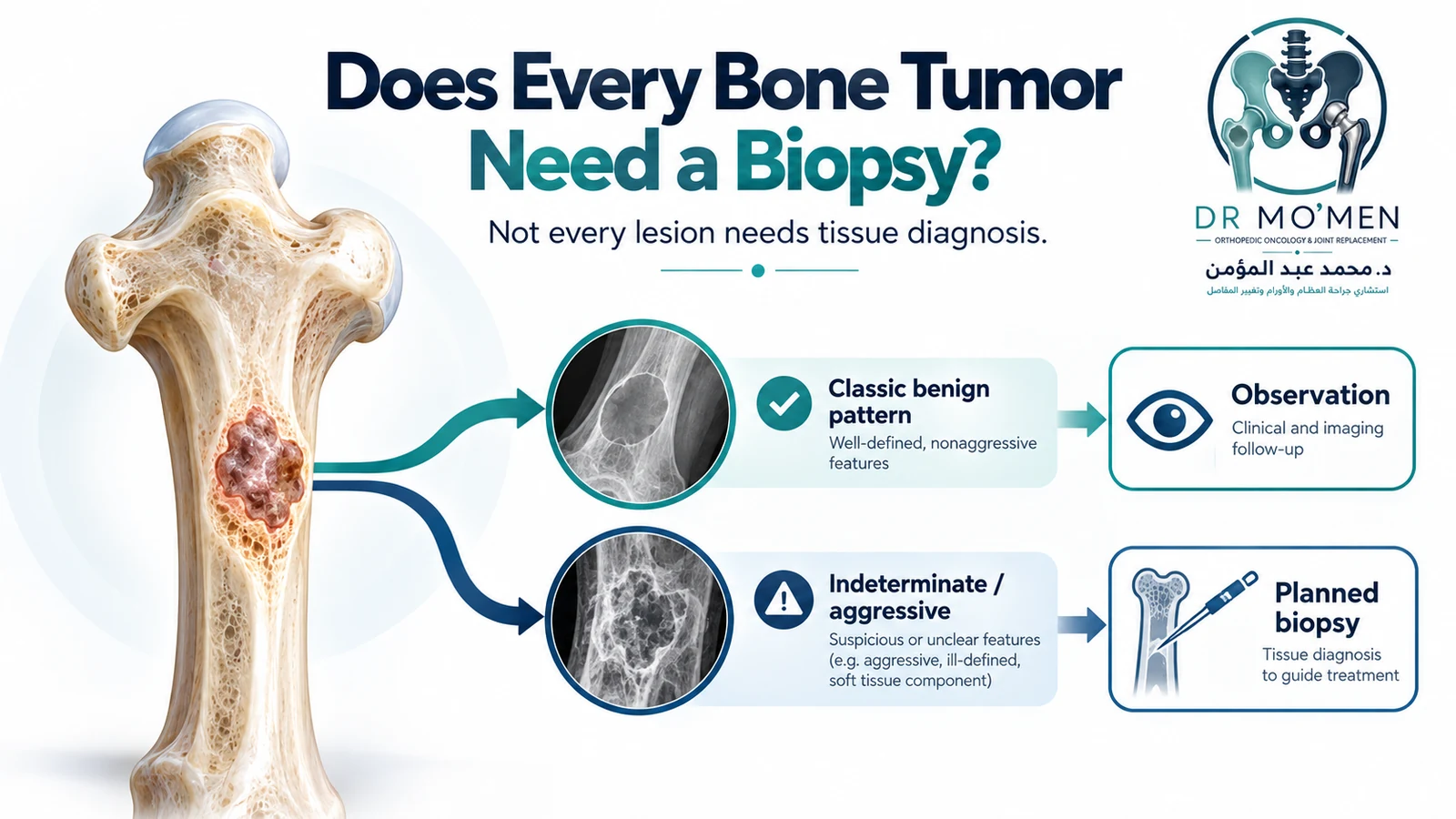
Finding a bone lesion on an X-ray or MRI often leads to an understandable question: “Do I need a biopsy?” The answer is no—not every bone lesion or bone tumor needs tissue sampling.
Some lesions have such characteristic benign imaging features that a biopsy may add little and expose the patient to an unnecessary procedure. Other lesions remain uncertain after imaging and may need follow-up or additional imaging. When a primary malignant bone tumor or another aggressive lesion remains a realistic possibility, however, tissue diagnosis is usually required before definitive treatment.
The important question is therefore not simply “Can this lesion be biopsied?” but “Does it need biopsy, and if so, how should the biopsy be planned so that it does not compromise definitive treatment?”
Why doesn't every bone lesion need a biopsy?
Bone lesions exist along a spectrum. Some have a classic benign radiographic appearance and biological behavior that can be recognized with high confidence. Examples can include a typical bone island, non-ossifying fibroma and selected cystic, fibrous or cartilage lesions without aggressive features.
If the patient's age, lesion location, margins, matrix and overall imaging pattern are all reassuring, observation or appropriate imaging follow-up may be safer and more useful than immediate tissue sampling.
A classic benign bone lesion may be diagnosed from its imaging pattern without biopsy.
When is observation better than biopsy?
- The lesion has characteristic benign imaging features.
- There are no aggressive radiographic findings.
- Symptoms are absent or clearly unrelated to the lesion.
- The bone is structurally stable.
- Interval imaging can safely confirm stability when needed.
A decision to observe does not mean the lesion is being ignored. It means the diagnostic value of biopsy is lower than the value of safe clinical or imaging surveillance.
When is a bone tumor biopsy necessary?
- Imaging remains indeterminate after appropriate work-up.
- A primary malignant bone tumor is suspected.
- An aggressive benign tumor is possible and histology would change treatment.
- Different possible diagnoses require substantially different treatments.
- Tumor type or grade would affect surgery, chemotherapy or radiotherapy.
- Pathology is needed before definitive oncological treatment.
- Imaging and clinical findings cannot provide a sufficiently secure diagnosis.
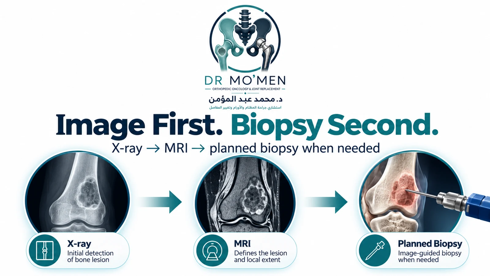
Should an aggressive bone lesion be biopsied immediately?
Usually not before the imaging work-up is complete. When radiographs show an indeterminate or aggressive lesion, MRI is typically obtained first because it defines local extent, identifies a representative area to sample and helps plan a biopsy route that is compatible with later surgery.
A useful sequence is: X-ray → MRI/CT when indicated → orthopedic oncology and radiology review → biopsy planning → tissue diagnosis → definitive treatment.
Abnormal X-ray → immediate unplanned biopsy is not the preferred sequence.
Why is MRI usually done before bone tumor biopsy?
MRI can show intramedullary extent, soft-tissue extension, relationship to nearby joints and neurovascular structures, viable and necrotic regions, and areas that may represent a higher-grade component.
This information helps select the most representative target and allows the biopsy team to plan a route that minimizes unnecessary contamination of normal tissue.
Why does the biopsy site matter?
A tumor biopsy is not simply a needle placed by the shortest route. The tract becomes part of the oncologic treatment plan.
- The route should avoid unnecessary crossing of uninvolved tissue planes.
- Major nerves and vessels should be protected.
- A joint should not be entered unnecessarily.
- The trajectory should be compatible with the planned definitive surgical approach.
- The tract should be identifiable and considered during definitive resection when appropriate.
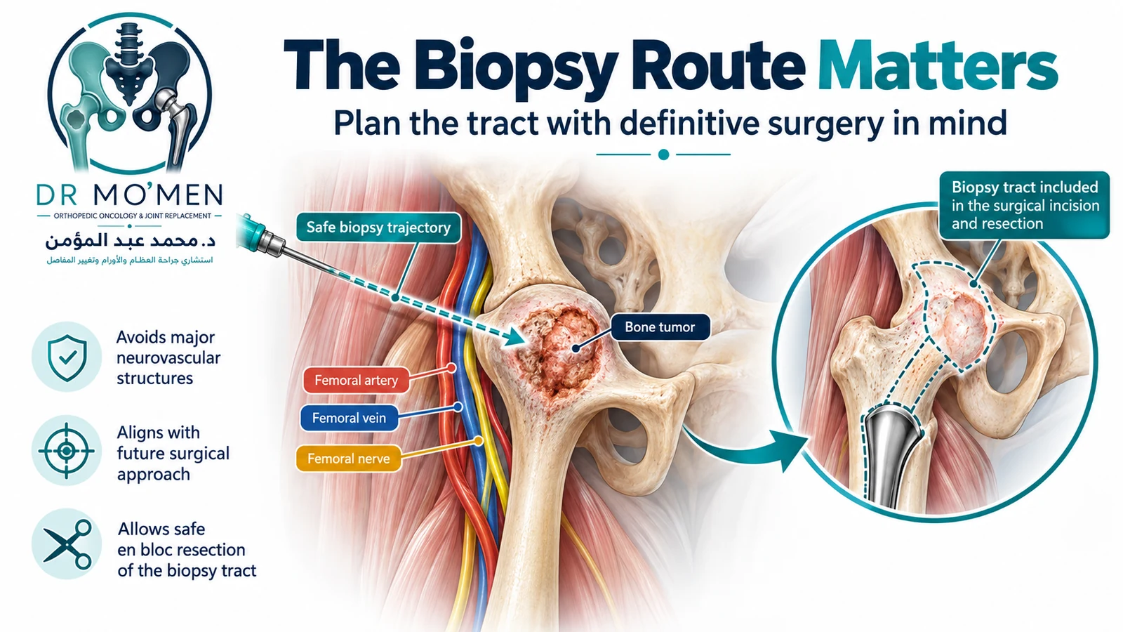
The route image in this package is a conceptual educational illustration. Actual biopsy trajectory depends on tumor location, compartments, imaging and the definitive surgical plan.
Can the wrong biopsy compromise limb-salvage surgery?
An unplanned or poorly positioned biopsy can make definitive surgery more difficult by involving tissues that otherwise might not have required resection. This does not mean every imperfect biopsy makes limb salvage impossible, but it is why biopsy planning is treated as part of definitive tumor treatment rather than as an isolated diagnostic procedure.
Who should plan a bone tumor biopsy?
When a primary malignant bone tumor is genuinely suspected, biopsy should ideally be planned with the specialist team responsible for definitive treatment. This often includes an orthopedic oncology surgeon, musculoskeletal or interventional radiologist, bone tumor pathologist and medical or pediatric oncologist when relevant.
What type of biopsy is usually used?
The two main methods are image-guided core needle biopsy and open incisional biopsy.
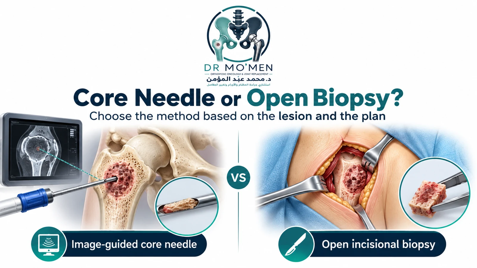
What is a core needle biopsy?
A core needle biopsy removes small cylinders of tissue using a biopsy needle. Depending on lesion location, it can be performed under CT, ultrasound, fluoroscopic or other image guidance.
Core needle biopsy is commonly used in specialist centers because it is less invasive than open biopsy and frequently provides enough material for histology and molecular testing when properly planned and sampled.
Why is image guidance useful?
- It helps target viable rather than necrotic tumor.
- It can target a specific solid or higher-grade-appearing component.
- It helps plan access to deep lesions such as those in the pelvis.
- It helps avoid major vessels, nerves and other critical structures.
Is core needle biopsy always enough?
No. It is sufficient in many cases, but a sample can be nondiagnostic or insufficient because too little tissue was obtained, a necrotic region was sampled, the lesion is heterogeneous, or additional architecture or molecular material is required.
When the result is inadequate or does not fit the imaging, further sampling may be required.
When is an open biopsy needed?
- Core needle biopsy is nondiagnostic or insufficient.
- Adequate tissue cannot safely be obtained percutaneously.
- The lesion's anatomy or suspected pathology requires a larger specimen.
- Repeated core sampling has not resolved the diagnosis.
- The specialist MDT determines that an open approach is more appropriate.
An open biopsy incision must be planned with particular care because the incision and underlying biopsy tract may need to be incorporated into definitive tumor surgery.
Is excisional biopsy appropriate for a bone tumor?
For a lesion that might represent a malignant primary bone tumor, simply removing the entire lesion without an established diagnosis is generally not the standard diagnostic approach.
Selected small lesions with a very strong benign diagnosis may be managed differently, but the decision should be based on the imaging diagnosis and an oncologically appropriate surgical plan—not on diagnostic uncertainty.
Can a bone tumor biopsy spread cancer?
This question causes considerable anxiety. The relevant oncologic issue is that a biopsy creates a tract that must be considered during later surgery. That is why specialist planning aims to minimize contamination of uninvolved tissues and place the tract where it can be incorporated into definitive treatment when appropriate.
A correctly indicated and properly planned biopsy is a standard part of diagnosing suspected malignant bone tumors. The greater concern is an unnecessary or poorly planned biopsy that complicates subsequent treatment.
What happens to the tissue after biopsy?
The specimen is evaluated by pathology. Depending on the suspected diagnosis, testing may include routine histology, immunohistochemistry, cytogenetic or molecular analysis, and microbiology when infection remains in the differential diagnosis.
The pathologist should have access to the patient's age, anatomical site, imaging findings and radiological differential diagnosis because bone tumor pathology cannot be interpreted optimally in isolation.
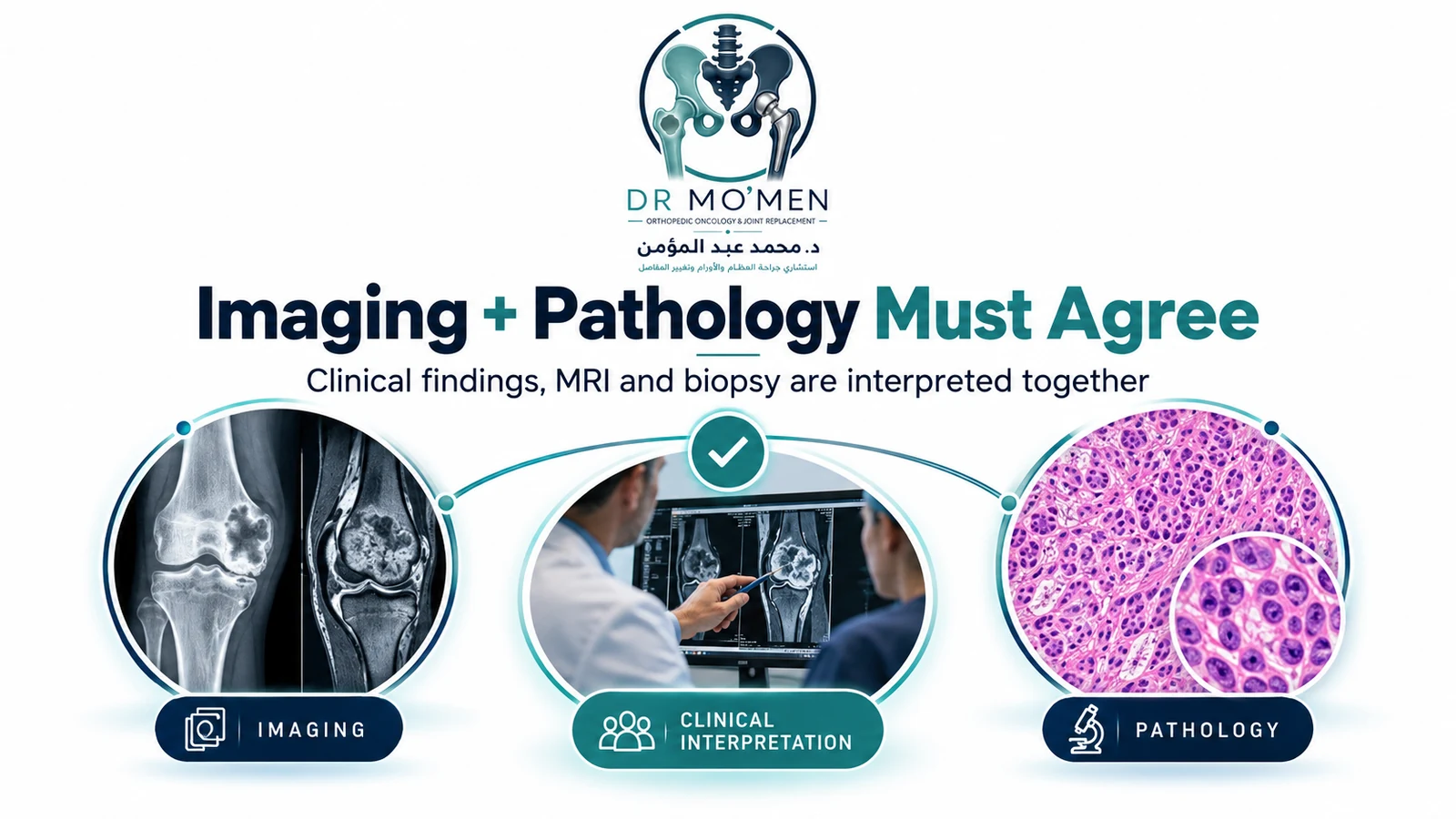
Why must imaging and pathology agree?
The strongest diagnosis comes from correlation among clinical findings, imaging and pathology. A pathology label that does not fit the X-ray or MRI should trigger reassessment rather than automatic acceptance.
Discordance can occur because of sampling error, tumor heterogeneity, sampling of a nonrepresentative area, an incorrect radiological assumption or an incorrect pathological interpretation.
When necessary, the next step may include specialist radiology review, pathology review, multidisciplinary discussion and repeat biopsy.
A bone tumor diagnosis should make sense clinically, radiologically and pathologically.
Can MRI diagnose bone cancer without biopsy?
MRI can strongly suggest whether a lesion is benign, aggressive or malignant and is essential for defining local extent, but it generally does not replace histological confirmation when a primary malignant bone tumor is suspected.
Imaging defines behavior and anatomy; pathology identifies the tissue diagnosis. The two are complementary.
What about cartilage tumors?
Cartilage tumors are a good example of why imaging and pathology must be interpreted together. Distinguishing a benign enchondroma from a low-grade chondrosarcoma can be difficult because imaging and histological features may overlap, and tumor grade can vary within the same lesion.
A small sample may not always represent the most biologically active part of a heterogeneous cartilage tumor, so multidisciplinary correlation is particularly important.
What about suspected metastatic bone disease?
Not every lesion in a patient with a known cancer needs biopsy. The decision depends on whether the primary cancer is known, whether the skeletal pattern is typical, whether the lesion is solitary or multiple, how long the disease-free interval has been, and whether tissue confirmation would change systemic or surgical management.
A solitary atypical bone lesion in a patient with previous cancer should not automatically be labelled a metastasis without appropriate review.
Should a pathological fracture be biopsied before fixation?
When a fracture occurs through an unexplained bone lesion, the diagnosis should ideally be established before definitive fixation whenever the patient's condition permits. Fixing an undiagnosed primary bone sarcoma can substantially complicate later oncologic surgery.
The usual pathway is local imaging and staging as appropriate → orthopedic oncology review → planned biopsy when required → definitive fracture and tumor treatment.
Can biopsy and definitive surgery be done at the same time?
For a suspected primary malignant bone tumor, the usual pathway is to obtain and review the biopsy diagnosis before definitive resection. This allows the multidisciplinary team to determine tumor type, grade where applicable, need for neoadjuvant treatment, required surgical margin and reconstruction strategy.
What if a biopsy was already done without oncology planning?
This does not automatically mean definitive treatment is impossible. The next step is careful specialist review of the biopsy tract, operative note, pathology slides and pre-biopsy imaging.
The important step is to avoid additional unplanned procedures until the case has been reviewed by the orthopedic oncology team.
Practical decision pathway
- Bone lesion identified.
- Clearly benign imaging appearance → no biopsy in many cases; observe or follow appropriately.
- Indeterminate lesion → MRI/CT and comparison with prior imaging.
- Aggressive or malignant-appearing lesion → orthopedic oncology review and complete local imaging.
- If tissue diagnosis will change treatment → plan biopsy trajectory with definitive surgery in mind.
- Image-guided core needle biopsy in many cases; open biopsy in selected situations.
- Specialist pathology review.
- Correlate clinical findings + imaging + pathology before definitive treatment.
When should you seek orthopedic oncology assessment before biopsy?
- The lesion looks aggressive on X-ray or MRI.
- A soft-tissue mass or cortical destruction is present.
- A pathological fracture has occurred.
- A primary malignant bone tumor is possible.
- The lesion is in the pelvis or another anatomically complex region.
- A biopsy has been recommended but no definitive treatment plan has been discussed.
- The biopsy result and MRI do not match.
- An open biopsy or excision has been proposed without a secure diagnosis.
Specialist bone tumor biopsy planning
Dr. Mohammed Abdelmoemen Abuelhadid evaluates bone and soft-tissue tumors and complex orthopedic-oncology cases where the appropriate biopsy strategy needs to be determined.
The objective is not to biopsy every abnormality. It is to decide whether tissue diagnosis is actually necessary—and when it is, to plan the procedure so that later limb-salvage or definitive tumor surgery is not compromised.
For patients outside Egypt, existing X-rays, CT, MRI images and reports can also be reviewed as part of an online second medical opinion before biopsy or travel when appropriate.
For appointments or imaging review: 01021690693