
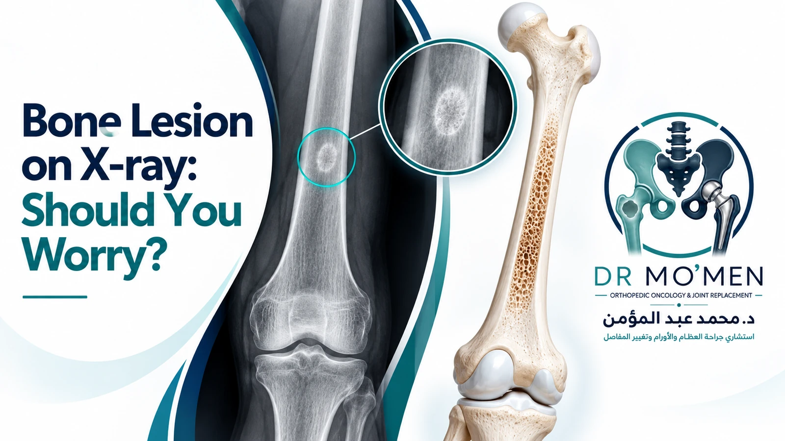
An X-ray taken after an injury, for joint pain, or for another unrelated reason may unexpectedly show an abnormal area inside a bone. The radiology report may use terms such as bone lesion, lucent or lytic lesion, sclerotic lesion, mixed lesion, cystic lesion, or incidental osseous abnormality.
Seeing the word “lesion” can be alarming, but a bone lesion does not automatically mean bone cancer. Many incidental bone findings are benign or tumor-like conditions that may need no treatment. Others need follow-up or additional imaging, and a smaller group needs orthopedic-oncology assessment and a carefully planned biopsy.
The next step depends on the patient’s age, symptoms, lesion location, radiographic pattern, previous cancer history and whether the bone appears structurally weakened—not on the word “lesion” alone.
Does a Bone Lesion on X-ray Mean Cancer?
No. “Bone lesion” is a descriptive imaging term, not a diagnosis. Some lesions have classic benign appearances, while others are indeterminate or show features that require further investigation.
The useful question is whether the lesion has reassuring imaging features or features that justify MRI, follow-up, orthopedic-oncology review or biopsy.
What Is an Incidental Bone Lesion?
An incidental bone lesion is an abnormal area found on imaging when the scan or X-ray was often performed for another reason—for example after a sports injury, during assessment of arthritis, or on a CT or MRI performed for unrelated symptoms.
The lesion may be unrelated to the patient’s original complaint. That is why the original images, not only the report, should be reviewed in the clinical context.
Does the Word “Lesion” Mean Tumor?
No. A lesion simply means that an area of bone looks different from the surrounding bone. Possible causes include benign bone tumors, tumor-like lesions, cysts, fibrous or cartilage lesions, infection, metabolic conditions, metastases and primary malignant bone tumors. See also benign vs malignant bone tumors.
Why Is the X-ray Important?
Plain radiographs are a key first investigation when a primary bone tumor is suspected. X-rays can show where the lesion sits within the bone, whether it is lucent, sclerotic or mixed, how sharply defined its margins are, how the cortex reacts, whether periosteal new bone is present, whether a soft-tissue component is suspected and whether there is a pathological fracture.
What X-ray Features Can Be Reassuring?
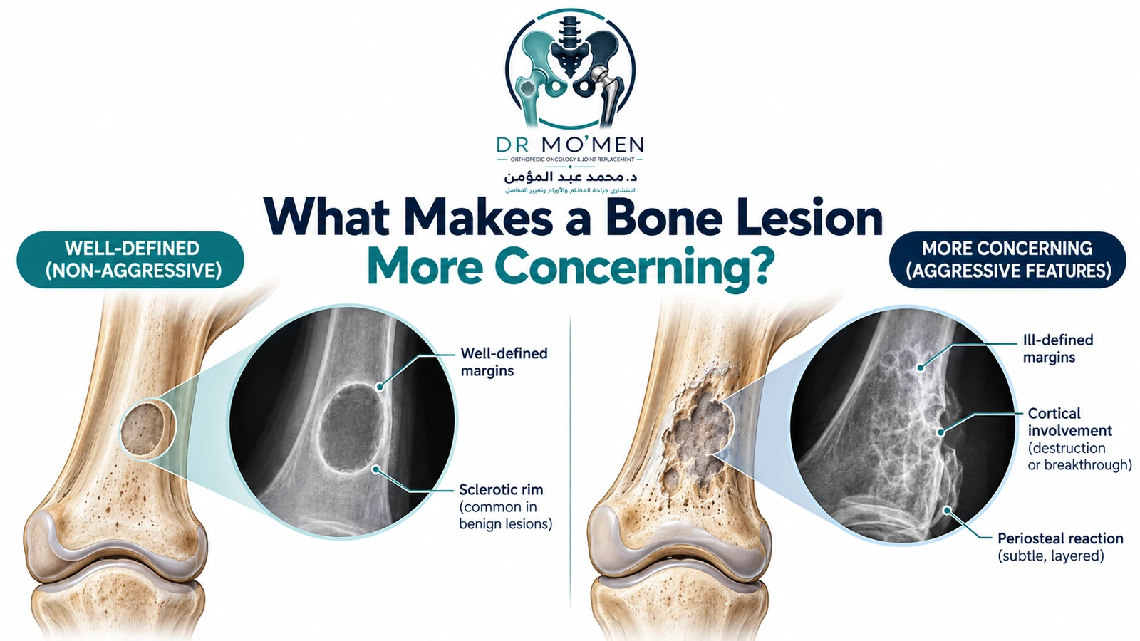
A well-defined lesion with a narrow zone of transition, a preserved cortex and no aggressive periosteal reaction may be reassuring when it matches a recognized benign pattern and the clinical setting is appropriate.
Examples that can sometimes have characteristic benign appearances include a bone island, non-ossifying fibroma, selected fibrous lesions and some cartilage lesions. Diagnosis depends on the complete imaging pattern rather than the name alone.
What X-ray Features Are More Concerning?
- Poorly defined or destructive margins / a wide zone of transition.
- Cortical destruction or significant cortical breakthrough.
- Aggressive periosteal reaction.
- A suspected soft-tissue mass.
- Pathological fracture or marked structural weakening.
- Progression or enlargement on serial imaging.
These features do not prove cancer. Infection and some benign but locally aggressive lesions can also look concerning. They mean that the lesion deserves a more complete diagnostic work-up.
Does Pain Make a Bone Lesion More Concerning?
It can. The important question is whether the pain is actually attributable to the lesion. A bone lesion discovered on an MRI for meniscal pain may be unrelated to the symptoms, while persistent focal pain arising from the same site as the lesion can increase the need for assessment.
What Does a Lytic Bone Lesion Mean?
A lytic or lucent lesion appears darker than surrounding bone on X-ray because that region is less mineralized. “Lytic” describes the imaging appearance; it does not by itself mean malignant.
Benign lesions, locally aggressive benign tumors, primary malignant tumors, metastases, myeloma and infection can all produce lytic appearances. Age, lesion location, margins, cortical response and associated imaging features help narrow the differential diagnosis.
What Does a Sclerotic Bone Lesion Mean?
A sclerotic lesion appears denser or whiter than surrounding bone. This can range from a benign bone island to metastatic or other pathological processes. The number of lesions, morphology, symptoms and previous cancer history are important.
Does Lesion Size Tell You Whether It Is Cancer?
Not reliably by itself. Size can contribute to assessment, but it should not be used in isolation. Some benign lesions can be large, and some malignant lesions can be relatively small. The complete clinical and imaging pattern is more informative.
What Happens After a Bone Lesion Is Found?
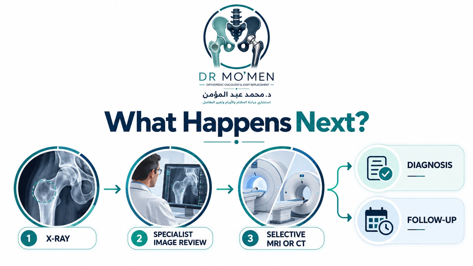
The next step usually falls into one of several pathways. A confidently benign lesion may need no further work-up. An incompletely characterized lesion may need comparison with previous imaging, dedicated radiographs, CT, MRI or interval follow-up. A lesion with aggressive or concerning features should be reviewed through an orthopedic-oncology pathway.
Do I Need an MRI for Every Bone Lesion?
No. A classic benign lesion may need no additional imaging. MRI becomes especially useful when radiographs are indeterminate or aggressive, when the lesion’s extent within the bone must be defined, when soft-tissue extension is suspected, or when a procedure such as biopsy or surgery is being planned.
For an indeterminate or aggressive-appearing lesion on radiographs, the 2024 ACR Appropriateness Criteria rate MRI of the area of interest as usually appropriate.
When Is CT Useful?
CT can be particularly useful for cortical detail, mineralization, matrix, calcification and subtle bone destruction. MRI is generally stronger for marrow involvement, soft-tissue extension, relationship to neurovascular structures and local tumor extent. The correct test depends on the question being asked.
Do I Need a Bone Scan or PET/CT?
Not every incidental bone lesion requires a bone scan or PET/CT. These investigations are used selectively—for example in staging certain confirmed or suspected malignancies, evaluating other skeletal sites, or in patients with a relevant cancer history. For a solitary incidental lesion, the first priority is usually to characterize the local lesion correctly.
What Is Bone-RADS?
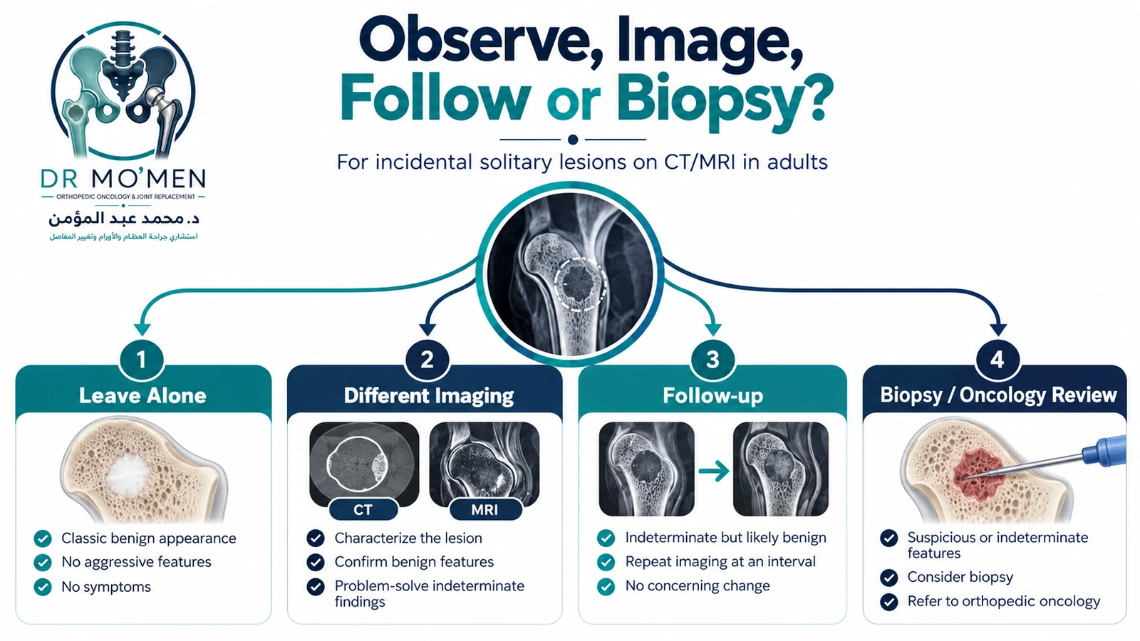
Bone-RADS is the Bone Reporting and Data System developed by the Society of Skeletal Radiology to standardize management of incidentally discovered solitary bone lesions on CT and MRI in adults.
It provides four broad management categories: leave alone, obtain a different imaging modality, follow with interval imaging, or biopsy and/or oncologic referral.
Does Every Bone Lesion Need a Biopsy?
No. Some lesions are sufficiently characteristic on imaging that biopsy is unnecessary. Others need additional imaging or follow-up before a biopsy decision can safely be made.
Biopsy becomes appropriate when tissue diagnosis is required to guide management—for example when malignancy or an aggressive lesion remains a realistic possibility and the pathology result will change treatment.
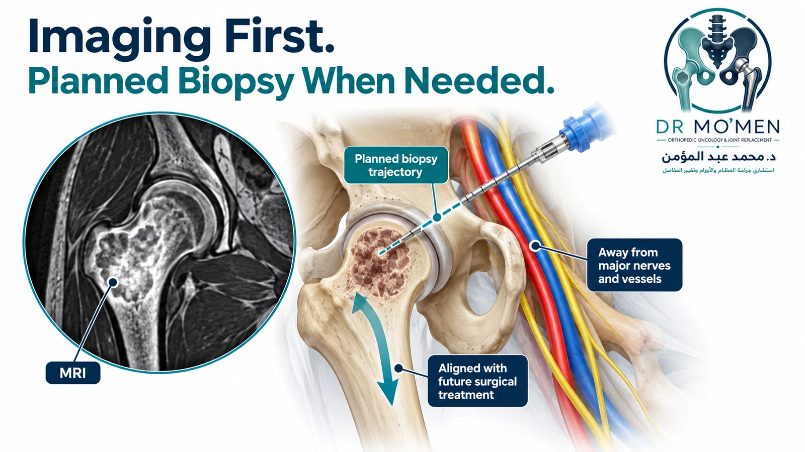
Why Should Imaging Be Completed Before a Bone-Tumor Biopsy?
Because biopsy is part of the treatment plan, not simply another test. Cross-sectional imaging should first define the lesion, identify the most representative area to sample and help determine a trajectory that minimizes contamination of normal tissues.
The 2024 UK bone-sarcoma guideline states that biopsy of a suspected primary malignant bone tumor should be performed after cross-sectional imaging and in consultation with the specialist surgical team so that the biopsy tract can be incorporated into definitive resection when required.
For a detailed comparison, see core needle vs open bone biopsy.
Should the Whole Involved Bone Be Assessed Before Biopsy?
When a primary malignant bone tumor is suspected, local MRI is used to define the extent of disease. Specialist protocols commonly include the whole involved bone and adjacent joints so that intramedullary extent, cortex, soft-tissue involvement and surgical anatomy can be assessed before biopsy and treatment planning.
What If I Already Had a Biopsy Elsewhere?
A pathology result should not be interpreted in isolation. Orthopedic-oncology diagnosis relies on correlation among the clinical picture, radiographs/CT/MRI and pathology. If these elements do not fit, review of the imaging and pathology—and occasionally repeat biopsy—may be necessary before definitive treatment.
What If the Lesion Is Found in a Child?
Children require age-specific interpretation because many pediatric bone lesions have strong relationships with skeletal maturity and location. Adult Bone-RADS algorithms are not designed for pediatric patients.
Does a Previous Cancer Change the Assessment?
Yes. A history of a cancer that commonly spreads to bone changes the level of concern and the imaging pathway. However, not every bone lesion in a patient with previous cancer is a metastasis; benign lesions remain common and the imaging pattern still matters.
When Should a Bone Lesion Be Reviewed by an Orthopedic Oncologist?
- The lesion is indeterminate after initial imaging.
- Aggressive radiographic features are present.
- The cortex is substantially involved or structurally weakened.
- A soft-tissue mass is present or suspected.
- A pathological fracture is present or there is concern about impending fracture.
- Pain appears attributable to the lesion.
- The lesion is enlarging or changing.
- Biopsy or surgery is being considered.
- Imaging and pathology do not fit together.
Referral to orthopedic oncology does not mean that the lesion is malignant. It helps make sure that imaging, biopsy and any surgery are performed in the correct sequence.
When Is Assessment More Urgent?
- Sudden severe pain suggesting pathological fracture.
- Inability to bear weight after minimal trauma.
- Rapidly enlarging swelling.
- Neurological symptoms from a spinal or pelvic lesion.
- A destructive lesion with marked cortical weakening.
- An aggressive radiological appearance with concern for primary malignancy.
A Practical Diagnostic Pathway
- Unexpected bone lesion on X-ray.
- Review the original images, not only the report.
- Assess age, symptoms, lesion location, radiographic pattern and cancer history.
- Clearly benign appearance → observation or no further work-up when appropriate.
- Indeterminate appearance → dedicated imaging, MRI/CT and/or interval follow-up.
- Aggressive/concerning appearance → orthopedic oncology review and appropriate cross-sectional imaging.
- Biopsy only when indicated and after imaging has been reviewed.
- Correlate radiology + pathology + clinical findings before definitive treatment.
Specialist Assessment of an Incidental Bone Lesion
Dr. Mohammed Abdelmoemen Abuelhadid evaluates bone and soft-tissue tumors, complex bone lesions and orthopedic-oncology cases, including incidental lesions where the diagnosis or next investigation remains uncertain.
A specialist review may help determine whether the lesion has a classic benign appearance, needs interval imaging, needs MRI or CT, requires fracture-risk assessment, should be discussed in a multidisciplinary team, or needs a planned biopsy.
For patients outside Egypt, existing X-rays, CT, MRI images and reports can also be reviewed as part of an online second medical opinion before travelling when appropriate. See the international-patient pathway for pre-travel review.
For appointments or imaging review: 01021690693
Evidence and Editorial Verification
- American College of Radiology. ACR Appropriateness Criteria: Suspected Primary Bone Tumors. Revised 2024.
- Chang CY, et al. Society of Skeletal Radiology white paper: Bone-RADS. Skeletal Radiology. 2022;51:1743–1764. PMID 35344076.
- UK guidelines for the management of bone sarcomas. British Journal of Cancer. Published 2024; issue 2025. PMCID PMC11723950.
Medical disclaimer: This article is for patient education and does not replace clinical examination, review of imaging and investigations, or individualized medical advice.