
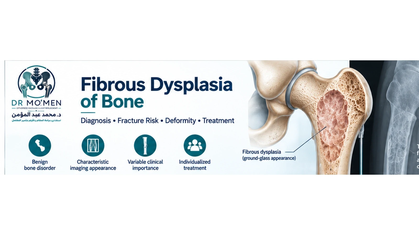
Being told that an X-ray shows fibrous dysplasia can be worrying, particularly when the report uses terms such as bone lesion, ground-glass appearance, or expansile lesion.
Fibrous dysplasia is a disorder in which part of normal bone is replaced by abnormal fibro-osseous tissue. It can involve one bone or multiple bones, and its clinical importance varies considerably between patients.
Many lesions remain stable and need observation rather than surgery.
Other cases can cause:
- pain
- repeated fractures
- progressive bowing or deformity
- weakness of a weight-bearing bone
- limb-length or alignment problems
- craniofacial complications
- uncertainty about whether the lesion truly represents fibrous dysplasia
The key question is therefore not simply:
“Is fibrous dysplasia present?”
It is:
“Is the diagnosis secure, how much bone is involved, and is the lesion causing or threatening a mechanical problem?”
What Is Fibrous Dysplasia?
Fibrous dysplasia is a benign skeletal disorder.
Normal bone and marrow are replaced to varying degrees by fibro-osseous tissue that does not have the same structural organization as healthy mature bone.
The condition results from a post-zygotic activating change in the GNAS gene. Because this genetic change occurs after conception and is present only in some cells, fibrous dysplasia is considered a mosaic condition rather than a conventional inherited disorder.
Fibrous dysplasia is therefore not usually passed from parent to child.
Monostotic vs Polyostotic Fibrous Dysplasia
Fibrous dysplasia is commonly divided according to how many bones are affected.
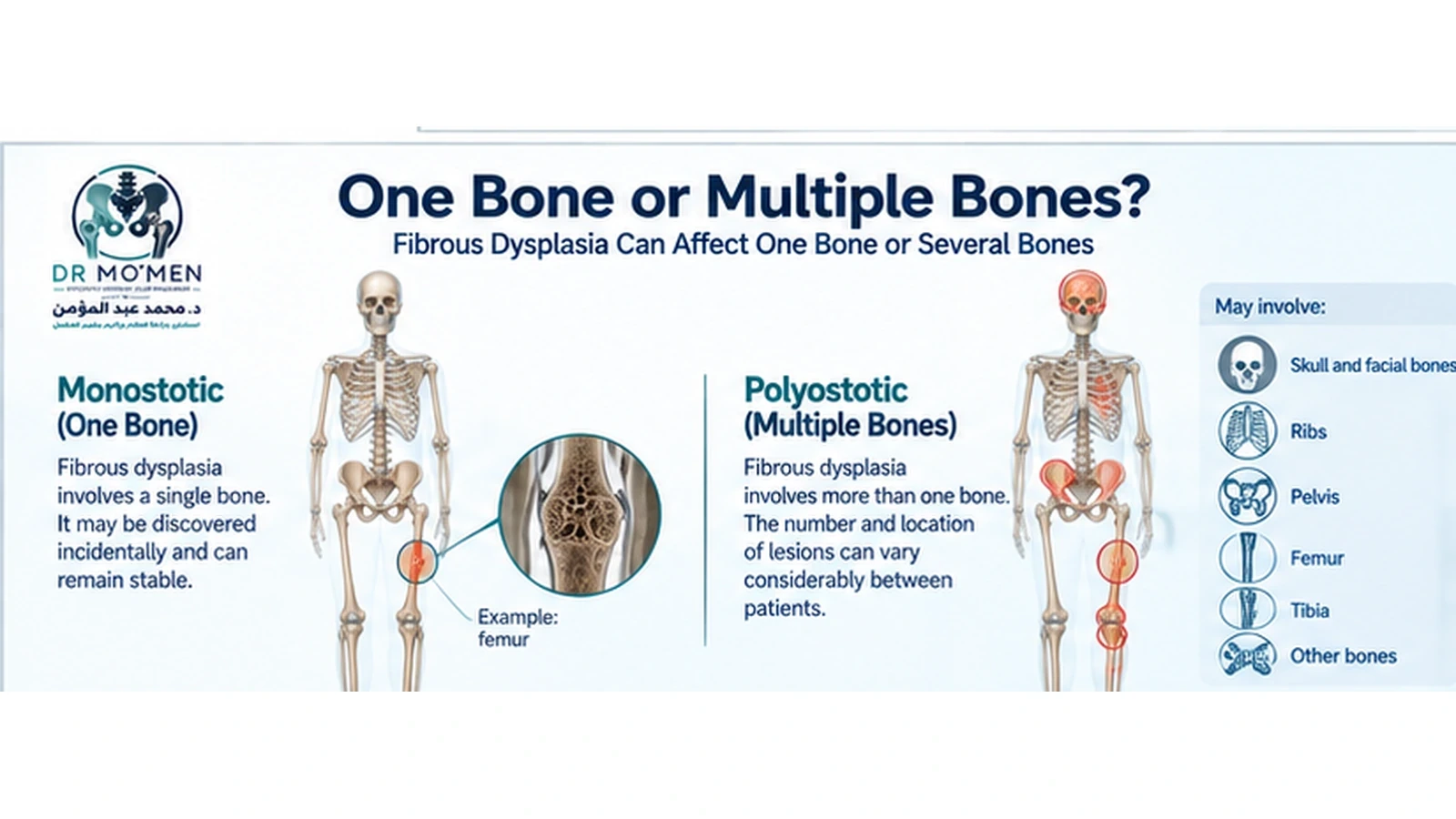
Monostotic fibrous dysplasia
Monostotic means that only one bone is involved.
This may be discovered incidentally when an X-ray is performed for another reason.
Possible sites include:
- femur
- tibia
- ribs
- humerus
- skull and facial bones
- pelvis
A small, characteristic, asymptomatic monostotic lesion may sometimes require only observation.
Polyostotic fibrous dysplasia
Polyostotic disease involves more than one bone.
The skeletal burden can range from a few lesions to extensive involvement.
Patients with more widespread disease require evaluation not only of individual lesions but also of:
- mechanical bone strength
- deformity
- previous fractures
- limb alignment
- spine
- mineral metabolism
- possible endocrine abnormalities
Is Fibrous Dysplasia a Bone Cancer?
Fibrous dysplasia itself is not a malignant bone tumor.
However, not every bone lesion that appears relatively benign on an initial X-ray is necessarily fibrous dysplasia.
This distinction matters.
Atypical features, unexpected progression, or a lesion that does not fit the usual clinical and imaging pattern may require further investigation.
Malignant transformation of true fibrous dysplasia is considered uncommon, but new rapid enlargement, major change in pain, cortical destruction, or development of a soft-tissue mass should not simply be attributed to the known diagnosis without reassessment.
What Symptoms Can Fibrous Dysplasia Cause?
The presentation varies according to:
- bone involved
- size of the lesion
- number of affected bones
- age
- fracture history
- deformity
- associated endocrine abnormalities
Many lesions cause no symptoms
Some patients discover fibrous dysplasia accidentally after an X-ray for:
- an injury
- back pain
- hip pain
- chest imaging
- another unrelated problem
This is especially common with limited disease.
Bone pain
Fibrous dysplasia can cause pain, although pain has many possible orthopedic causes.
Pain should therefore be assessed carefully rather than automatically attributed to the radiographic lesion.
A patient with fibrous dysplasia of the proximal femur, for example, may still have another cause of hip pain.
Fracture
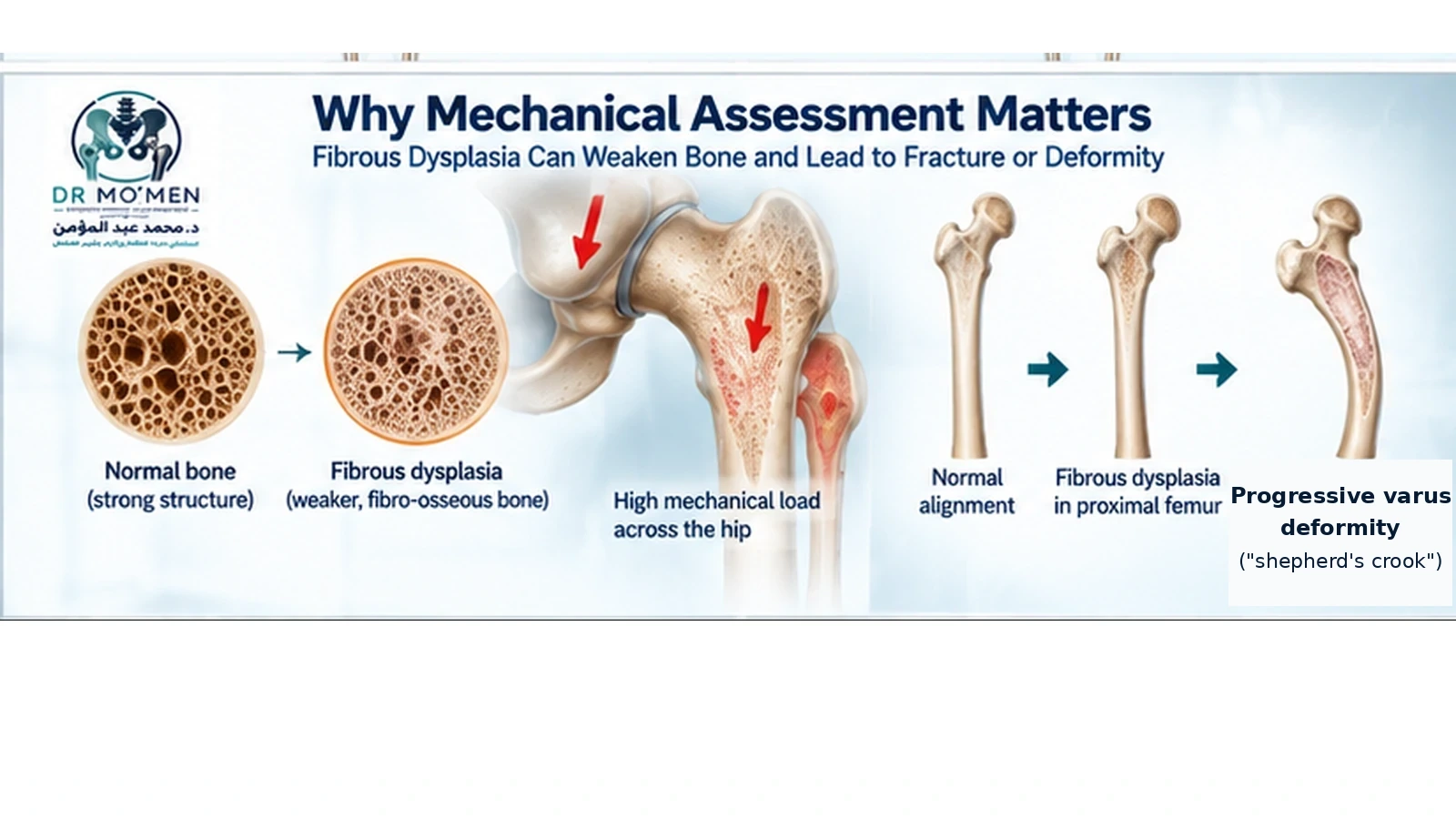
Affected bone may be mechanically weaker.
A fracture may occur through a significant lesion, particularly in a weight-bearing bone.
The consequences depend on:
- lesion extent
- fracture pattern
- bone quality
- deformity
- age
- previous operations
Progressive deformity
When extensive fibrous dysplasia affects a load-bearing bone, repeated abnormal loading may gradually lead to deformity.
The classic example is the proximal femur, where progressive varus deformity can develop in severe disease.
This is sometimes described as a “shepherd's crook” deformity.
The important issue is not the name of the deformity but its mechanical consequences:
- altered hip biomechanics
- shortening
- limp
- recurrent fracture risk
- increasing reconstruction difficulty
What Does Fibrous Dysplasia Look Like on X-Ray?
Fibrous dysplasia can have a characteristic radiographic appearance, but it does not always look identical.
One commonly described feature is a ground-glass matrix.
Other features may include:
- expansion of the bone
- relatively well-defined margins
- cortical thinning
- deformity
- varying degrees of sclerosis or lucency
Radiographic appearance also depends on:
- anatomical site
- age
- extent of disease
- previous fracture
- previous surgery
Therefore, the words “ground glass” can support the diagnosis but should not be treated as an automatic diagnosis in isolation.
What Tests Are Used to Diagnose Fibrous Dysplasia?
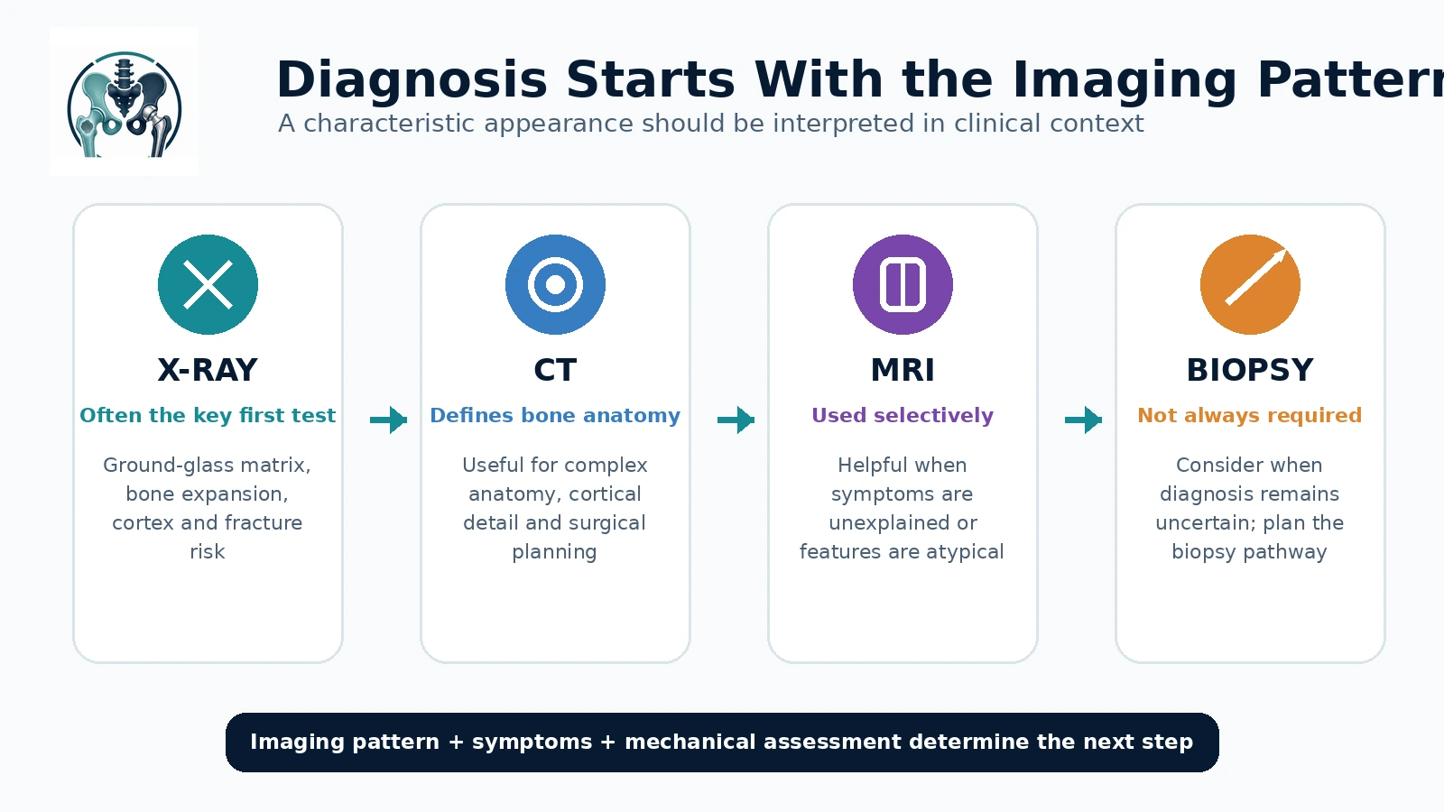
1. Plain X-rays
X-rays are often the most important first imaging investigation for a skeletal lesion.
They help evaluate:
- lesion location
- matrix
- margins
- cortical integrity
- deformity
- fracture
- relationship to the joint
2. CT scan
CT can be especially useful for:
- craniofacial fibrous dysplasia
- complex anatomy
- cortical assessment
- surgical planning
- selected indeterminate lesions
3. MRI
MRI is not always required for a classic uncomplicated lesion.
It may be useful when:
- symptoms are unexplained
- the lesion has atypical features
- a fracture is suspected
- another diagnosis is being considered
- soft-tissue extension needs evaluation
- surgery is being planned
Fibrous dysplasia can have variable MRI signal characteristics, so MRI findings should be interpreted together with radiographs and the complete clinical context.
4. Whole-skeleton assessment
When polyostotic disease is suspected, additional imaging may be used to determine how much of the skeleton is affected.
The exact imaging strategy should be individualized, particularly in children.
Does Every Fibrous Dysplasia Lesion Need a Biopsy?
No.
A lesion with a highly characteristic appearance in the correct clinical context may not always require biopsy.
However, biopsy becomes more important when:
- the diagnosis remains uncertain
- imaging is atypical
- there is unexpected progression
- another benign or malignant bone lesion remains possible
- surgery is being considered and diagnostic confirmation is required
Biopsy planning matters
If a bone tumor diagnosis is uncertain, biopsy should not be considered a casual procedure.
The biopsy approach should ideally be planned by, or coordinated with, the orthopedic oncology team that would manage the lesion if definitive surgery became necessary.
An incorrectly positioned biopsy tract can complicate later treatment.
Related: Bone Tumor Biopsy
Could Fibrous Dysplasia Be Confused With Another Bone Lesion?
Yes.
Depending on the patient's age, location and imaging appearance, the differential diagnosis may include other fibro-osseous, cystic or neoplastic lesions.
The purpose of specialist assessment is not simply to attach a name to the X-ray.
It is to determine whether:
- the imaging is genuinely characteristic of fibrous dysplasia,
- the lesion behaves as expected,
- further imaging is needed,
- biopsy would change management.
This is particularly important before curettage or another operation is performed on a lesion whose diagnosis is uncertain.
What Is McCune-Albright Syndrome?
Fibrous dysplasia can occur as part of the broader fibrous dysplasia/McCune-Albright syndrome spectrum.
In addition to skeletal disease, some patients can have endocrine abnormalities or characteristic skin pigmentation.
Potential associated endocrine problems may include abnormalities involving:
- puberty
- thyroid function
- growth hormone
- phosphate metabolism
The presence of multiple skeletal lesions, childhood presentation, unusual growth patterns or other clinical features may therefore prompt endocrine assessment.
Why Are Phosphate and Vitamin D Important?
Some patients with fibrous dysplasia can develop abnormalities of phosphate metabolism.
Low phosphate can contribute to:
- impaired mineralization
- bone pain
- increased skeletal weakness
- fracture risk
Vitamin D status and other aspects of bone metabolism may also require assessment.
This becomes particularly important before considering medications directed at bone pain or major orthopedic reconstruction.
Does Fibrous Dysplasia Always Need Treatment?
No.
Treatment depends on symptoms and mechanical risk, not merely the presence of a lesion.
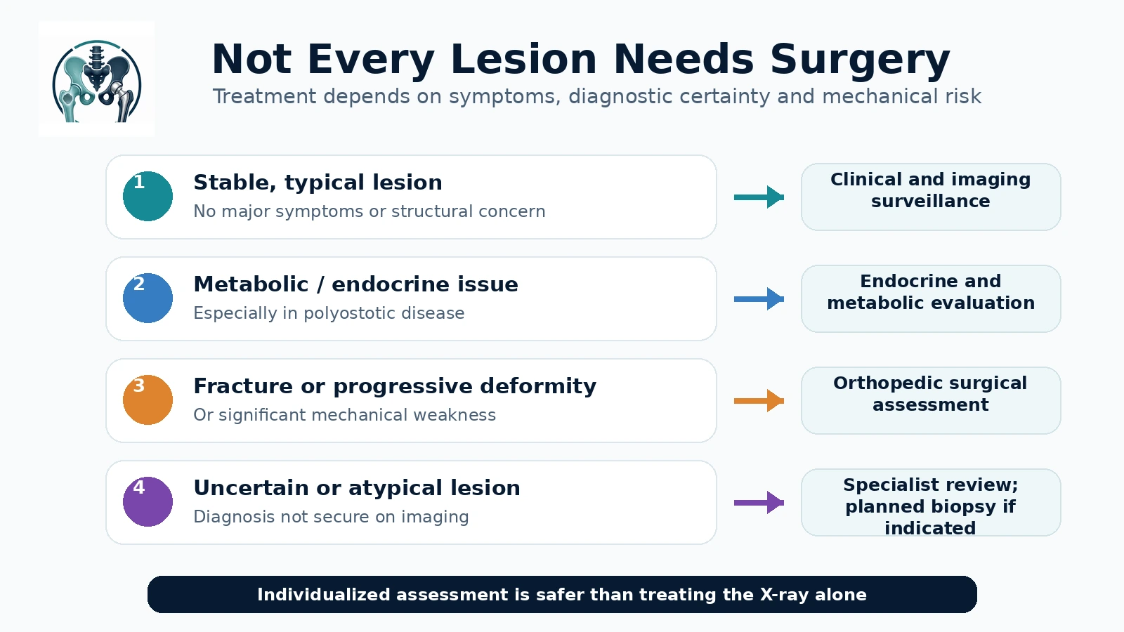
Observation may be appropriate when:
- diagnosis is secure
- lesion is stable
- there is no significant pain attributable to the lesion
- there is no fracture
- no progressive deformity is present
- the bone remains mechanically satisfactory
Observation may involve periodic clinical review and imaging according to the patient's age, lesion and symptoms.
When Might Surgery Be Needed?
Surgery may become appropriate for selected patients with:
- pathological fracture
- recurrent fracture
- progressive deformity
- significant mechanical weakness
- functional impairment
- selected painful structural lesions
- complex proximal femoral disease
- uncertainty requiring tissue diagnosis
- problems caused by previous inadequate surgery
The operation should be tailored to the mechanical problem.
Is Curettage and Bone Grafting the Standard Treatment?
Not necessarily.
Simply curetting the fibrous dysplasia and filling the cavity with graft does not reliably solve all forms of the disease, particularly extensive lesions.
Orthopedic treatment should focus on fracture management, deformity prevention/correction and durable mechanical reconstruction when needed.
The correct operation depends heavily on:
- location
- lesion extent
- deformity
- fracture pattern
- remaining bone stock
- patient age
Fibrous Dysplasia of the Proximal Femur
The proximal femur deserves particular attention because it carries substantial mechanical load.
Extensive disease may lead to:
- repeated fractures
- varus deformity
- shortening
- limp
- progressive loss of mechanical alignment
Treatment planning may therefore require assessment of the entire femur and lower-limb mechanics, not simply the visible lesion.
In more complex cases, reconstruction may involve stabilization and correction of deformity rather than simple removal of abnormal tissue.
Are Bisphosphonates Used for Fibrous Dysplasia?
Bisphosphonates have been used in selected patients with significant fibrous-dysplasia-related bone pain.
However, they should not be described as a treatment that removes the lesion or reliably stops disease progression.
Their use may be considered for selected patients with persistent moderate-to-severe bone pain after metabolic abnormalities such as hypophosphatemia and vitamin D problems have been addressed.
Medication decisions should therefore be made by clinicians experienced in metabolic bone disease/fibrous dysplasia rather than used routinely for every radiographic lesion.
What About Denosumab?
Denosumab is not routine first-line treatment for fibrous dysplasia.
Specialist sources advise considerable caution because of potential metabolic complications.
For most patients, management continues to center on diagnosis, surveillance, metabolic optimization and treatment of mechanical complications.
When Should a Patient With Fibrous Dysplasia Be Reassessed Promptly?
Specialist reassessment is appropriate when there is:
- sudden major increase in pain
- new inability to bear weight
- suspected fracture
- progressive deformity
- rapidly enlarging swelling
- unexpected radiographic progression
- cortical destruction
- a new soft-tissue mass
- new neurological symptoms
- uncertainty about the original diagnosis
These findings do not automatically mean malignancy, but they should not simply be attributed to stable fibrous dysplasia without evaluation.
What About Fibrous Dysplasia of the Skull and Face?
Craniofacial fibrous dysplasia requires a different assessment from disease in the long bones.
Potential concerns include:
- facial asymmetry
- sinus involvement
- hearing problems
- visual symptoms
- dental/maxillofacial issues
Management may involve a multidisciplinary craniofacial team.
New visual or hearing symptoms require appropriate specialist assessment.
Can Patients With Fibrous Dysplasia Exercise?
Physical activity should be individualized according to:
- location of lesions
- bone strength
- previous fractures
- deformity
- symptoms
- skeletal disease burden
Exercise may help maintain strength and function.
However, patients with significant structural skeletal involvement may need to avoid activities with a high fracture risk.
The objective is generally safe activity rather than unnecessary restriction.
When Is Orthopedic Oncology Assessment Useful?
Specialist orthopedic oncology review may be useful when:
- the diagnosis is uncertain
- imaging is atypical
- another bone tumor remains possible
- biopsy is being considered
- a large weight-bearing bone is substantially affected
- fracture or deformity is present
- previous surgery has failed
- the lesion is changing unexpectedly
- complex reconstruction may be required
For uncertain lesions, the safest sequence is often:
Clinical assessment → Appropriate imaging → Diagnostic review → Planned biopsy if genuinely necessary → Treatment according to diagnosis and mechanical risk
rather than immediate unplanned curettage.
The Role of Dr. Mo’men in Complex Fibrous Dysplasia Cases
Dr. Mohammed Abdelmoemen Abuelhadid evaluates bone lesions and complex orthopedic oncology cases with particular attention to:
- confirming whether imaging is consistent with fibrous dysplasia
- distinguishing typical lesions from those requiring further investigation
- reviewing X-rays, CT and MRI
- determining whether biopsy is necessary
- planning biopsy appropriately when diagnosis is uncertain
- assessing fracture and deformity risk
- evaluating proximal femoral and other weight-bearing lesions
- planning reconstruction when mechanical complications have developed
- reviewing patients after previous unsuccessful surgery
The aim is not to operate on every fibrous dysplasia lesion.
The treatment plan depends on diagnostic certainty, symptoms, bone strength, deformity, fracture risk and functional impact.
Patients outside Egypt may also send their available X-rays, MRI/CT images and reports for an initial specialist review before deciding whether in-person assessment is appropriate.
Orthopedic Oncology: /en/services/orthopedic-oncology/
Bone Biopsy: /en/services/bone-biopsy/
Second Opinion: /en/second-opinion/
International Patients: /en/international-patients/
For appointments or imaging review: 01021690693