Patients comparing benign and malignant bone lesions may also find this symptom guide useful: Night Bone Pain: Causes and Red Flags.

Finding a bone lesion on an X-ray, MRI, or CT scan can be alarming. Many patients immediately worry about cancer, but not every bone tumor is malignant. Some lesions are benign, slow-growing, and require observation or planned treatment. Others are aggressive or cancerous and need prompt specialist evaluation.
The difference cannot be judged safely from symptoms alone. Accurate diagnosis combines clinical examination, imaging review, and pathology when biopsy is indicated.
What Are Bone Tumors?
A bone tumor is an abnormal growth of cells within or involving bone. Primary bone tumors may arise from bone-forming, cartilage-forming, fibrous, vascular, or marrow-related tissues. Soft-tissue tumors can also secondarily involve adjacent bone.
International review for suspicious bone lesions
If a bone lesion has been found on imaging and you are outside Egypt, specialist review may help decide whether staging, biopsy planning or clinic assessment is needed.
Bone lesions are generally discussed in three clinically important groups:
- Benign bone tumors: non-cancerous lesions that do not spread to distant organs.
- Primary malignant bone tumors: cancers originating in bone or related tissues.
- Metastatic and hematologic malignancies involving bone: cancers such as metastases or multiple myeloma that affect bone but are not primary bone sarcomas.
Benign Bone Tumors
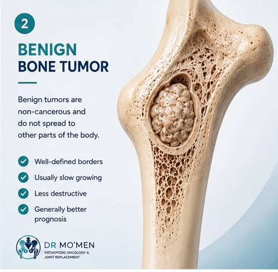
Benign bone tumors are often discovered incidentally during imaging for pain, trauma, or another orthopedic problem. Some remain silent for years, while others become symptomatic.
Examples include osteochondroma, enchondroma, non-ossifying fibroma, simple bone cyst, aneurysmal bone cyst, osteoid osteoma, fibrous dysplasia, and giant cell tumor of bone. Giant cell tumor is generally classified as benign but can be locally aggressive.
Benign does not always mean “ignore.” Monitoring may be sufficient for a small, asymptomatic, stable lesion. Planned surgery may be considered when a lesion is painful, growing, close to a joint, causing deformity, or increasing fracture risk. Specialist orthopedic oncology evaluation helps determine the appropriate pathway.
Malignant Bone Tumors and Other Malignancies Involving Bone
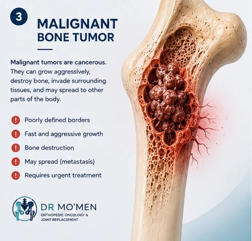
Primary malignant bone tumors include osteosarcoma, chondrosarcoma, Ewing sarcoma, and primary lymphoma of bone. These conditions differ in age distribution, imaging appearance, pathology, and response to chemotherapy or radiotherapy.
Metastatic bone disease occurs when cancer from another organ—such as breast, lung, prostate, kidney, or thyroid—spreads to bone. Multiple myeloma is a hematologic malignancy that can produce destructive skeletal lesions. These are malignant conditions involving bone but are clinically distinct from primary bone sarcomas.
Symptoms may include persistent or progressive pain, night pain, swelling, a growing mass, or fracture after minor trauma. Treatment may include surgery, chemotherapy, radiotherapy, systemic oncology therapy, stabilization, or reconstruction depending on the diagnosis. Learn more about metastatic bone disease treatment.
Key Differences Between Benign and Malignant Bone Tumors
| Feature | Benign bone tumors | Malignant bone tumors |
|---|---|---|
| Nature | Non-cancerous | Cancerous |
| Growth | Often slow, but exceptions occur | May be faster or more aggressive |
| Spread | Do not metastasize | Some can spread to other sites |
| Bone destruction | Often more contained | May be destructive or ill-defined |
| Soft-tissue mass | Less typical, although locally aggressive lesions can extend | A concerning feature when present |
| Treatment | Observation or planned local treatment | Specialist oncologic treatment based on type and stage |
| Urgency | Depends on symptoms and imaging | Prompt specialist evaluation and staging |
This comparison is general. Some benign tumors behave aggressively, and some malignant tumors initially appear subtle. Imaging and pathology must be interpreted in clinical context.
Symptoms That Require Specialist Evaluation
- Persistent bone pain lasting more than a few weeks or continuing to worsen.
- Night pain that repeatedly wakes the patient.
- Visible swelling, a deep mass, or an enlarging lump.
- Pain without a clear injury.
- A fracture after minimal trauma.
- Limping, restricted movement, weakness, or reduced function.
For a symptom-focused explanation, read early warning signs of bone tumors.
How Doctors Diagnose the Difference
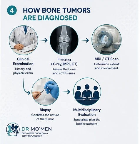
- Clinical evaluation: age, pain pattern, symptom duration, trauma history, previous cancer, swelling, limb function, and neurological findings.
- Plain X-rays: lesion location, margins, bone destruction, matrix, periosteal reaction, and fracture risk.
- MRI: marrow extent, soft-tissue extension, joint involvement, and relationship to nerves and vessels.
- CT: cortical detail, mineralized matrix, complex anatomy, and selected surgical planning.
- Staging: when malignancy is suspected, chest imaging, PET-CT, bone scan, or other studies may be selected according to the tumor type and clinical context.
- Biopsy: tissue diagnosis when imaging and clinical findings indicate that pathology is required.
Why Biopsy Planning Is Critical
Biopsy is part of the treatment strategy, not an isolated sampling procedure. The route should avoid unnecessary tissue contamination and should align with the future definitive surgical approach. Inappropriate biopsy placement can complicate limb-salvage surgery or require a larger resection.
Before biopsy, the team considers the required imaging, safest route, future incision, possible infection or other mimics, and whether image-guided core needle biopsy or open biopsy is appropriate. Patients should avoid unplanned excision or random biopsy of a suspicious mass before specialist review. See the planned bone biopsy service.
Treatment Options
Some benign lesions require observation only. Others may be treated with curettage, local adjuvants, bone graft, bone substitute, cement, or internal fixation according to the diagnosis and fracture risk.
Malignant tumors may require wide resection with an oncologic margin. Reconstruction after tumor removal can involve an endoprosthesis, graft, fixation, or joint replacement. Some tumor types require chemotherapy, radiotherapy, targeted systemic therapy, or combined treatment coordinated through a multidisciplinary team. When feasible and oncologically appropriate, limb-salvage surgery aims to remove the tumor while preserving useful function.
Why Multidisciplinary Care Matters

Bone tumor care may involve an orthopedic oncology surgeon, musculoskeletal radiologist, pathologist, medical oncologist, radiation oncologist, rehabilitation team, and pain or palliative specialists in selected metastatic cases. Coordination helps ensure diagnosis and treatment occur in the correct sequence.
When to Seek Urgent Consultation
Prompt review is warranted for rapidly increasing pain, night pain with swelling, a growing mass, fracture after minor trauma, known cancer with new bone pain, weakness or numbness, difficulty walking, spinal pain with neurological symptoms, or an imaging report describing an aggressive lesion, cortical destruction, soft-tissue mass, or suspected malignancy.
Urgent review does not mean the diagnosis is definitely cancer. It means assessment should not be delayed.
Dr. Mo’men’s Role in Complex Bone Tumor Cases
Dr. Mohammed Abdelmoemen Abuelhadid focuses on orthopedic oncology, limb-salvage surgery, bone and soft-tissue tumors, metastatic bone disease, and complex orthopedic second opinions. Patients may request review when imaging shows a suspicious lesion, biopsy is being considered, or surgery has been proposed.
For patients outside Egypt, preliminary online review of available X-rays, MRI, CT, PET-CT reports, pathology, and previous treatment details may help determine the appropriate next step before travel. Use the second-opinion pathway for details.

Medically reviewed by Dr. Mohammed Abdelmoemen Abuelhadid
Lecturer of Orthopedic Surgery at Ain Shams University, with clinical focus on orthopedic oncology, limb salvage, and complex reconstruction. About the doctor · ORCID profile
How this article was reviewed
This patient-education article was reviewed for clinical accuracy, patient safety, and decision-making clarity by Dr. Mohammed Abdelmoemen Abuelhadid. It is intended to support, not replace, clinical examination and review of the actual imaging and reports.
Authoritative medical sources
Request an appointment or imaging review
For specialist evaluation, a clinic appointment, or preliminary review of available scans and reports, contact the clinic.