What is the problem?
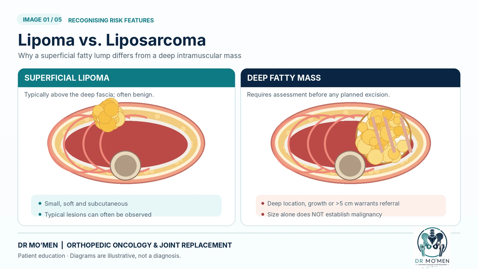
A benign lipoma is a common collection of mature fat cells and is usually superficial, soft, and slow-growing. An atypical lipomatous tumor (ALT) is a low-grade but locally active fatty tumor, while liposarcoma is a malignant fatty tumor that requires specialist orthopedic oncology evaluation and treatment planning.
Warning signs that deserve attention
These findings prompt further evaluation; they do not diagnose cancer. Some sarcomas are painless, and smaller but rapidly growing masses may also need assessment.
- A mass 5 cm or larger.
- A mass located deep to the fascia or within the muscles.
- Progressive enlargement or a change in character.
- Recurrence after previous excision.
- Symptoms suggesting pressure on nerves or vessels.
What is the difference between lipoma, ALT, and liposarcoma?
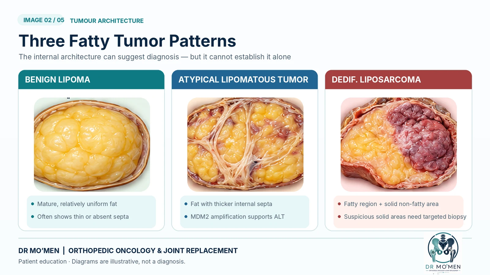
1) Benign Lipoma
- Usually small and superficial.
- Made of homogeneous mature fat.
- Does not metastasize.
2) Atypical Lipomatous Tumor (ALT)
- Often larger and deeper.
- May contain thick or irregular fibrous septa.
- Requires careful surgical planning and may need biopsy in selected cases.
Terminology note: ALT and well-differentiated liposarcoma (WDLPS) are names for the same pathological entity; the preferred term depends on anatomical site and whether complete excision is feasible. ALT in a resectable limb site generally behaves differently from dedifferentiated or myxoid liposarcoma.
3) Liposarcoma
- Includes subtypes such as myxoid, dedifferentiated, and pleomorphic liposarcoma.
- May contain non-fatty solid components.
- Requires multidisciplinary specialist care.
Diagnosis and investigations
Clinical assessment
Clinical examination helps determine size, depth, mobility, and the relationship of the mass to surrounding tissues.
MRI
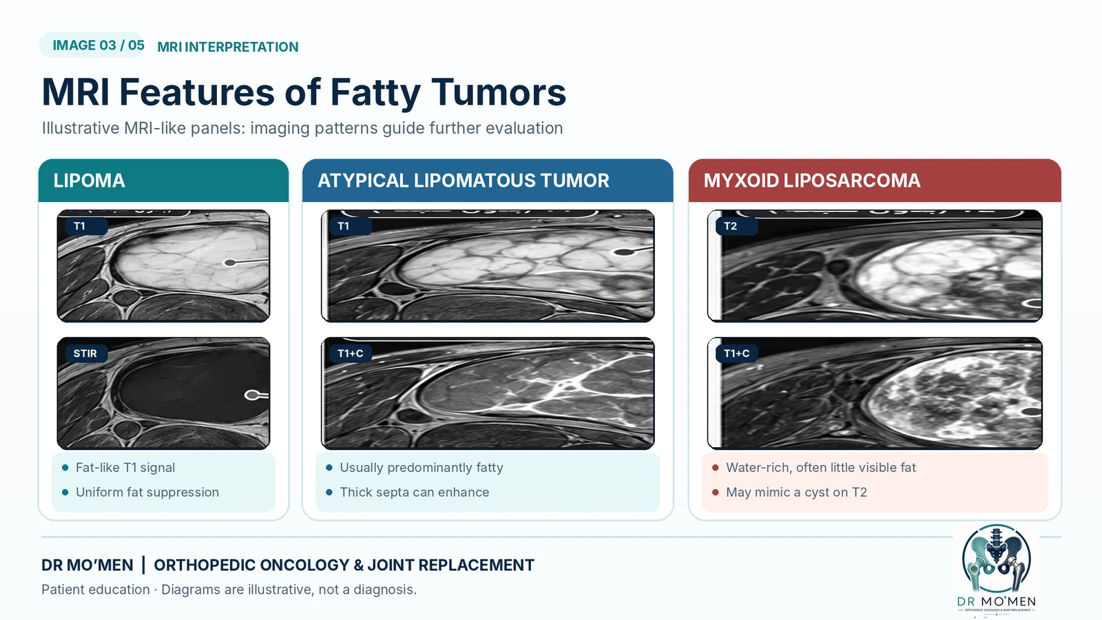
MRI is the key imaging study for large or deep fatty masses. Important features include:
- Fat homogeneity.
- Septal thickness.
- Non-fatty nodules or solid areas.
- Contrast enhancement.
- Relationship to muscles, vessels, and nerves.
Core needle biopsy
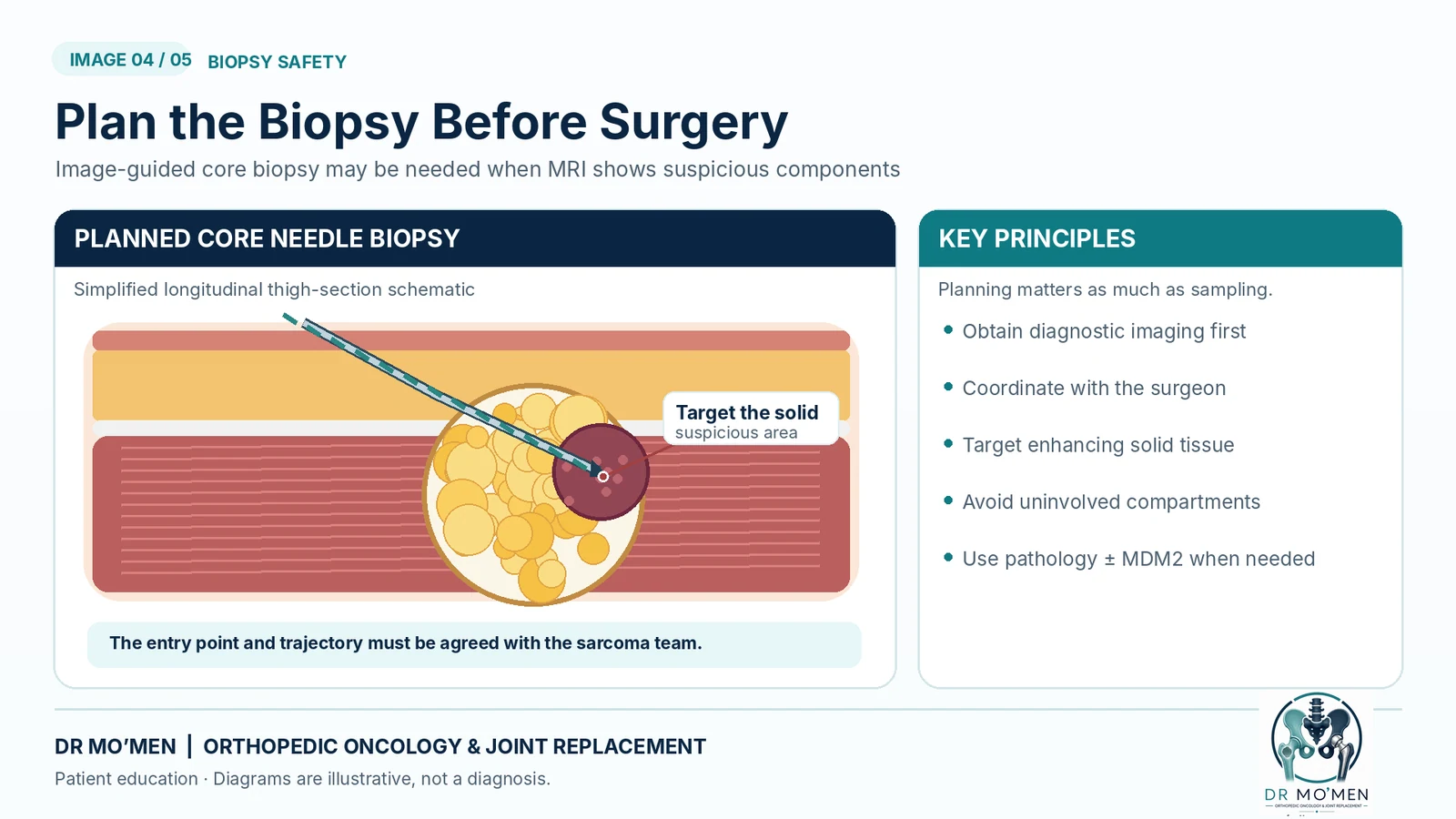
An image-guided core needle biopsy may be required for large, deep, or atypical fatty tumors, especially when thick septa or non-fatty solid areas are present. The biopsy tract should be planned with the orthopedic oncology surgeon.
MRI can raise or lower suspicion but does not establish the histologic diagnosis. Fat content varies between liposarcoma subtypes, so no single imaging sign is definitive.
MDM2 testing
MDM2 testing may help distinguish some fatty tumors in selected cases, but it should not be interpreted in isolation. It must be correlated with imaging, pathology, and sample adequacy.
Treatment options
Benign lipoma
- Observation may be appropriate if the lesion is asymptomatic.
- Excision may be performed for symptoms, diagnostic uncertainty, or patient concern.
Atypical lipomatous tumor (ALT)
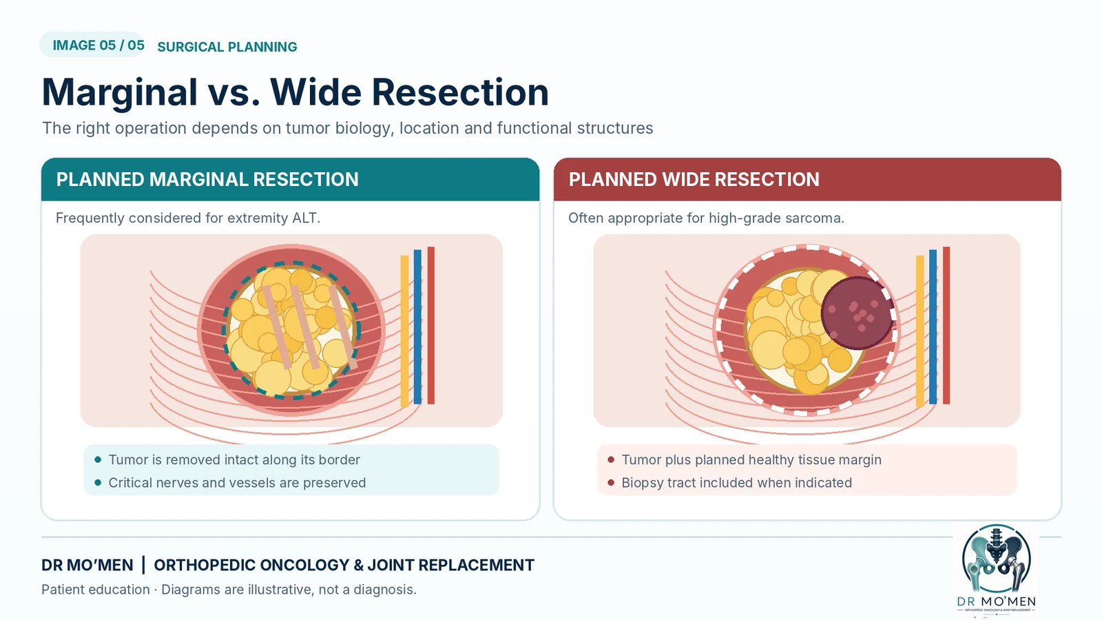
- Management depends on size, location, and relation to important structures.
- Many cases are treated with planned function-preserving excision.
- In selected situations, MRI surveillance may be part of the decision-making process.
Liposarcoma
- Usually requires planned oncologic resection, sometimes with radiotherapy or other adjunctive treatment depending on subtype, grade, and stage.
- Treatment planning may require multidisciplinary discussion.
Why should unplanned excision be avoided?
Removing a deep fatty mass as if it were a “simple lipoma” without proper imaging or planning may complicate treatment if the mass later proves to be a sarcoma. That is why suspicious lesions should be properly evaluated before surgery.
Read why unplanned excision of a suspected soft-tissue sarcoma can complicate definitive treatment.
See also our soft-tissue sarcoma diagnostic guide and orthopedic oncology services.
When should you seek specialist assessment?
You should seek specialist evaluation if the mass is:
- 5 cm or larger.
- Deep to the fascia.
- Increasing in size.
- Recurrent after previous surgery.
- Associated with neurological or vascular symptoms.
Dr. Mohammed Abdelmoemen’s role in complex cases
Dr. Mohammed Abdelmoemen Abuelhadid focuses on the evaluation and management of bone and soft-tissue tumors, including:
- Reviewing MRI findings and determining the need for biopsy.
- Proper biopsy-tract planning.
- Avoiding unplanned surgery.
- Treating complex cases and limb-salvage situations when appropriate.
- Providing specialist second opinions for patients in Egypt and abroad.
For booking or imaging review: 01021690693
Medical evidence and references
- European Society of Musculoskeletal Radiology — Imaging of adult soft-tissue tumors (2023)
- UK Guidelines for Management of Soft-Tissue Sarcomas (2024)
- WHO classification terminology: ALT / well-differentiated liposarcoma
- Mayo Clinic Laboratories — MDM2 amplification testing and interpretation
For patient education only; not a substitute for examination, imaging and specialist pathology. Draft pending physician approval before publication.