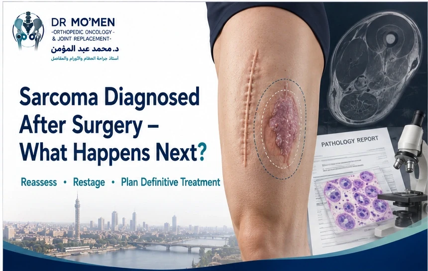
A lump in the thigh, shoulder, arm, buttock, or another part of the body may initially appear harmless. Sometimes it is removed as a presumed cyst, lipoma, hematoma, or another benign mass.
Then the pathology report unexpectedly says: soft-tissue sarcoma.
Patients understandably become concerned: Was the first operation a mistake? Has the tumor spread because it was removed without planning? Do I need another operation? Can the limb still be preserved?
The most important point is this: do not rush into another operation before the case has been properly reassessed.
An operation performed without prior oncological planning is commonly described as an unplanned excision. It may also be informally called a “whoops procedure.” This situation does not automatically mean that the final outcome will be poor or that limb salvage is impossible. However, it can make definitive treatment more complicated because microscopic tumor may remain in the operative bed and tissues outside the original tumor may have been exposed during surgery.
The next steps should usually involve specialist review of the pathology, original imaging, operative details and current surgical area before definitive treatment is decided.
Key safety message
After an unexpected sarcoma diagnosis, the next priority is structured reassessment—not another routine “simple removal.”
What Is an Unplanned Excision?
An unplanned excision occurs when a soft-tissue tumor is removed without the preoperative workup and surgical planning normally used for a suspected sarcoma. This may happen when a mass is thought to be benign before surgery.
- lipoma
- cyst
- hematoma
- benign fibrous mass
- muscle injury
- abscess
- other non-malignant soft-tissue lesion
The unexpected diagnosis is discovered only after histopathological examination.
Why Is Soft-Tissue Sarcoma Surgery Different From Removing a Benign Lump?
A benign superficial mass can often be removed directly. Sarcoma surgery is different. The operation has to consider tumor boundaries, anatomical compartments, nearby nerves and blood vessels, muscles and fascia, the biopsy or previous surgical tract, planned oncological margins, and reconstructive options.
The surgeon should ideally know what the lesion is and how extensive it is before definitive removal. This allows the incision and resection to be designed around the tumor rather than discovering the diagnosis afterwards.
Does an Unplanned Excision Mean the Surgeon Did Something Wrong?
Not necessarily. Some sarcomas can look deceptively benign clinically or even on imaging. Small superficial lesions may be particularly difficult to distinguish from benign masses.
The priority after an unexpected diagnosis is not to assign blame. It is to determine what tumor was removed, what tissues may have been exposed, whether residual disease remains, and what definitive treatment is now required.
Which Soft-Tissue Masses Need More Careful Assessment Before Removal?
Not every lump requires extensive tumor investigations. However, a mass deserves careful assessment when it is progressively enlarging, deep to the fascia, unusually firm, painful without another clear explanation, recurrent after previous removal, clinically atypical, or associated with concerning imaging features.
Size is also relevant, but a small lesion is not automatically benign. When the clinical picture is uncertain, appropriate imaging before surgery can reduce the risk of an unplanned excision.
What Should Happen After the Pathology Says “Sarcoma”?
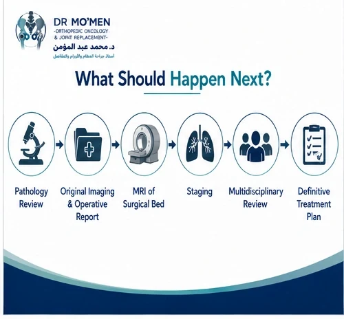
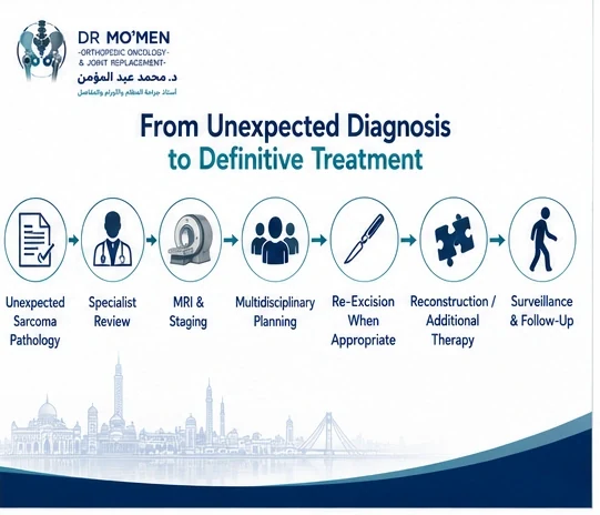
The next step is usually not another immediate excision. A structured reassessment is needed. This may include pathology review, review of preoperative imaging, review of the operative report, MRI of the surgical area, appropriate staging, multidisciplinary sarcoma review, and planning of definitive treatment.
Each step can influence the extent of additional surgery.
Step 1: Confirm the Pathology
Soft-tissue sarcoma includes many different histological subtypes. Treatment can differ according to subtype, grade, tumor biology, anatomical site and margin status.
In an unexpected diagnosis, specialist pathology review may be appropriate, particularly if terminology is unusual, the tumor is rare, the grade is uncertain, the diagnosis will significantly alter treatment, or molecular testing may be relevant.
This may involve review of the pathology report, slides, paraffin blocks, immunohistochemistry, and molecular testing where appropriate. The goal is to make sure the definitive treatment is based on the correct diagnosis.
Step 2: Obtain the Original Operative Information
The original operative report can be extremely valuable. Important details include the incision location, depth of the tumor, whether the tumor was removed intact, whether the capsule was disrupted, whether the mass was fragmented, muscles or fascia entered, and drains placed.
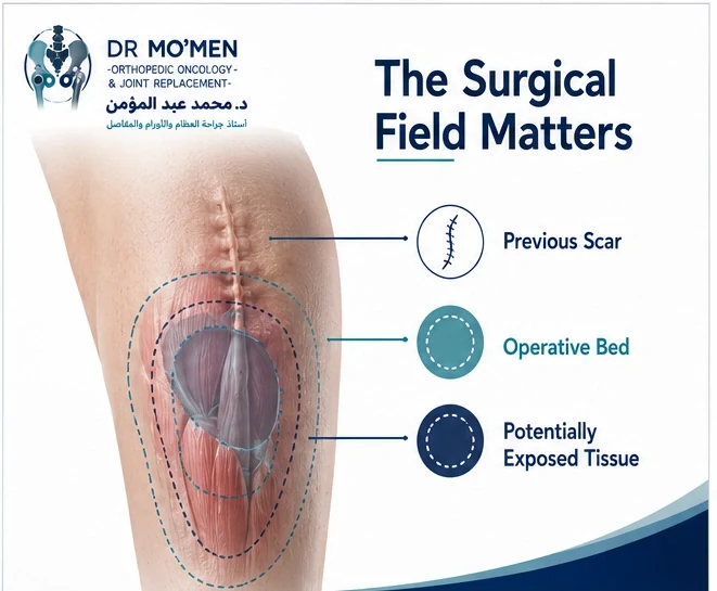
The operative scar is not always the only area that matters. The previous surgical field may influence the definitive resection.
Why Can a Drain Site Matter?
If a drain was used after an unplanned tumor excision, its pathway may need to be considered during definitive planning. The principle is similar to biopsy planning: tissues exposed during the initial procedure may need to be assessed as part of the potentially contaminated field. The significance depends on the individual operation.
Step 3: MRI of the Surgical Area
MRI is often important after an unplanned excision. It can help assess residual mass, the operative cavity, surrounding edema, hematoma, scar tissue, relationship to muscles and fascial planes, major nerves and vessels, and adjacent bone.
However, postoperative MRI can be difficult to interpret. Scar tissue, edema and postoperative change may mimic residual tumor. MRI findings should therefore be interpreted together with the original pathology and operative information.
Can MRI Prove That No Tumor Is Left?
Not always. A postoperative MRI that does not show a clear mass does not necessarily prove that all microscopic tumor has been removed. Residual sarcoma may be microscopic and therefore not reliably visible on imaging. This is one of the key reasons why management cannot be based on MRI alone.
Step 4: Review the Original Imaging
If MRI, ultrasound or CT was performed before the first operation, those images should be obtained whenever possible. They may help establish original tumor size, location, depth, anatomical compartment, relation to neurovascular structures, whether the tumor was superficial or deep, and whether aggressive features were already present.
Preoperative imaging can significantly improve reconstruction of what happened during the first procedure.
Step 5: Restaging
When soft-tissue sarcoma is confirmed, appropriate staging may be required. The exact staging strategy depends on sarcoma subtype, grade, anatomical location and clinical context. The aim is to determine whether disease appears localized or has spread elsewhere.
Step 6: Multidisciplinary Review
A newly diagnosed sarcoma after unplanned surgery should ideally be discussed within a sarcoma multidisciplinary pathway. Depending on the case, the team may include orthopedic oncology, surgical oncology, musculoskeletal radiology, pathology, medical oncology, radiation oncology, plastic or reconstructive surgery, vascular surgery and rehabilitation.
The team considers the diagnosis, grade, original tumor location, previous operation, margins, current imaging, staging and reconstructive options.
What Does a Positive Margin Mean?
A positive surgical margin means tumor cells are identified at the edge of the removed specimen. This suggests that microscopic disease may remain.
However, even when the initial pathology report describes negative margins, an unplanned excision may still require specialist review because the specimen and surgical field were not necessarily designed according to sarcoma oncological principles. The significance of margins should therefore be interpreted in context.
Is Another Operation Usually Needed?
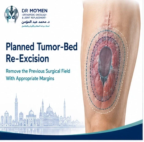
Many patients with an unplanned excision require consideration of planned re-excision of the previous tumor bed. The purpose is to remove tissue that may contain residual microscopic tumor. This is often called tumor-bed re-excision or wide re-excision.
The exact need and extent of re-excision depend on pathology, grade, margins, tumor location, original operative field, imaging, patient factors and the expected morbidity of further surgery. Therefore, not every case should be treated by an identical operation.
What Does Tumor-Bed Re-Excision Involve?
The surgeon may need to remove the previous scar, operative cavity, tissues around the previous tumor location, involved fascia, selected muscle, a drain tract where relevant, and other tissues believed to be part of the exposed field.
The amount removed may be larger than the original tumor itself. This is why the first operation can influence the complexity of definitive treatment.
Can the Re-Excision Be More Extensive Than the First Operation?
Yes. The first surgeon may have removed only a relatively small mass. After diagnosis, definitive oncological surgery may need to remove a wider area because the previous operation may have exposed adjacent tissues.
The resulting defect can sometimes require complex wound closure, skin graft, local flap, muscle flap, reconstructive plastic surgery, or vascular reconstruction in selected cases. Multidisciplinary planning can therefore be particularly important.
Can the Limb Still Be Saved?
In many cases, yes. An unplanned excision does not automatically mean amputation. Limb-preserving surgery may still be possible if adequate local control can be achieved, major neurovascular structures can be preserved or reconstructed when appropriate, the resulting defect can be reconstructed, and expected function remains reasonable.
However, the operation can become more complex if the initial procedure has extensively exposed surrounding tissue. For wider reconstruction principles, see limb salvage surgery for bone tumors.
When Might Amputation Be Considered?
Amputation may be considered in selected complex circumstances, such as when complete resection cannot reasonably be achieved with limb preservation, major neurovascular structures are extensively involved, repeated recurrence has compromised the limb, severe infection complicates the surgical field, or reconstruction would result in very poor function.
It should not be assumed to be necessary simply because the first excision was unplanned.
Does Every Patient Need Radiotherapy?
No. The role of radiotherapy depends on sarcoma subtype, grade, tumor size, anatomical location, margins, local recurrence risk, ability to obtain further surgical margins and previous radiation exposure. Radiotherapy decisions should be individualized with the sarcoma team.
Does Every Patient Need Chemotherapy?
No. Chemotherapy has different roles across different soft-tissue sarcoma subtypes and clinical scenarios. Some sarcomas are more sensitive to systemic therapy than others. The decision depends on histological subtype, grade, stage, patient age, overall health, metastatic risk and multidisciplinary recommendation.
What If the Pathology Says “Low Grade”?
Low-grade sarcomas may behave differently from high-grade tumors, but they still require appropriate assessment. The treatment decision depends on more than grade alone, including local extent, subtype, margin, anatomy, recurrence risk and morbidity of re-excision.
What If the Tumor Was Very Small?
Small sarcomas can still require oncological management. Tumor size matters, but it is not the only factor. A small malignant mass removed through an inappropriate plane can still create a larger surgical field requiring specialist assessment.
What If the Tumor Was Removed in Pieces?
Fragmentation can make subsequent assessment more difficult. It may complicate evaluation of true tumor size, surgical margin, orientation and exact relationship to surrounding tissues. Detailed operative information and multidisciplinary review become particularly important.
What If the Pathology Report Is Uncertain?
Do not proceed directly to major definitive treatment without clarifying the diagnosis. Specialist pathology review may be particularly helpful when the report includes uncertain malignant potential, unusual spindle-cell tumor, atypical lipomatous lesion, undifferentiated neoplasm, uncertain grading, complex immunohistochemistry, or rare sarcoma terminology.
Different diagnoses can require very different treatment strategies.
What If the Lump Has Already Grown Back?
A recurrent mass after previous excision should be reassessed carefully. Evaluation may include MRI, pathology review, staging, biopsy if required, and assessment of the previous operative field.
Repeated simple excision without oncological planning can make later definitive surgery progressively more difficult. See also recurrent tumor assessment.
Why You Should Avoid Another “Simple Removal”
Once sarcoma has been diagnosed or strongly suspected, another routine excision without oncological planning should generally be avoided. The next operation should answer three questions:
- What tissue must be removed for local disease control?
- What important structures can safely be preserved?
- How will the resulting defect be reconstructed?
This is fundamentally different from simply removing a visible lump.
What Records Should You Collect?
Patients should obtain copies of the pathology report, pathology slides or blocks where possible, original ultrasound, original MRI or CT, operative report, discharge summary, current MRI, staging imaging, and oncology reports.
Do not rely only on a verbal summary of the first operation. The original documents can significantly improve treatment planning.
What If the Surgery Was Performed Abroad or at Another Hospital?
That does not prevent proper reassessment. The most useful approach is to collect original pathology, imaging files, operative documentation and current imaging. If some information cannot be retrieved, specialist assessment can still begin using the available records.
Warning Signs That Need Prompt Assessment
Patients should seek timely evaluation if they develop a rapidly enlarging new mass, recurrent swelling at the surgical site, progressive pain, neurological symptoms, unexplained wound problems, or new functional impairment.
Urgent local care is appropriate for severe bleeding, acute neurovascular symptoms, serious infection or other emergency problems.
When Should You Seek an Orthopedic Oncology or Sarcoma Second Opinion?
A specialist opinion is particularly valuable when sarcoma was diagnosed only after the mass had been removed, the pathology diagnosis is uncertain, another operation has been recommended without MRI or staging, amputation has been proposed, wide re-excision is being discussed, a major nerve or blood vessel may be involved, the mass has already recurred, multiple previous operations have been performed, or radiotherapy or chemotherapy recommendations are unclear.
A second opinion does not necessarily mean the previous treatment was incorrect. It can help reconstruct what happened during the first operation and plan the safest next step. Request an orthopedic oncology second opinion.
International Patients: What Should You Send Before Travelling?
If you live outside Egypt and soft-tissue sarcoma was unexpectedly diagnosed after surgery, send as much original information as possible. Useful records include pathology report, pathology slides or blocks if available, original preoperative imaging, current MRI, operative report, staging studies, oncology reports, radiotherapy records, chemotherapy records, and discharge summary.
The combination of preoperative imaging, operative report, pathology and current MRI can be particularly valuable.
Send Your Records Before You Travel
A preliminary review may help determine whether pathology review is needed, whether current MRI is adequate, whether staging is required, whether tumor-bed re-excision should be assessed, whether limb preservation appears technically feasible, whether plastic or vascular reconstruction may be needed, and whether travel for specialist evaluation is reasonable.
Remote review does not replace physical examination, pathology confirmation, staging or multidisciplinary sarcoma decision-making.
For appointments or medical-record review: +20 102 1690693
International Patients Request an Orthopedic Oncology Second Opinion
About Dr. Mohammed Abdelmoemen Abuelhadid
Dr. Mohammed Abdelmoemen Abuelhadid is an Orthopedic Oncology & Joint Replacement Surgeon and Lecturer of Orthopedic Surgery at Ain Shams University.
His clinical areas include orthopedic oncology, bone and soft-tissue tumors, limb-salvage surgery, tumor resection and reconstruction, recurrent tumors, complex orthopedic reconstruction, metastatic bone disease, megaprosthetic reconstruction, and second medical opinions for complex orthopedic cases.
For an unexpectedly diagnosed sarcoma, the priority is accurate reassessment and definitive treatment planning rather than rushing into another procedure.
Frequently Asked Questions
What is a “whoops procedure” in sarcoma surgery?
It is an informal term used for an unplanned removal of a soft-tissue sarcoma before the tumor was recognized and treated according to sarcoma surgical principles.
Does an unplanned excision mean cancer has spread?
No. An unplanned excision does not automatically mean metastatic spread. The main concern is that residual microscopic tumor may remain locally or that additional tissue planes may have been exposed during surgery. Appropriate staging is still required according to the diagnosis.
Do I always need another operation?
Not every case is identical, but many patients require consideration of planned tumor-bed re-excision. The decision depends on pathology, margins, grade, MRI findings, the original surgical field and expected morbidity.
Can MRI tell if all the sarcoma was removed?
MRI can identify visible residual tumor, but it cannot reliably exclude microscopic residual disease. MRI therefore cannot be used alone to decide that no further treatment is necessary.
Can limb salvage still be possible after unplanned surgery?
Yes, frequently. However, the definitive operation may be more extensive because the previous surgical field may need to be included in the resection.
Should pathology be reviewed again?
Specialist pathology review may be valuable when the diagnosis is unexpected, rare, uncertain, or likely to substantially change treatment planning.
Should I have another biopsy?
Not automatically. If the original pathology is definitive, a repeat biopsy may not be necessary. If the diagnosis is uncertain or a new suspicious lesion has appeared, biopsy may be considered after specialist planning.
How this article was reviewed
This patient-education article was reviewed for clinical accuracy, patient safety, and decision-making clarity by Dr. Mohammed Abdelmoemen Abuelhadid. It is intended to support, not replace, specialist sarcoma assessment, pathology review, staging, imaging review, or multidisciplinary treatment planning.

Author and medical reviewer
Dr. Mohammed Abdelmoemen Abuelhadid — Orthopedic Oncology & Joint Replacement Surgeon and Lecturer of Orthopedic Surgery at Ain Shams University.
Medical Disclaimer: This article is for patient education and does not replace clinical examination, specialist pathology review, imaging, staging investigations or individualized multidisciplinary sarcoma care.