
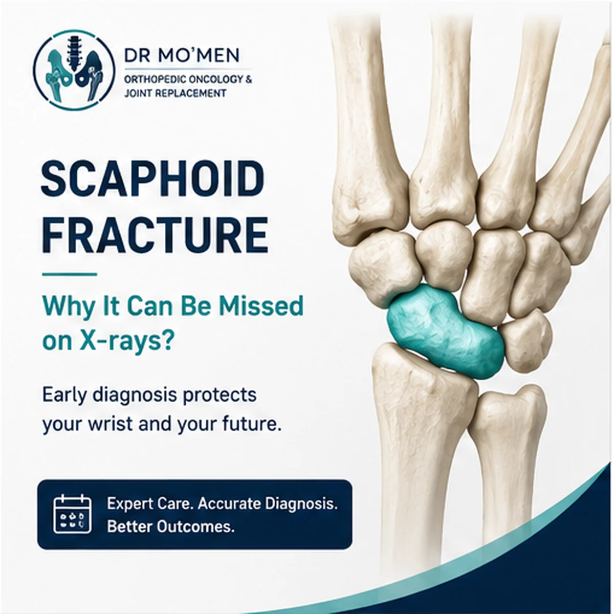
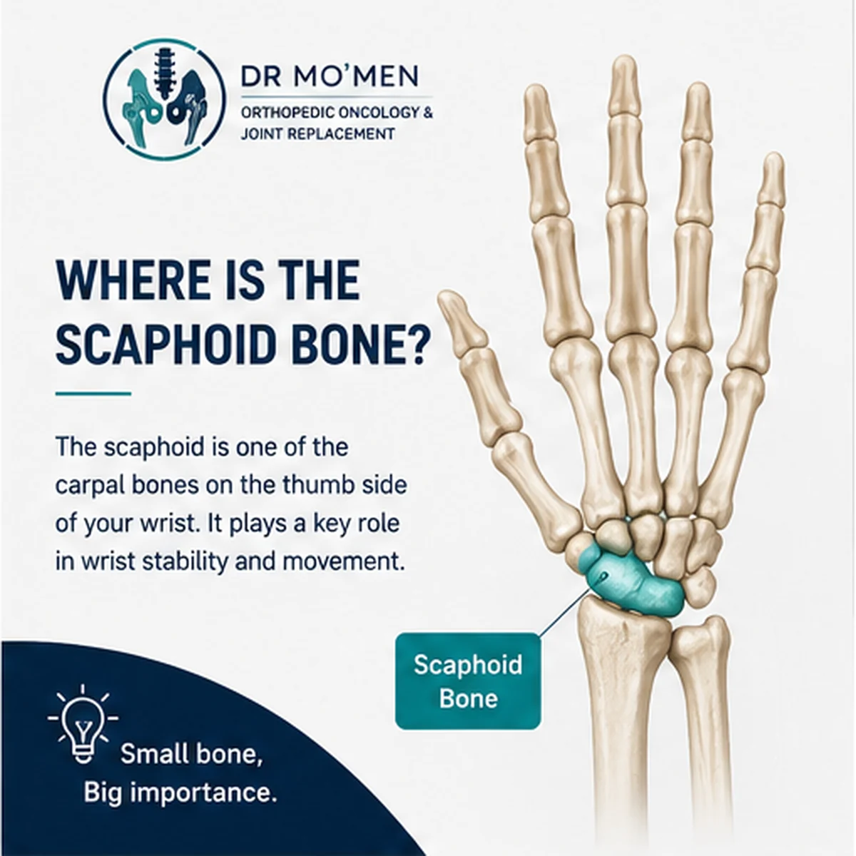
What Is the Scaphoid Bone?
The scaphoid is one of the eight small carpal bones that form the wrist. It sits on the thumb side and helps connect the two rows of carpal bones. This position makes it important for wrist stability, coordinated movement, and the transfer of load through the hand.
The bone also has a distinctive blood supply. In some fracture patterns—particularly those involving the proximal part of the scaphoid—the blood flow to one fragment may be vulnerable. Delayed diagnosis, displacement, and instability can increase concern about delayed union, nonunion, or osteonecrosis in selected cases.
How Does a Scaphoid Fracture Happen?
The typical mechanism is a fall onto an outstretched hand, with force travelling through the palm and wrist. The injury may occur during football, running, cycling, contact sports, a simple slip, or a road-traffic incident.
A major deformity is uncommon, so the wrist may not look “broken” even when a fracture is present.
Symptoms and Warning Signs
These symptoms can overlap with a wrist sprain. Being able to move the fingers or having only mild swelling does not reliably exclude a fracture.
- Pain or tenderness at the base of the thumb or on the radial side of the wrist.
- Tenderness in the anatomical snuffbox—the small hollow that appears when the thumb is extended.
- Pain when gripping, twisting, pushing up from a chair, or loading the wrist.
- Mild swelling, reduced grip strength, or pain with wrist movement.
- Persistent focal pain after a fall despite an initially normal X-ray.
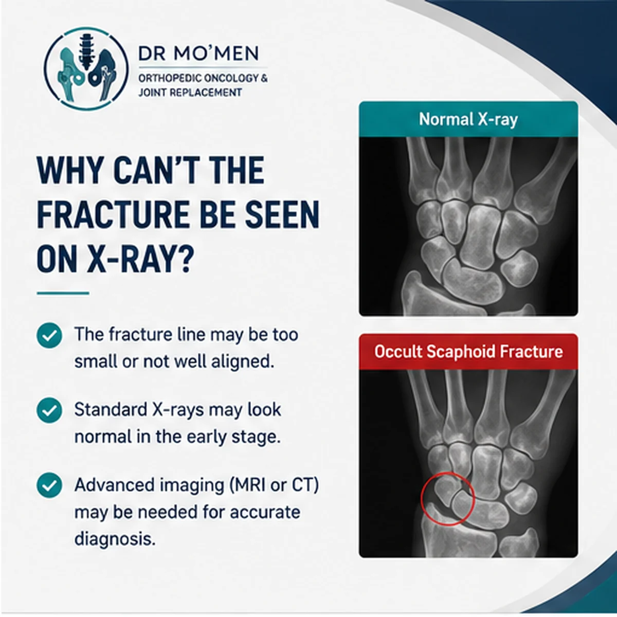
Why Can the Initial X-ray Look Normal?
A true scaphoid fracture may be occult on the first radiographs. The fracture line may be very fine, may not be aligned with the X-ray beam, or may be obscured by the overlapping carpal bones. Early bone changes can also be too subtle to identify immediately.
When clinical suspicion remains high, the wrist may be protected while further assessment is arranged. MRI can help detect an occult fracture and associated soft-tissue injury. CT can provide detailed information about the fracture line, displacement, alignment, healing, and surgical planning in selected patients.
How Is a Scaphoid Fracture Diagnosed?
Injury History
The clinician considers how the injury occurred, when it happened, where the pain is located, and whether the patient has continued to use the wrist. Previous immobilization, repeated injuries, and work or sports demands are also relevant.
Clinical Examination
Examination usually includes localized palpation around the scaphoid and anatomical snuffbox, wrist movement, grip-related pain, and assessment of circulation and sensation. No single test should be interpreted in isolation.
Imaging
- Dedicated scaphoid radiographs are usually the initial imaging test.
- MRI may help confirm or exclude an occult fracture when initial radiographs are negative or indeterminate.
- CT may help evaluate displacement, stability, union, deformity, and operative planning.
MRI or CT After a Normal Scaphoid X-ray?
| MRI | CT |
|---|---|
| Detects occult fracture and marrow injury | Defines fracture line and bony geometry in detail |
| Useful when early X-rays are negative or indeterminate | Useful for displacement, alignment and deformity |
| Can show associated soft-tissue injury | Useful for assessing union/nonunion in selected follow-up cases |
| Helps confirm or exclude an early hidden fracture | Helps operative planning when fixation is being considered |
Treatment Options
Treatment is individualized according to the fracture site, displacement, stability, time since injury, evidence of healing, and the patient’s functional needs. A professional athlete, a manual worker, and a patient with a delayed presentation may require different discussions even when the X-ray appearances seem similar.
For broader fracture-care context, see the Fracture & Trauma Surgery service.
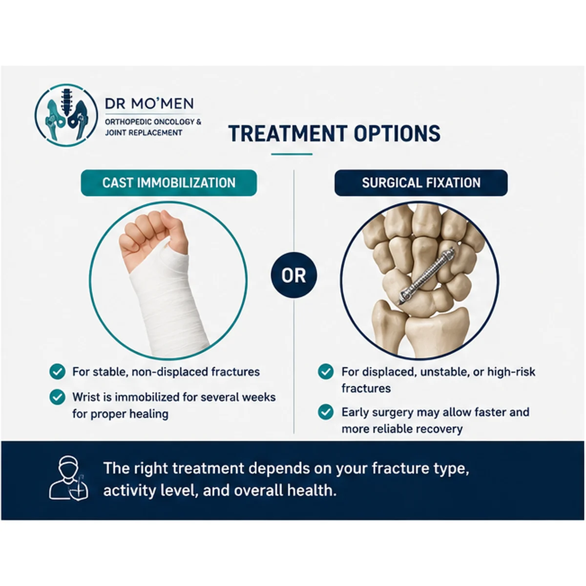
Cast or Brace Immobilization
Some recent, stable, non-displaced fractures can be managed without surgery. The clinician selects the type and duration of immobilization and arranges follow-up imaging when needed. The patient should keep the wrist protected, avoid premature loading, and attend follow-up until satisfactory healing is demonstrated.
Related cast and lower-limb injury reading: Foot Swelling After a Cast and Ankle Sprain: Cast or Physiotherapy?.
Surgical Fixation
- Surgery may be considered for displaced or unstable fractures, fractures in higher-risk locations, established nonunion, or situations in which fixation is expected to improve alignment and the probability of healing. A headless compression screw is commonly used, while delayed cases may require additional procedures depending on bone quality, deformity, and vascularity.
- Not every scaphoid fracture requires an operation, and surgery does not guarantee an immediate return to activity. Potential benefits must be balanced against risks such as infection, stiffness, injury to surrounding structures, persistent symptoms, and the need for rehabilitation and imaging follow-up.
What Can Happen If Diagnosis or Treatment Is Delayed?
Selected patients may develop delayed union, nonunion, compromised blood supply to a fragment, altered wrist mechanics, or post-traumatic arthritis. These complications do not occur in every case, but they explain why persistent thumb-side wrist pain after a fall should not be dismissed solely because an early X-ray was reported as normal.
When Should You Seek Urgent Assessment?
- Severe or worsening pain after a recent injury with marked difficulty using the hand.
- Numbness, cold fingers, pallor, blue discoloration, or reduced circulation.
- Major swelling, deformity, an open wound, or a high-energy injury.
- Focal scaphoid pain despite a normal initial X-ray.
- Increasing pain, pressure, numbness, or colour change while wearing a cast or splint.
The Role of Dr. Mo’men in Complex Cases
Delayed presentations, persistent pain with unclear imaging, suspected nonunion, and fractures with displacement or deformity may require detailed review of radiographs, CT, or MRI alongside a focused clinical examination.
Patients outside Egypt may request an online second-opinion review of their scans and reports. Acute injuries and red-flag symptoms still require timely local examination, because circulation, sensation, swelling, and skin condition cannot be assessed fully online.
For appointments or X-ray review: 01021690693