
A 50-year-old man presented with a swelling at the inferior scapular region, pain during shoulder movement, and marked scapulothoracic crepitus. This pattern raises suspicion for elastofibroma dorsi, a benign deep soft-tissue lesion classically situated between the inferior scapula and the posterior chest wall.
The case in one minute
Elastofibroma dorsi may present as a shoulder problem rather than an obvious tumor. A mass at the inferior scapular border with pain or scapulothoracic crepitus should prompt examination of the infrascapular region and appropriate imaging.
- Clinical category
- Benign deep soft-tissue lesion
- Anatomical site
- Between the inferior scapula and posterior chest wall
- Symptoms
- Swelling, painful shoulder motion, and marked crepitus/snapping
- Decision illustrated
- Surgical excision for a symptomatic painful mass with mechanical crepitus
Related specialist resources
Why the Diagnosis Can Be Difficult
Elastofibroma dorsi is typically deep and slow growing. The mass may be subtle when the arm is resting, while the dominant symptoms can be pain, discomfort, snapping, crepitus, stiffness, or pain during shoulder motion. These complaints overlap with far more prevalent conditions, so the lesion can be missed if the assessment is limited to the glenohumeral joint.
What Can It Be Mistaken For?
The following are important clinical mimics and do not imply that this particular patient received each of these diagnoses. In undetected cases, symptoms may initially be attributed to:
- Rotator cuff disease.
- Subacromial impingement or bursitis.
- Scapulothoracic bursitis and other causes of snapping scapula.
- Periscapular muscular pain.
- Cervical referred pain or cervical degenerative disease.
- Other benign chest-wall/soft-tissue masses; atypical lesions also require exclusion of more aggressive soft-tissue tumors.
Accordingly, some patients may undergo analgesic treatment, physiotherapy, or shoulder-directed treatment before the infrascapular source is recognized. Persistent symptoms associated with a palpable swelling, scapular prominence, or reproducible crepitus should prompt examination of the scapulothoracic region and targeted cross-sectional imaging.
Clinical Examination: Look Beyond the Shoulder Joint
Examination should include the shoulder, scapula, and posterior chest wall. The lesion may become more apparent as the scapula moves away from the thoracic cage or with specific arm positions. An inferior-scapular mass associated with painful scapulothoracic motion or snapping strongly supports the diagnostic pathway toward elastofibroma dorsi.
Imaging
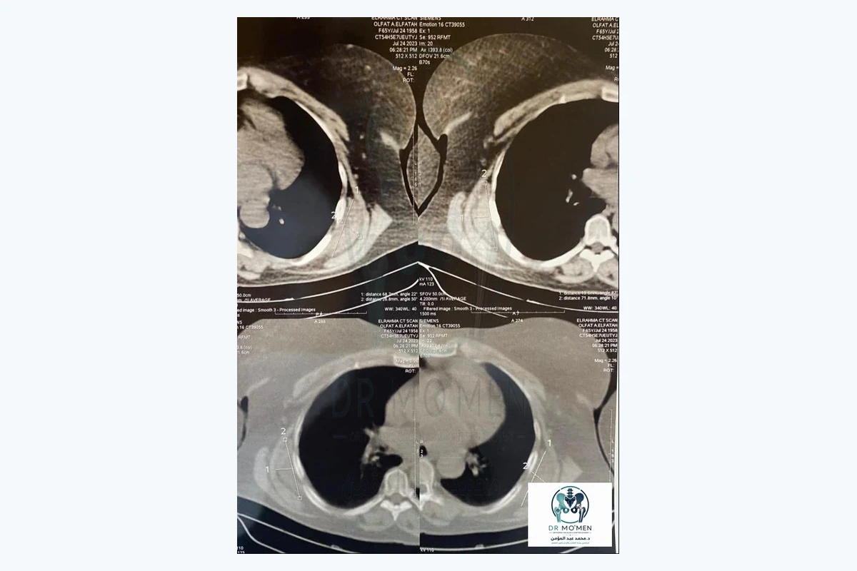
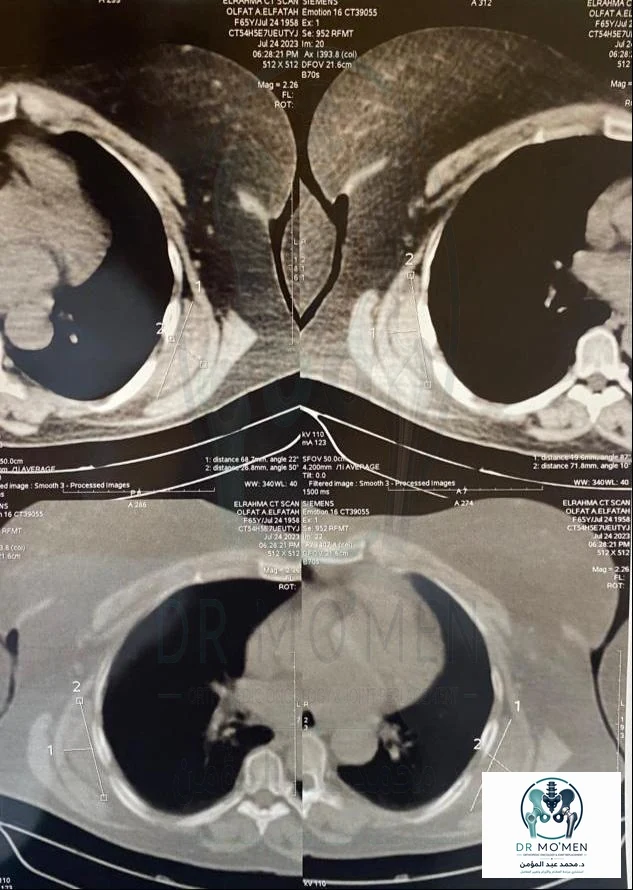
In this case, CT demonstrated a deep infrascapular soft-tissue mass adjacent to the chest wall. The characteristic location and internal fibrofatty appearance are important diagnostic clues. MRI is particularly useful when the lesion is atypical, when tissue characterization is needed, or when the relationship to adjacent muscles and the chest wall requires further definition.
Why Surgery Was Chosen
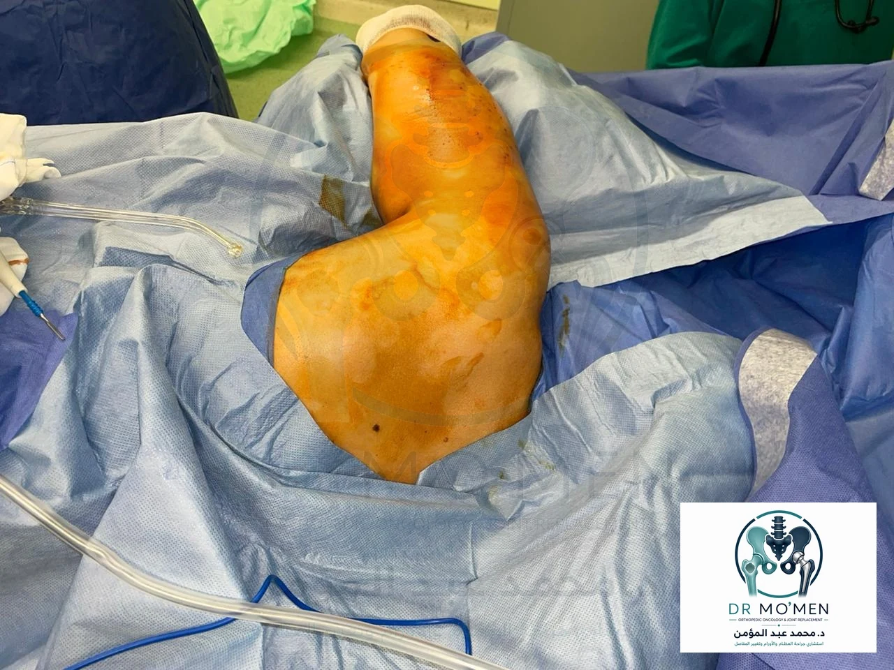
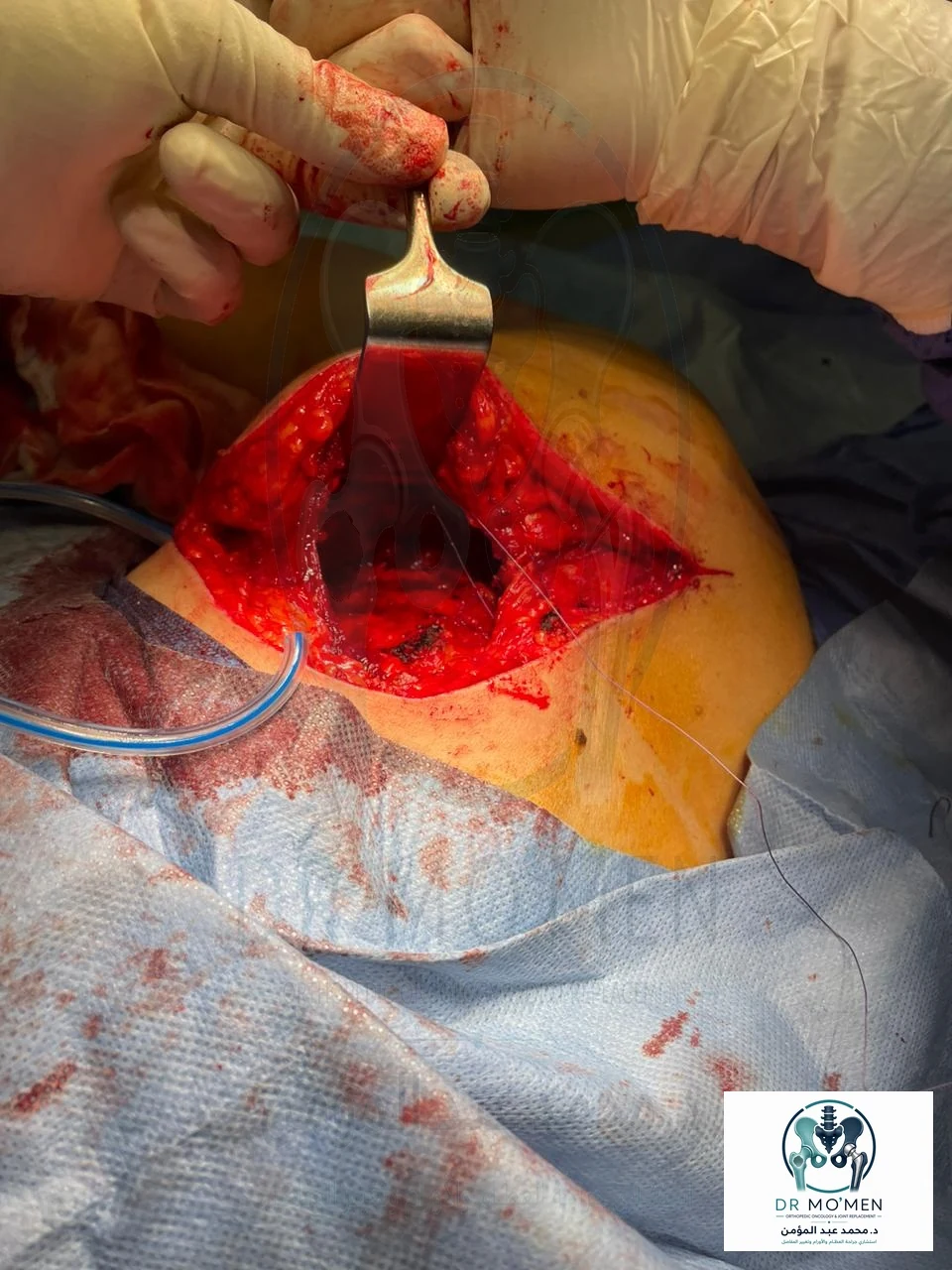
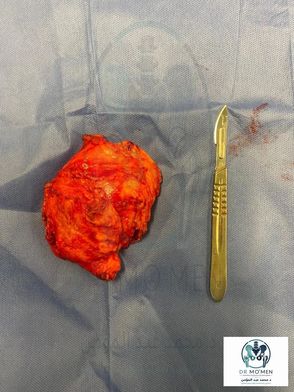
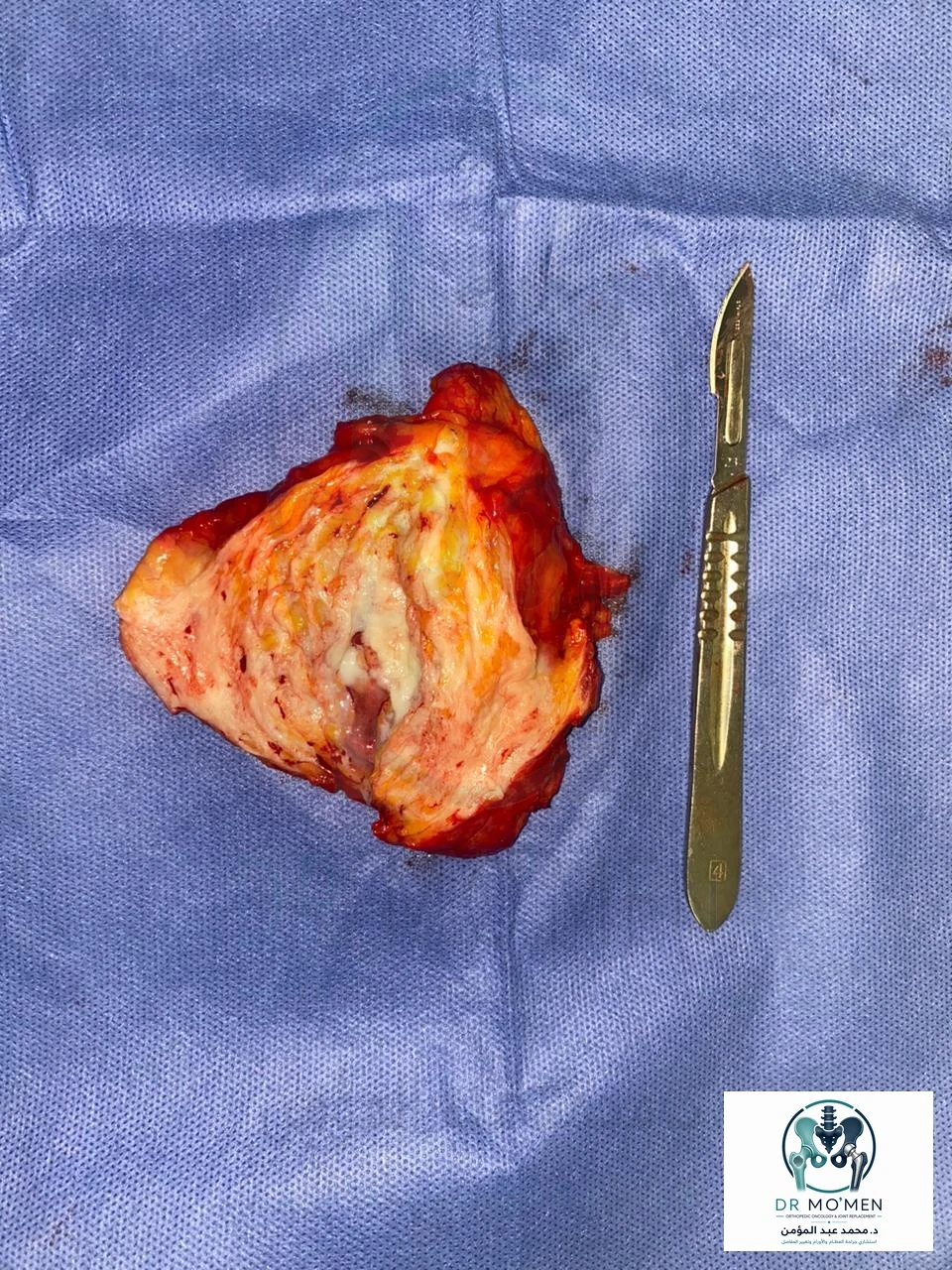
Not every radiologically identified elastofibroma dorsi requires resection. Management is primarily driven by symptoms and diagnostic certainty. This patient had a painful mass with pain on shoulder motion and pronounced mechanical crepitus; therefore, surgical excision was performed. The specimen was submitted for histopathologic assessment as part of standard surgical practice.
Operative and Imaging Figures
Educational Message
Not all shoulder pain and crepitus originate inside the shoulder joint. Pain around the inferior scapula associated with swelling, snapping, or reproducible crepitus should trigger assessment of the scapulothoracic space.
Elastofibroma dorsi is a useful diagnostic lesson because a benign deep chest-wall lesion can clinically mimic common shoulder or cervical disorders. Recognition depends on combining the symptom pattern with a dynamic scapular examination and the characteristic infrascapular appearance on CT or MRI. This approach reduces the risk of prolonged, non-targeted treatment.
Selected References
- Deveci MA, et al. Elastofibroma dorsi: Clinical evaluation of 61 cases and review of the literature. Acta Orthop Traumatol Turc. 2017;51(1):7-11. doi:10.1016/j.aott.2016.10.001.
- Smith HG, et al. Elastofibroma dorsi: The clunking tumour that need not cause alarm. Ann R Coll Surg Engl. 2016;98(3):208-211. doi:10.1308/rcsann.2016.0064.
- Daigeler A, et al. Elastofibroma dorsi - differential diagnosis in chest wall tumours. World J Surg Oncol. 2007;5:15.
Need specialist review of an infrascapular or soft-tissue mass?
Assessment depends on the clinical examination and full imaging review, especially when shoulder pain is associated with swelling or reproducible scapulothoracic crepitus.
Request specialist review