
Key points
Patient summary
Elastofibroma dorsi can mimic common shoulder conditions because the lesion is deep and symptoms are often mechanical. A clear diagnosis should come before biopsy or surgery.
- A deep infrascapular mass can be subtle at rest.
- Classic imaging may support observation, while atypical lesions require further assessment.
Contents
A deep lump beneath the shoulder blade can be difficult to recognize. Some patients notice swelling, while others mainly complain of aching, rubbing, or a snapping sensation when the shoulder moves. Because these symptoms overlap with common shoulder and scapular problems, elastofibroma dorsi may not be considered at the first visit. The key is not to assume that every posterior shoulder symptom comes from a tendon or muscle problem: a persistent infrascapular mass deserves proper clinical and imaging assessment.

What is the problem?
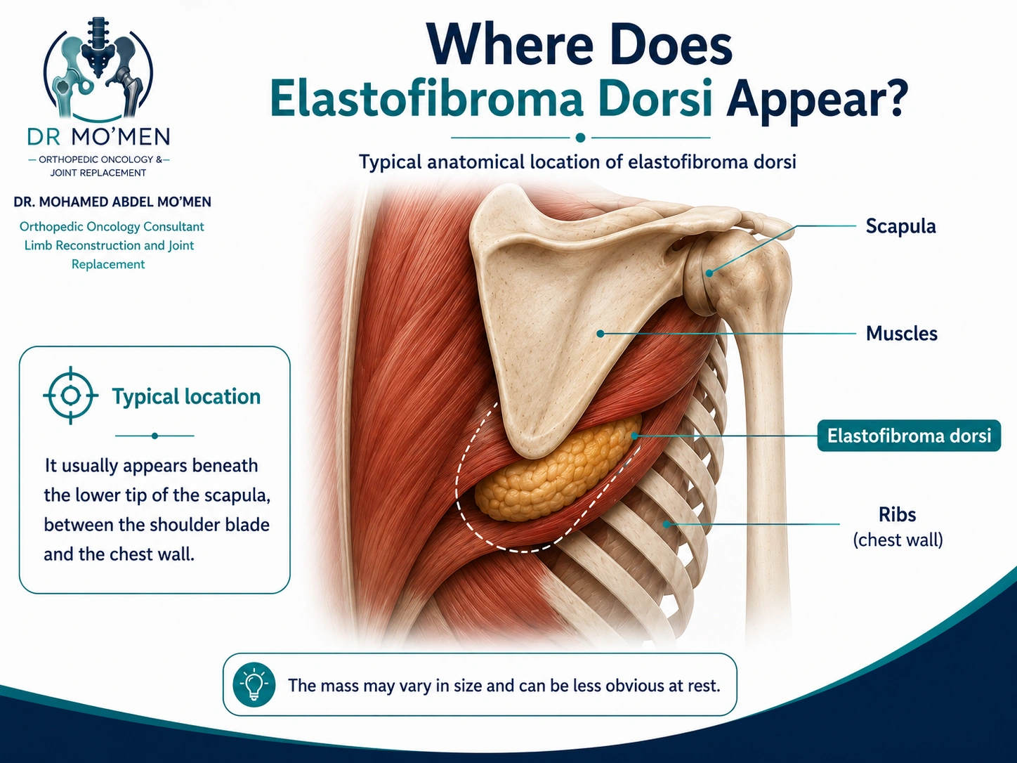
Elastofibroma dorsi is a benign, slow-growing soft-tissue lesion composed of fibrous, elastic, and fatty tissue. It classically develops deep to the lower scapular region, between the shoulder blade and the chest wall. It may occur on one side or on both sides.
The clinical challenge is not aggressive tumor behavior; it is recognition. The lesion may be deep, poorly defined on palpation, and less obvious when the arm is at rest. For that reason, symptoms can be attributed to rotator-cuff disease, muscle strain, scapular dyskinesis, mechanical back pain, or other causes of snapping scapula before the mass is identified.
A new or deep soft-tissue mass should still be assessed on its own merits. Atypical location, rapid change, or unusual imaging should prompt specialist orthopedic oncology evaluation rather than an assumption that the lesion is elastofibroma.
Symptoms and red flags
Some lesions are found incidentally and cause no symptoms. When symptomatic, the problem is often mechanical.
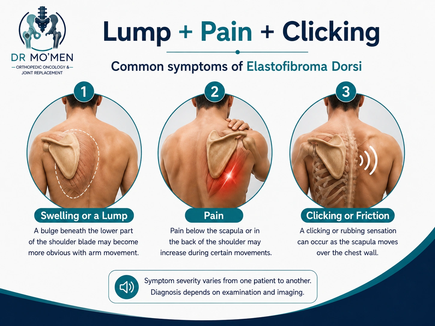
- A swelling or lump below the shoulder blade, sometimes more visible with arm movement.
- A dull ache, pressure, or discomfort behind the shoulder or beneath the scapula.
- Clicking, rubbing, or crepitus as the scapula moves over the chest wall.
- Reduced confidence in shoulder movement because certain positions reproduce discomfort.
Features that deserve prompt reassessment include rapid growth, unexpectedly severe or progressive pain, a hard fixed mass, neurological symptoms, or imaging that does not show the typical pattern. These features do not automatically indicate cancer, but they reduce the certainty of a benign clinical diagnosis. General warning patterns are reviewed in Bone Tumor Symptoms and Red Flags.
What causes elastofibroma dorsi?
The exact cause is not established. Repetitive scapulothoracic friction and chronic mechanical stress have been proposed as contributing factors, but no single mechanism explains every case. The lesion is reported more often in older adults and women, although it can also occur in men.
These demographic patterns can support clinical suspicion, but they should never replace examination and imaging.
Why can the diagnosis be delayed?
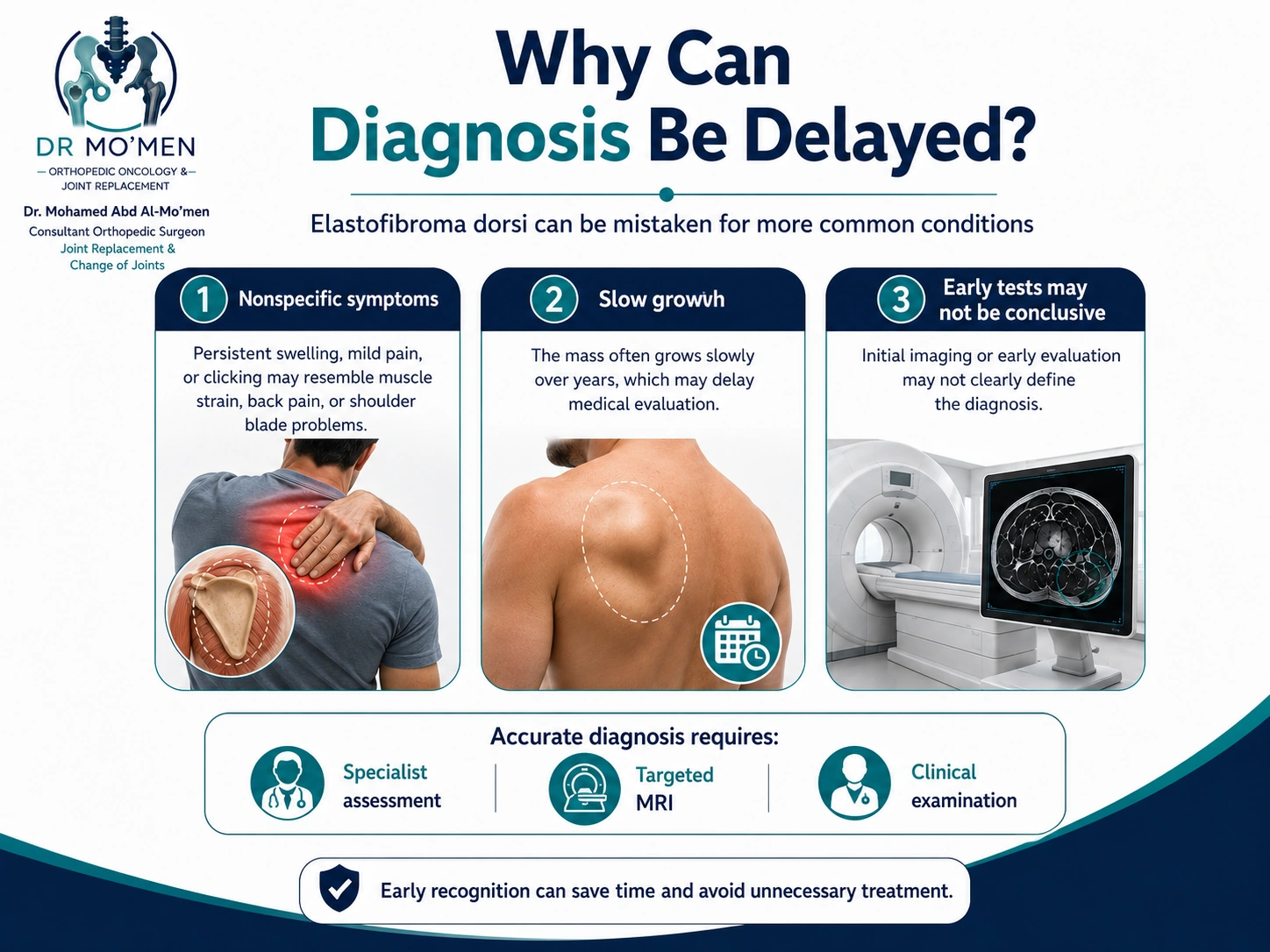
Elastofibroma dorsi is a good example of a lesion that can remain hidden behind common musculoskeletal symptoms. The mass may be deep and slow growing, while pain or clicking is mild. Temporary improvement with pain medication or physiotherapy can reinforce the impression of an ordinary shoulder disorder.
If symptoms persist, especially when a lump is palpable beneath the inferior scapular angle, the diagnostic question should be revisited rather than simply repeating the same treatment. Specialist examination and targeted imaging can clarify whether the symptoms are coming from the shoulder joint, the scapulothoracic interface, or a soft-tissue mass.
Diagnosis and imaging
Assessment begins with the history and a focused examination, ideally checking the mass with the arm in different positions. The clinician assesses its location, relationship to scapular movement, tenderness, crepitus, shoulder range of motion, and any findings that are atypical for elastofibroma.
MRI is usually the most informative imaging test for a deep infrascapular lesion. In a classic location, elastofibroma dorsi often shows a characteristic mixture of fibrous tissue and interspersed fat. Ultrasound or CT may also contribute in selected cases or when previous imaging is already available.
When the location, MRI appearance, and clinical findings are classic, tissue biopsy is not required in every patient. If the lesion is atypical, the diagnosis remains uncertain, or another soft-tissue tumor must be excluded, biopsy may be appropriate.
Biopsy should be planned by the team responsible for definitive management. An unplanned biopsy or excision of an undiagnosed soft-tissue mass can complicate later surgery if the lesion turns out to be something other than elastofibroma. See the Bone Biopsy Service.
Treatment options
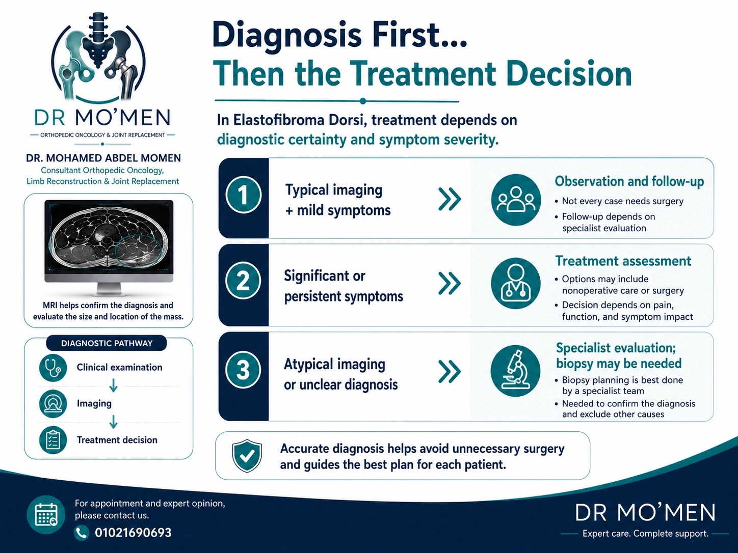
Not every elastofibroma dorsi needs an operation. Management is based on diagnostic certainty, symptom severity, functional impact, lesion size and location, and the patient's priorities.
Observation: A patient with classic imaging and minimal or no symptoms may be followed after specialist assessment.
Non-operative symptom management: Measures aimed at pain control and shoulder mechanics may be useful for mild symptoms. They may improve comfort but do not remove the lesion.
Surgical excision: Surgery may be considered when pain, persistent crepitus, mechanical limitation, or a large symptomatic mass significantly affects daily activity. Diagnostic uncertainty should be addressed before definitive excision rather than treated with an unplanned operation.
Potential surgical complications include seroma, hematoma, wound problems, infection, and postoperative pain. Recovery varies with lesion size, surgical approach, and the patient's overall health, so the expected benefit and risks should be discussed individually.
Related educational case: See the Elastofibroma Dorsi painful snapping-scapula surgical case in the Case Library. The general Blog article and the anonymized clinical case remain separate.
When should you seek specialist assessment?
- A deep or enlarging lump beneath the shoulder blade.
- Persistent pain or snapping despite treatment for a common shoulder condition.
- An MRI report describing a soft-tissue mass or an unclear infrascapular lesion.
- A recommendation for biopsy or excision before a clear diagnosis has been established.
- A need for a second opinion before surgery or before arranging international travel for care.
Patients who need a structured review before treatment or travel can use the Second Medical Opinion pathway.
Dr. Mohammed Abdelmoemen Abuelhadid's role in complex cases
For a mass around the scapula, the first question should not be “How do we remove it?” The first questions are: What is the diagnosis? Are the imaging features truly typical of a benign elastofibroma? Is biopsy needed? If biopsy is required, how should it be planned?
Dr. Mohammed Abdelmoemen Abuelhadid works in orthopedic oncology, soft-tissue and bone tumors, limb-salvage surgery, and joint reconstruction. Complex or unclear imaging can be reviewed before deciding whether the next step should be observation, additional imaging, planned biopsy, or surgery. Patients outside Egypt can also request an online second opinion before making travel arrangements.
For appointments or imaging review: 01021690693
Medical disclaimer: This article is for patient education and does not replace a clinical examination or review of imaging and investigations.