
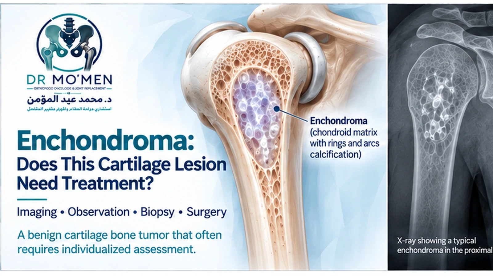
An enchondroma is a benign cartilage-forming tumor that develops inside bone.
Many enchondromas are found accidentally when an X-ray or MRI is performed for another reason. In these cases, the most important question is often not how to remove the lesion—it is whether the imaging confidently represents a harmless enchondroma or whether the lesion requires additional evaluation.
This distinction matters because central cartilage lesions exist on a spectrum ranging from a typical benign enchondroma to an atypical cartilaginous tumor (ACT) and, less commonly, higher-grade chondrosarcoma.
For many patients with a characteristic, stable enchondroma, observation is more appropriate than surgery.
The decision should be based on:
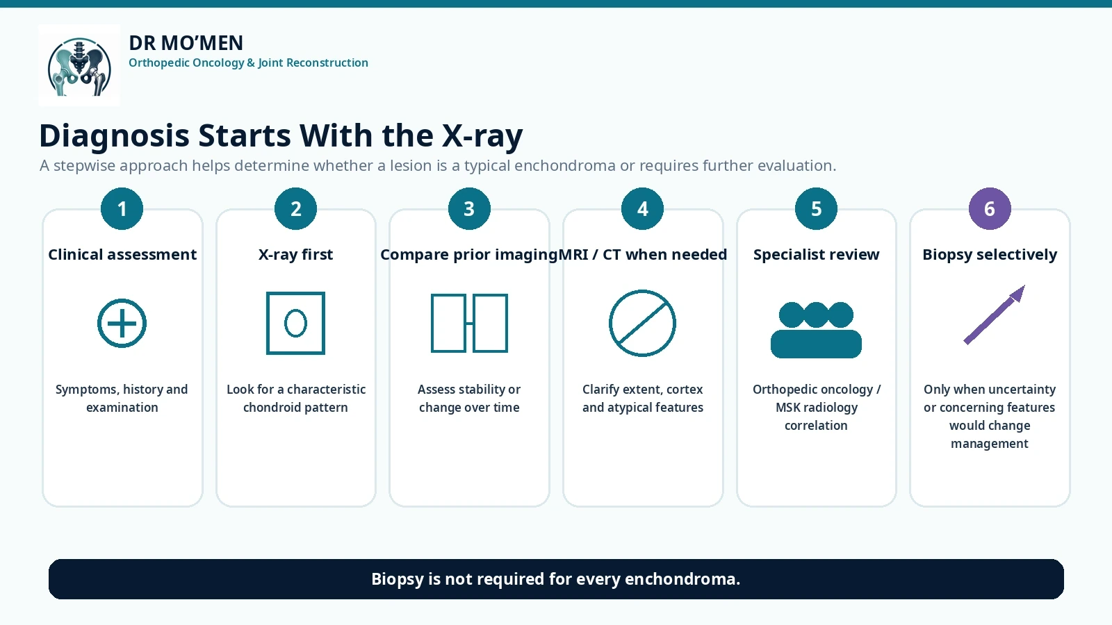
Symptoms → X-ray pattern → cortical behavior → lesion location → comparison with previous imaging → additional imaging when necessary → specialist review if uncertain.
What Is an Enchondroma?
An enchondroma is a benign tumor composed of mature hyaline cartilage located within the medullary cavity of bone.
It typically develops in bones formed through endochondral ossification.
Common locations include:
- bones of the hands and fingers
- proximal humerus
- femur
- tibia
- other long bones
Enchondromas are particularly common incidental findings around the shoulder and knee because MRI examinations are frequently performed in these regions for unrelated joint problems.
Is an Enchondroma Cancer?
No.
A typical enchondroma is a benign cartilage tumor.
However, differentiating an enchondroma from an atypical cartilaginous tumor can sometimes be challenging, especially in the long bones.
This does not mean that every enchondroma is “precancerous.”
Most characteristic solitary enchondromas behave benignly.
The important clinical issue is identifying the smaller group of cartilage lesions that demonstrate concerning biological or radiological behavior.
What Is an Atypical Cartilaginous Tumor?
An atypical cartilaginous tumor, or ACT, is a locally aggressive cartilage tumor of the appendicular skeleton.
It lies biologically between a clearly benign enchondroma and more aggressive chondrosarcoma.
The distinction between enchondroma and ACT is important, but it can sometimes be difficult even when radiographs, MRI and pathology are available.
This is one reason why treatment should not be based on a single phrase in an MRI report.
How Is an Enchondroma Usually Discovered?
Many enchondromas cause no symptoms.
A patient may undergo imaging because of:
- knee pain
- shoulder pain
- trauma
- suspected meniscal injury
- rotator cuff symptoms
- another orthopedic problem
The radiology report then unexpectedly mentions:
“Enchondroma”
or
“Chondroid lesion”
or
“Cartilaginous lesion.”
The next step is to determine whether the lesion has typical benign imaging features.
What Does an Enchondroma Look Like on X-Ray?
Typical enchondromas often appear as centrally located medullary lesions.
Cartilage mineralization may produce characteristic:
- rings
- arcs
- stippled calcification
Other features may include:
- well-defined margins
- relatively limited cortical remodeling
- absence of aggressive periosteal reaction
- absence of a soft-tissue mass
The appearance depends on the anatomical site.
For example, an enchondroma in a finger can look considerably different from one in the proximal humerus.
Why Are “Rings and Arcs” Important?
Cartilage tumors often mineralize in a characteristic chondroid matrix pattern.
On X-rays or CT, the calcification can resemble:
- rings
- arcs
- small stippled calcifications
These findings support a cartilage-forming lesion.
However, they do not independently prove that the lesion is benign.
The overall behavior of the lesion remains more important.
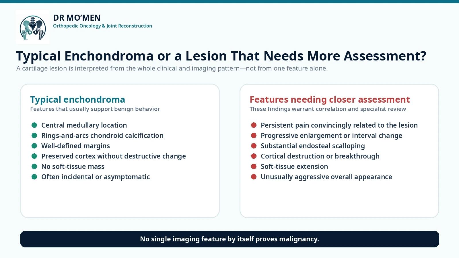
What Features Make a Cartilage Lesion More Concerning?
No single imaging feature should be interpreted in isolation.
Features that may prompt closer assessment include:
- unexplained lesion-related pain
- progressive enlargement
- substantial endosteal scalloping
- cortical destruction
- cortical breakthrough
- aggressive periosteal reaction
- soft-tissue extension
- unusually large or aggressive-appearing lesion
- change compared with previous imaging
What Is Endosteal Scalloping?
An enchondroma grows within the medullary cavity.
As it enlarges, it may produce some remodeling of the inner surface of the cortex.
This is called endosteal scalloping.
Limited scalloping may occur in benign lesions.
More extensive scalloping, particularly when associated with additional aggressive features, can raise concern for ACT or another more active cartilage tumor.
The interpretation depends on the entire radiographic pattern.
Does Pain Mean an Enchondroma Is Malignant?
No.
This is a particularly important point.
Many patients undergo an MRI precisely because they already have pain.
The MRI then discovers an enchondroma.
The cartilage lesion may therefore be incidental rather than the source of the patient's symptoms.
For example, shoulder pain might instead arise from:
- rotator cuff disease
- frozen shoulder
- arthritis
- impingement
Knee pain may arise from:
- osteoarthritis
- meniscal pathology
- ligament injury
- patellofemoral disease
Pain should therefore be correlated carefully with the lesion before deciding that an enchondroma requires surgery.
Persistent pain that appears genuinely attributable to the lesion deserves further evaluation, particularly if imaging is also atypical.
Enchondroma vs Atypical Cartilaginous Tumor
This is one of the most common diagnostic questions.
Features favoring a typical enchondroma may include:
- incidental discovery
- characteristic cartilage matrix
- stable appearance
- absence of cortical destruction
- absence of soft-tissue extension
- lack of clearly lesion-related symptoms
Features raising concern for ACT may include:
- convincing lesion-related pain
- more substantial endosteal scalloping
- cortical remodeling or involvement
- interval growth
- other radiologically active features
However, there is substantial overlap.
Both imaging and pathology have limitations in distinguishing some enchondromas from ACTs, which is why specialist musculoskeletal radiology and orthopedic oncology review can be valuable.
Do You Need MRI for an Enchondroma?
Not every characteristic enchondroma requires MRI.
MRI may be useful when:
- X-rays are not sufficiently diagnostic
- the lesion is incompletely visualized
- symptoms appear potentially related
- aggressive features are suspected
- marrow extent needs clarification
- soft-tissue extension is questioned
- previous imaging suggests change
MRI may demonstrate:
- lobulated cartilage architecture
- high-water-content cartilage signal
- surrounding marrow reaction when present
- cortical relationship
- soft-tissue involvement
The findings must still be interpreted together with X-rays.
When Is CT Useful?
CT can be helpful when better assessment is needed of:
- cartilage calcification
- cortical integrity
- endosteal scalloping
- complex anatomical sites
It may be particularly useful when the chondroid mineralization pattern is difficult to evaluate on plain radiographs.
Is PET/CT Needed?
Usually not for a typical incidental enchondroma.
Advanced imaging such as PET/CT may occasionally contribute in selected indeterminate cartilage tumors, but it should not routinely replace careful radiographic and MRI assessment.
The correct investigation depends on the specific diagnostic problem.
Does Every Enchondroma Need a Biopsy?
No.
Many characteristic enchondromas do not require biopsy.
In fact, unnecessarily biopsying every incidental cartilage lesion may create more uncertainty rather than less.
Biopsy may become appropriate when:
- imaging remains indeterminate
- aggressive characteristics are present
- higher-grade disease is suspected
- tissue diagnosis would alter management
Why Can Biopsy Be Difficult in Cartilage Tumors?
Cartilage tumors can be heterogeneous.
A needle biopsy samples only part of the lesion.
In addition, microscopic differences between enchondroma and ACT can sometimes be subtle.
Therefore, pathology should not be interpreted in isolation.
The most reliable assessment often requires correlation between:
Clinical findings + radiographs + MRI/CT + pathology when required.
This is a classic example of why multidisciplinary musculoskeletal tumor assessment matters.
Why Does Biopsy Planning Still Matter?
If biopsy is necessary, the route should be planned carefully.
The biopsy tract may need to be removed during definitive surgery if the lesion proves malignant.
Therefore, biopsy should ideally be coordinated with the orthopedic oncology team responsible for definitive treatment.
Related:
Bone Tumor Biopsy: Core Needle vs Open Biopsy
Does Every Enchondroma Need Follow-Up?
Not necessarily.
Follow-up depends on:
- imaging certainty
- anatomical location
- lesion size
- symptoms
- skeletal maturity
- comparison with previous imaging
- institutional protocol
There is no universally accepted follow-up schedule for every solitary enchondroma.
Some typical, clearly benign lesions may eventually require little or no continuing surveillance after specialist assessment.
Others may benefit from interval imaging to confirm stability.
What Does Growth Mean?
Growth deserves reassessment, but growth alone does not automatically establish malignancy.
The importance of growth depends on:
- magnitude of change
- time interval
- patient's skeletal maturity
- cortical changes
- symptoms
- other imaging characteristics
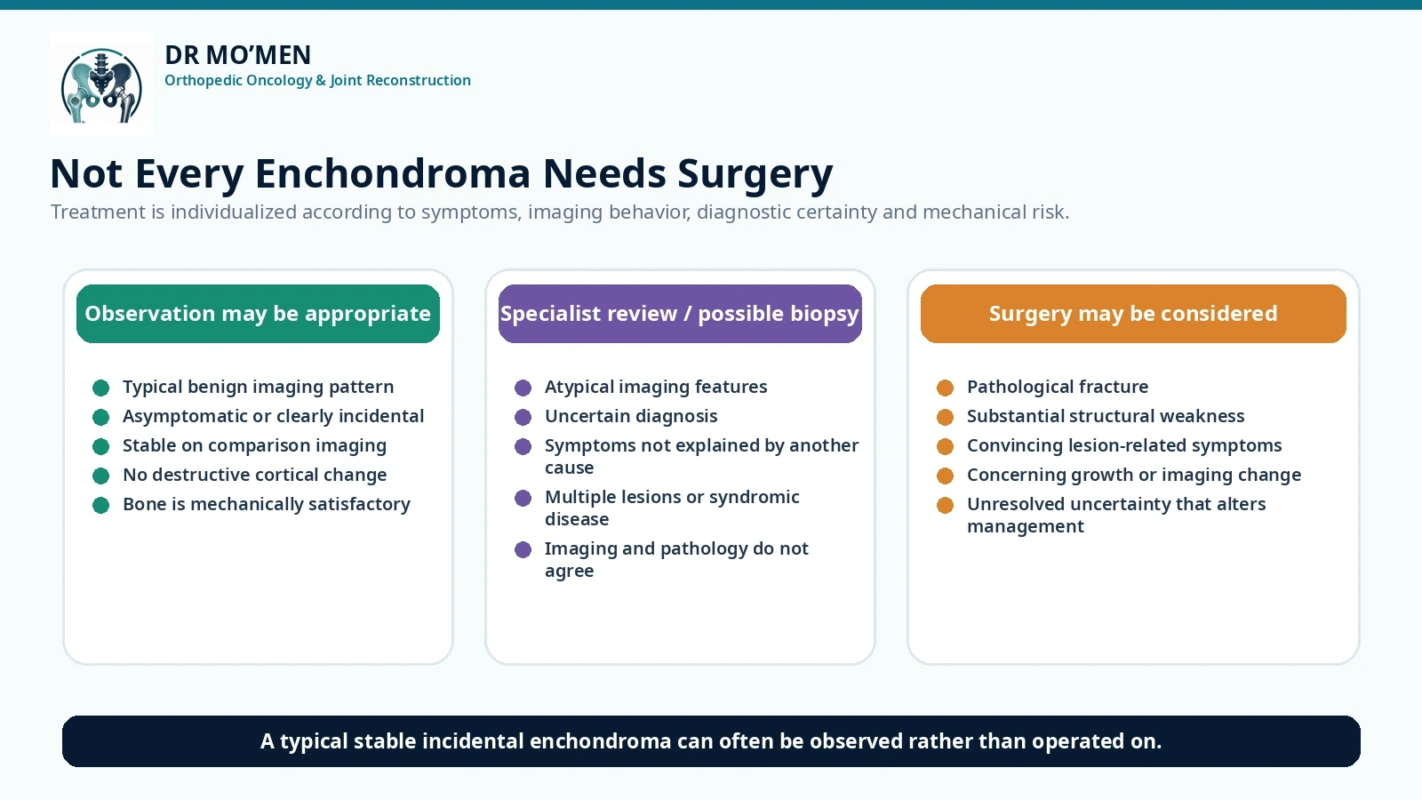
When Can an Enchondroma Be Observed?
Observation is often appropriate when the lesion:
- has characteristic benign imaging features
- is asymptomatic or clearly incidental
- has no destructive cortical change
- has no soft-tissue extension
- shows no concerning progression
- has no immediate mechanical complication
In such situations, surgery may create unnecessary morbidity without clear benefit.
When Might Surgery Be Recommended?
Surgery may be considered when there is:
- pathological fracture
- substantial structural weakness
- convincing symptoms attributable to the lesion
- concerning growth or imaging change
- diagnostic uncertainty that cannot otherwise be resolved
- another clinically significant mechanical problem
The reason for surgery should be clearly defined before the operation.
How Is an Enchondroma Treated Surgically?
When surgery is appropriate, a common technique is intralesional curettage.
This means the cartilage lesion is removed from inside the bone.
Depending on the defect and location, the remaining cavity may be managed with:
- bone graft
- bone substitute
- other reconstruction strategies
Additional fixation may occasionally be required if the bone is mechanically weak or fractured.
The exact technique depends heavily on:
- anatomical location
- lesion size
- fracture risk
- cortical integrity
- final diagnosis
What About Enchondroma in the Hand?
The small bones of the hand are a common location for enchondroma.
Patients may present because of:
- incidental imaging
- swelling
- deformity
- pathological fracture
Because the bones are small, even a benign lesion may occupy a substantial proportion of the bone and weaken it.
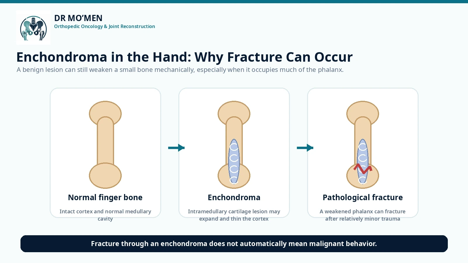
Treatment decisions therefore consider mechanical stability as well as tumor biology.
A fracture through a finger enchondroma does not automatically mean aggressive tumor behavior.
Management may involve fracture treatment followed by tumor surgery, or combined management in selected situations.
What Is Ollier Disease?
Ollier disease is characterized by multiple enchondromas.
The distribution is often asymmetric.
Because patients have multiple cartilage lesions, surveillance and clinical assessment differ from those for a solitary enchondroma.
Patients with Ollier disease have greater concern for malignant transformation than patients with a typical solitary lesion and therefore require specialist follow-up.
What Is Maffucci Syndrome?
Maffucci syndrome involves:
- multiple enchondromas
- vascular lesions
It is rare.
Like Ollier disease, it carries a greater tumor risk than an isolated enchondroma and generally warrants specialist multidisciplinary surveillance.
A patient with numerous cartilage lesions should therefore not be managed in exactly the same way as a patient with one incidental lesion.
Can an Enchondroma Become Chondrosarcoma?
Malignant transformation of a solitary typical enchondroma is uncommon.
The risk is substantially more clinically relevant in conditions involving multiple enchondromas, such as Ollier disease and Maffucci syndrome.
Rather than focusing on a single percentage, patients should understand the warning features that deserve reassessment:
- increasing lesion-related pain
- new enlargement
- progressive cortical destruction
- enlarging soft-tissue component
- significant radiographic change
What Is the Difference Between ACT and Chondrosarcoma?
Terminology depends partly on anatomical location.
A low-grade central cartilage tumor in the appendicular skeleton is generally termed atypical cartilaginous tumor.
Similar histology in certain axial locations is treated differently because local behavior and surgical implications differ.
Higher-grade chondrosarcoma is biologically more aggressive and requires a different treatment strategy.
Therefore, the words “cartilage tumor” do not describe one single disease.
What Happens if the Lesion Is an ACT Rather Than an Enchondroma?
Management is increasingly individualized.
For selected ACTs of the long bones, contemporary practice may include active surveillance, while surgery remains appropriate for selected lesions based on symptoms, imaging, progression and mechanical factors.
When surgery is indicated, intralesional curettage is commonly used for appendicular ACT rather than automatically performing wide resection.
This is another reason why accurate diagnosis matters before surgery.
When Should You Seek Specialist Orthopedic Oncology Review?
Specialist assessment is particularly useful when:
- the lesion is painful without another clear explanation
- imaging is not typical of a simple enchondroma
- substantial cortical scalloping is present
- cortical destruction is suspected
- there is a soft-tissue component
- the lesion has changed on serial imaging
- pathology and imaging do not agree
- biopsy has been recommended
- surgery is being proposed for an uncertain cartilage lesion
- multiple enchondromas are present
- a previous procedure was performed without a clear diagnosis
A Common Mistake: Operating on the MRI Report
A radiology report may state:
“Possible enchondroma.”
or:
“Enchondroma versus low-grade chondroid lesion.”
That does not automatically mean the patient requires curettage.
The correct sequence is:
Review symptoms → Examine the patient → Review X-rays → Review MRI/CT when required → Compare old imaging → Determine biological behavior → Biopsy only if necessary → Treat according to diagnosis and mechanical risk.
This avoids unnecessary surgery while still identifying lesions that genuinely need treatment.
The Role of Dr. Mo’men in Cartilage Bone Lesions
Dr. Mohammed Abdelmoemen Abuelhadid evaluates enchondromas and other cartilage-forming bone lesions with attention to:
- confirming whether the lesion has a typical benign pattern
- reviewing the actual X-rays and MRI rather than relying only on the written report
- assessing endosteal scalloping and cortical integrity
- identifying features that may suggest ACT or chondrosarcoma
- determining whether pain genuinely arises from the lesion
- comparing current and previous imaging
- deciding whether observation is appropriate
- determining whether biopsy is genuinely necessary
- planning biopsy correctly when required
- assessing fracture and mechanical risk
- planning curettage or reconstruction when intervention is justified
The objective is not to operate on every cartilage lesion.
The objective is to distinguish patients who can be safely observed from those who require further investigation or treatment.
Patients outside Egypt may send their X-rays, MRI/CT studies and reports for an initial specialist review before deciding whether in-person assessment is appropriate.
Orthopedic Oncology: /en/services/orthopedic-oncology/
Bone Biopsy: /en/services/bone-biopsy/
Second Opinion: /en/second-opinion/
International Patients: /en/international-patients/
For appointments or imaging review: 01021690693