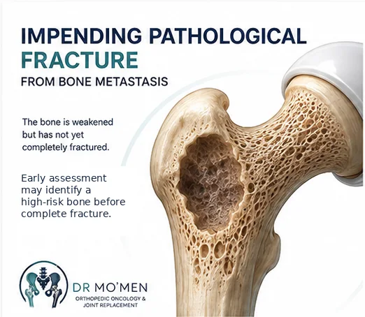
Bone metastases can weaken a long bone gradually.
Sometimes the first warning is pain. In other patients, an X-ray or CT scan shows that a substantial portion of the bone has already been destroyed even though the bone has not yet completely fractured.
This situation may be described as an impending pathological fracture.
For the patient, the important question is: “Should we stabilize the bone now, or can we safely treat the metastasis without surgery?”
There is no single answer for every lesion. The decision combines symptoms, lesion location, amount of bone destruction, fracture-risk assessment, cancer type, overall health and the expected benefit from surgery.
What is a pathological fracture?
A pathological fracture occurs when a bone breaks because it has been weakened by disease rather than by normal high-energy trauma. In metastatic bone disease, tumor cells can replace or destroy normal bone and reduce its mechanical strength.
A fracture may therefore occur while walking, standing from a chair, turning in bed, after a minor fall, or occasionally without a memorable injury.
What does “impending fracture” mean?
An impending fracture means that the bone has not yet fully broken, but the lesion is considered sufficiently weak that fracture risk has become clinically important. This is not a perfectly binary diagnosis, and no single X-ray measurement predicts every fracture correctly.
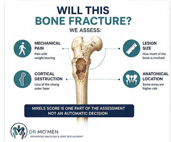
Orthopedic oncologists therefore combine symptoms, lesion location, cortical destruction, overall imaging, cancer biology and patient factors.
Why identify an impending fracture before the bone breaks?
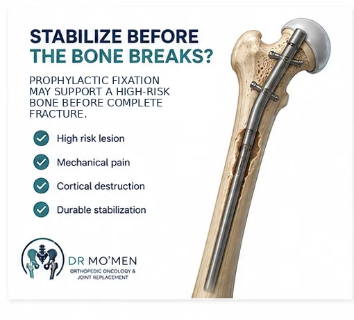
When a very high-risk metastatic lesion is identified before fracture, prophylactic stabilization may allow the bone to be supported before mechanical collapse occurs.
Retrospective clinical studies of metastatic femoral lesions have reported advantages for selected patients treated before completed fracture, including less operative burden and better early functional outcomes. These data support timely assessment, but they do not mean every metastatic lesion should be operated on.
Which bones are most important?
Weight-bearing bones are particularly important because they experience repeated mechanical load. Common sites include the proximal femur, femoral shaft, distal femur, proximal tibia and humerus.
The proximal femur, especially the peritrochanteric region, deserves particular attention because high forces pass through the hip during standing and walking.
What symptoms suggest mechanical instability?
Pain is one of the most useful clinical clues. Of particular concern is functional or mechanical pain that becomes markedly worse with standing, walking, transferring weight, climbing stairs or turning through the affected limb.
Pain at rest can occur with bone metastases, but pain specifically triggered by loading raises greater concern about mechanical weakness.
Does severe pain automatically mean the bone will fracture?
No. Pain is important but not sufficiently specific on its own. It may result from tumor activity, inflammation, microfracture, marrow involvement, nearby soft-tissue disease or true mechanical instability. Symptoms must therefore be interpreted together with imaging.
The Mirels score
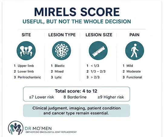
Mirels score is one of the most widely used clinical tools for estimating fracture risk in long-bone metastases. It combines four domains: anatomical site, radiographic lesion type, lesion size and pain.
| Domain | Traditional scoring |
|---|---|
| Site | Upper limb = 1; lower limb = 2; peritrochanteric = 3 |
| Lesion type | Blastic = 1; mixed = 2; lytic = 3 |
| Size | <1/3 of bone diameter = 1; 1/3–2/3 = 2; >2/3 = 3 |
| Pain | Mild = 1; moderate = 2; functional = 3 |
| Traditional interpretation | Total 4–12; ≤7 lower risk, 8 borderline, ≥9 historically considered higher risk |
The traditional thresholds are useful for communication, but they should not be treated as an automatic surgical command.
Does Mirels score decide who needs surgery?
No. Mirels is a decision-support tool. Reviews of fracture prediction show that it has limitations, particularly false-positive classification of some lesions that would not fracture. A high score should therefore trigger careful evaluation rather than automatic fixation.
Current fracture-risk assessment is best viewed as multifactorial and may incorporate additional imaging or biomechanical methods when available.
What does the surgeon look for on X-ray?
Plain radiographs remain an important first-line mechanical assessment. The surgeon looks at lesion type, location, size, cortical thinning or destruction, actual cortical breach, fracture lines and the alignment of the bone.
Predominantly lytic lesions generally weaken bone more than purely sclerotic lesions of similar size, although the mechanical effect varies between tumors and anatomical sites.
Why is cortical destruction important?
The cortex is one of the main load-bearing structural components of a long bone. A lesion that destroys a substantial portion of the cortex is generally more concerning mechanically than a small medullary lesion with intact cortex.
Historical thresholds such as lesion length or percentage cortical involvement can help frame risk, but modern practice uses them together with symptoms, site and overall imaging rather than as isolated absolute rules.
When can CT help?
CT may clarify cortical destruction, lesion geometry, circumferential involvement and the amount of remaining bone when plain radiographs are insufficient.
Research tools such as CT-based rigidity analysis and finite-element analysis can estimate mechanical strength more quantitatively and may outperform simpler clinical scores in selected settings, but they are not universally available in routine practice.
MRI or CT: which is more important?
They answer different questions. MRI is better for marrow involvement, soft-tissue extension and neurovascular relationships. CT is often more useful for cortical integrity and detailed osseous structure. The imaging strategy should match the clinical question.
The most important safety rule: make sure it is really a metastasis
A dangerous mistake is to see a destructive bone lesion and immediately insert a nail or plate without confirming the diagnosis when the clinical setting is uncertain.
This is particularly important for a solitary destructive lesion, which can represent metastasis, multiple myeloma, lymphoma, a primary bone sarcoma or another bone disorder.
A solitary lesion should undergo appropriate staging and diagnostic evaluation before fixation. In selected cases, biopsy is required before definitive surgery so that an unrecognized primary bone sarcoma is not contaminated by unplanned internal fixation.
Does every patient with known cancer need a bone biopsy?
No. In a patient with a known carcinoma, multiple typical skeletal lesions and imaging fully consistent with widespread metastatic disease, histological confirmation of every lesion may not be necessary.
Biopsy becomes particularly important when the lesion is solitary, the primary cancer is unknown, imaging is atypical, behavior is unexpected, or a primary bone tumor or hematological malignancy remains possible.
When biopsy is required, diagnostic staging and biopsy planning should be coordinated with the team that will perform definitive treatment.
What other factors determine whether surgery is worthwhile?
Fracture risk is only one part of the decision. The team also considers overall cancer burden, expected response to systemic therapy, radiosensitivity, functional status, medical comorbidities, anticipated survival, current mobility and goals of care.
A technically possible operation is not necessarily the best treatment if the surgical burden is unlikely to provide meaningful benefit.
Does radiotherapy prevent pathological fracture?
Radiotherapy is highly important for pain relief and local tumor control, but it does not instantly restore mechanical strength to a severely weakened long bone. If the bone is already at high risk of mechanical failure, radiotherapy alone may not provide adequate structural protection.
Can systemic therapy make surgery unnecessary?
Sometimes systemic treatment can produce excellent tumor control. However, tumor response and structural bone integrity are different questions. A femur that has already lost substantial cortical support may remain at risk during the time required for oncological treatment to work.
The orthopedic question is therefore: “Will the bone remain mechanically safe while the cancer treatment takes effect?”
When is prophylactic fixation considered?
Preventive stabilization becomes increasingly reasonable when several high-risk features occur together, such as significant functional pain, large lytic destruction, substantial cortical involvement, a mechanically critical location, progressive radiographic destruction, a high fracture-risk score and sufficient expected functional benefit from surgery.
No individual factor should be used in isolation.
What is prophylactic stabilization?
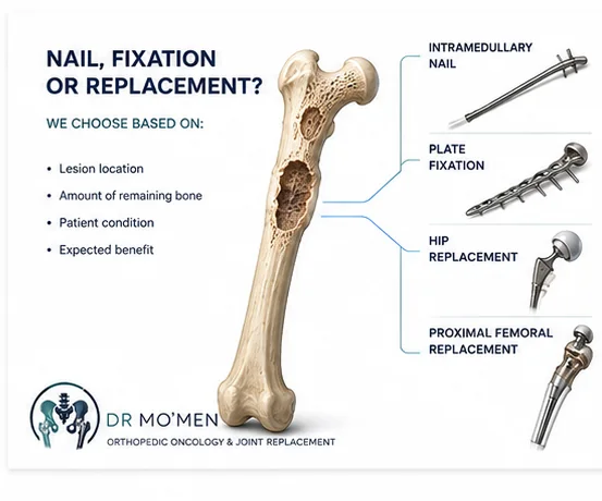
It means surgically supporting the bone before a complete pathological fracture occurs. Depending on the lesion, options may include intramedullary nailing, plate-and-screw fixation, arthroplasty or endoprosthetic reconstruction.
The correct operation depends primarily on where the lesion is, how much usable bone remains and what reconstruction is likely to remain durable.
Intramedullary nail or plate?
Intramedullary nailing
Intramedullary fixation is often considered for appropriate lesions in the femoral shaft, subtrochanteric region and selected peritrochanteric locations when enough bone remains for useful fixation.
Plate fixation
Plate-and-screw constructs may be useful in selected lesions depending on anatomical location, residual bone, joint proximity and whether cement augmentation is needed.
The fixation should ideally be selected to remain mechanically useful for the patient’s expected lifetime rather than relying on fracture healing alone.
When might hip replacement be better than a nail?
A nail depends on sufficient bone to hold fixation. Lesions involving the femoral head, femoral neck or extensively destroyed proximal femur may be better reconstructed with arthroplasty or endoprosthetic replacement rather than internal fixation through mechanically nonviable bone.
When might proximal femoral replacement be needed?
Extensive metastatic destruction of the proximal femur can occasionally leave insufficient bone for reliable internal fixation. In that situation, proximal femoral replacement may be considered according to the extent of destruction, hip involvement, tumor type, remaining bone, medical fitness and expected benefit.
Does surgery remove all metastatic disease?
Usually not. Surgery for metastatic bone disease is generally designed to achieve durable mechanical stability, pain relief, preservation or restoration of mobility, and safe continuation of oncological treatment.
It is usually one part of a multidisciplinary cancer-treatment plan rather than isolated tumor surgery.
Why should reconstruction be durable?
Metastatic bone may not heal like a normal traumatic fracture. The reconstruction therefore needs to remain mechanically useful even if biological healing is limited.
What about very vascular metastases?
Some cancers, particularly renal cell and thyroid carcinoma, can produce highly vascular skeletal metastases. When major surgery is planned, the multidisciplinary team may consider whether additional vascular planning or preoperative embolization is appropriate. This is individualized and is not necessary for every patient.
What happens after stabilization?
Management may include early mobilization when fixation allows, analgesia, venous-thrombosis prevention, physiotherapy, radiotherapy when indicated and continuation or modification of systemic cancer treatment. Coordination between orthopedics, medical oncology and radiation oncology is important.
What if the bone is already fractured?
A completed pathological fracture changes the mechanical problem. Treatment must address the fracture, metastatic lesion, bone defect, limited biological healing potential and durability of the reconstruction. Surgery after completed fracture can be more complex than prophylactic stabilization of a carefully selected high-risk lesion.
Impending metastatic fracture is not the same as fracture through a primary bone tumor
Metastatic bone disease arises from a malignancy that has spread to bone. A primary bone tumor originates in the bone itself. A pathological fracture through a suspected primary bone sarcoma requires a fundamentally different diagnostic and surgical pathway.
Never treat an unexplained destructive lesion simply as metastasis and fix it before appropriate oncological workup.
When does the patient need urgent assessment?
- rapidly increasing weight-bearing pain
- sudden inability to walk normally
- pain with every step
- new deformity
- an audible crack or sudden severe pain
- a large destructive lesion on imaging
- progressive cortical destruction
- new neurological or vascular symptoms.
If a fracture has already occurred, urgent clinical evaluation is required.
When is a second opinion useful?
- preventive fixation has been proposed
- Mirels score is high but symptoms are mild
- imaging and symptoms do not agree
- the lesion is solitary
- biopsy has not been performed despite diagnostic uncertainty
- the lesion involves the proximal femur
- the choice is between nail, arthroplasty and endoprosthetic reconstruction
- the benefit of major surgery is uncertain in extensive metastatic disease.
Role of Dr. Mohammed Abdelmoemen Abuelhadid in metastatic bone disease
The decision to stabilize a metastatic bone lesion requires more than simply calculating Mirels score. Assessment may include:
- Reviewing the primary cancer and current oncological status.
- Confirming whether the bone lesion is truly metastatic when necessary.
- Comparing current and previous imaging.
- Assessing mechanical pain.
- Evaluating cortical destruction and fracture risk.
- Determining whether biopsy is needed before fixation.
- Selecting fixation versus replacement according to location and bone stock.
- Coordinating surgery with systemic therapy and radiotherapy.
- Planning a reconstruction intended to remain durable for the patient’s expected needs.
Dr. Mohammed Abdelmoemen Abuelhadid evaluates metastatic bone lesions, impending pathological fractures and complex reconstruction decisions, including patients seeking a second opinion before prophylactic stabilization.
Patients outside Egypt may send imaging, oncology reports and relevant investigations for preliminary review before travelling when appropriate.
For appointments or imaging review: +20 102 1690693
For complex decisions, especially a solitary lesion or a choice between fixation and replacement, you can use the specialist second-opinion pathway.
Frequently Asked Questions
Does every bone metastasis need surgery?
No. Many bone metastases are treated with systemic therapy, radiotherapy, bone-targeted therapy or observation. Surgery is mainly considered when mechanical instability, fracture or another specific indication is present.
Does Mirels 9 automatically mean surgery?
No. A score of 9 or more has historically represented higher fracture risk, but the score has limited specificity. Symptoms, imaging, cortical destruction, prognosis and expected benefit must also be considered.
Is it better to operate before the bone fractures?
For appropriately selected high-risk lesions, prophylactic stabilization can avoid the additional mechanical consequences of a completed fracture. It is not necessary for every lesion.
Can radiotherapy strengthen the bone enough to avoid surgery?
Radiotherapy can reduce pain and tumor activity, but a severely mechanically compromised bone may still require stabilization because radiation does not provide immediate structural support.
Should a solitary bone lesion be biopsied before fixation?
Often yes when the diagnosis is uncertain. A solitary destructive lesion should be investigated carefully to exclude a primary bone tumor before unplanned internal fixation.
How this article was reviewed
This patient-education article was reviewed for clinical accuracy, patient safety and decision-making clarity by Dr. Mohammed Abdelmoemen Abuelhadid. It supports, but does not replace, clinical examination, oncology review, current imaging, fracture-risk assessment or biopsy planning when indicated.
Considering travel to Egypt for a metastatic bone lesion or impending fracture?
Before arranging travel, send the available X-rays, CT or MRI, oncology diagnosis or pathology when available, current cancer treatment, and a brief note about pain and weight-bearing. Preliminary specialist review can help determine whether urgent in-person assessment, further investigations, stabilization, or another pathway should be considered.

Author and medical reviewer
Dr. Mohammed Abdelmoemen Abuelhadid — Orthopedic Oncology & Joint Replacement Surgeon and Lecturer of Orthopedic Surgery at Ain Shams University.
Medical Disclaimer: This article is for patient education and does not replace clinical examination, oncology assessment, review of current imaging, fracture-risk evaluation or biopsy planning when indicated.