Being told that a scan shows a bone tumor can immediately raise frightening questions. Is it cancer? Will the tumor have to be removed? Will I lose my arm or leg?
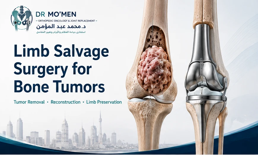
Modern orthopedic oncology is often able to remove a bone tumor while preserving the limb. This approach is known as limb salvage surgery or limb-sparing surgery.
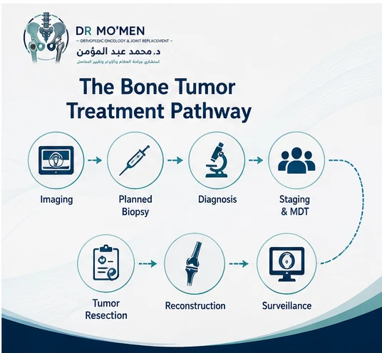
However, limb salvage is not simply an operation to “remove the mass.” Successful treatment requires careful diagnosis, appropriately planned biopsy, accurate staging, assessment of the tumor's relationship to major nerves and blood vessels, and a reconstruction strategy designed around the amount of bone and soft tissue that must be removed.
Patients with a suspected primary bone tumor should ideally be evaluated by an orthopedic oncology team before an unplanned biopsy or surgery is performed.
What Is Limb Salvage Surgery?
Limb salvage surgery aims to remove a bone or soft-tissue tumor with an appropriate oncological margin while preserving a functional arm or leg whenever this can be done safely.
The operation usually has two major components:
- Tumor resection — removing the tumor together with an appropriate margin of surrounding tissue.
- Reconstruction — rebuilding the resulting bone, joint, or soft-tissue defect.
Depending on the location and extent of the tumor, reconstruction may involve a tumor megaprosthesis, joint replacement, internal fixation, biological reconstruction, bone graft, allograft in selected settings, combined prosthetic and biological reconstruction, or soft-tissue reconstruction.
Related article: Megaprosthesis after bone tumor resection.
The most appropriate method depends on the diagnosis, tumor location, age, bone quality, remaining anatomy, treatment plan, and expected functional demands.
Before Any Biopsy or Surgery: Planning Matters
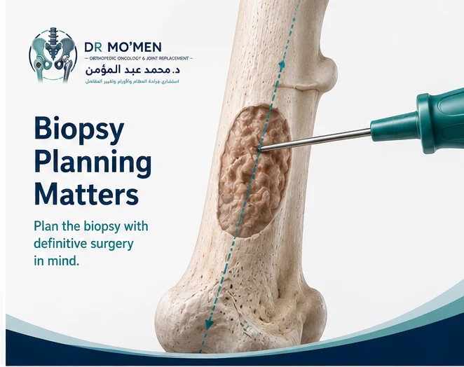
One of the most important principles in orthopedic oncology is that biopsy should be planned as part of the definitive treatment pathway.
A biopsy is not merely a diagnostic test. The biopsy tract may potentially become contaminated by tumor cells and may therefore need to be removed during definitive tumor surgery. For this reason, the location and direction of the biopsy can affect later surgical options.
Whenever possible, a suspected primary malignant bone tumor should undergo appropriate imaging and specialist planning before biopsy. An inadequately planned biopsy can complicate later limb-salvage surgery.
Patients who have already undergone an unplanned biopsy or operation should not assume that limb salvage is impossible, but the case may require more extensive reassessment.
Read more about why bone tumor biopsy planning matters.
Which Bone Tumors May Require Limb Salvage Surgery?
Limb-salvage principles may be considered in several types of locally aggressive or malignant bone tumors, including selected cases of:
- Osteosarcoma
- Chondrosarcoma
- Ewing sarcoma
- Giant cell tumor of bone
- Other aggressive primary bone tumors
- Selected metastatic bone lesions
- Recurrent bone tumors
Learn more in the dedicated giant cell tumor of bone treatment guide.
Not every tumor requires the same operation. Some benign tumors can be treated with curettage rather than wide resection. Others require removal of a segment of bone or an entire joint. Treatment depends on the precise diagnosis and biological behavior of the lesion.
Can Every Limb With a Bone Tumor Be Saved?
No. Limb preservation should never compromise adequate tumor control.
Whether limb salvage is appropriate depends on tumor size, anatomical location, involvement of major nerves or blood vessels, extent of soft-tissue involvement, pathological fracture, infection or contamination, response to systemic treatment where relevant, previous surgery or biopsy, ability to achieve an adequate surgical margin, available reconstruction options, and expected function after reconstruction.
The goal is not simply to preserve the limb anatomically. The goal is oncologically appropriate tumor removal plus a useful, functional limb whenever feasible.
In selected situations, amputation may still provide the safer oncological or functional solution.
Why Accurate Diagnosis Comes Before Surgery
Different bone tumors behave differently. A treatment appropriate for one tumor may be completely inappropriate for another.
- Some benign lesions can be observed.
- Some are treated by curettage.
- Some require wide surgical resection.
- Some require chemotherapy as part of treatment.
- Some are primarily treated surgically.
- Some may require radiotherapy in specific circumstances.
Treatment should not begin simply because an X-ray shows a destructive lesion. The diagnostic process may include plain X-rays, MRI, CT, staging imaging, laboratory investigations, planned biopsy, and histopathological diagnosis. The sequence matters.
Imaging Before Bone Tumor Surgery
X-rays
Plain radiographs remain extremely important in the initial assessment of bone tumors. They may demonstrate whether the lesion is lytic or sclerotic, cortical destruction, periosteal reaction, matrix mineralization, pathological fracture, and anatomical location within the bone.
MRI
MRI is particularly important for defining the local extent of the tumor. It can help assess intramedullary extent, soft-tissue extension, relationship to the joint, neurovascular structures, muscle compartments, surrounding edema, and possible skip lesions in appropriate cases. MRI should ideally be obtained before biopsy when a primary bone malignancy is suspected.
CT and Staging Studies
CT may provide additional information regarding cortical bone, mineralized tumor matrix, complex anatomy, and chest staging where clinically appropriate. The staging pathway depends on the suspected diagnosis.
Why Multidisciplinary Care Matters
Management of malignant bone tumors often involves more than one specialty, including orthopedic oncology, musculoskeletal radiology, pathology, medical oncology, radiation oncology, plastic or reconstructive surgery, vascular surgery, rehabilitation, and physiotherapy.
Multidisciplinary discussion helps ensure that biopsy, staging, systemic treatment, surgery, and reconstruction follow a coordinated plan.
What Does “Wide Resection” Mean?
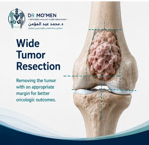
Patients often hear the term wide resection when discussing malignant bone tumors. This means removing the tumor together with a planned margin of surrounding tissue that has not been directly involved by the tumor.
The exact margin required depends on tumor type, anatomical barriers, previous procedures, response to treatment, and relationship to surrounding structures. A wide resection can sometimes require removal of a significant segment of bone, muscle, tendon, or joint. That is why reconstruction planning is essential before surgery begins.
How Is the Limb Reconstructed After Tumor Removal?
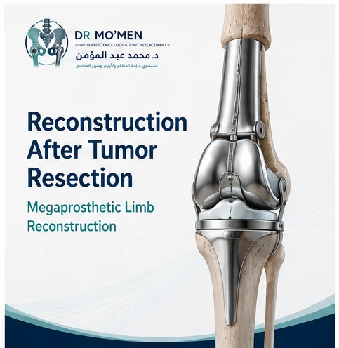
Once the tumor-bearing segment has been removed, the surgeon must reconstruct the resulting defect. The reconstruction strategy depends largely on where the tumor is located and how much tissue has been removed.
Megaprosthetic Reconstruction
A megaprosthesis is a large modular orthopedic implant designed to replace major segments of bone and, frequently, the adjacent joint. It may be used after resection of tumors involving areas such as the distal femur, proximal tibia, proximal femur, proximal humerus, and other selected anatomical regions.
Megaprosthetic reconstruction can allow relatively early structural stability and mobilization. However, it is a major reconstruction and carries potential long-term complications such as infection, mechanical failure, loosening, instability, dislocation depending on location, soft-tissue failure, periprosthetic fracture, and possible future revision surgery.
Biological Reconstruction
In selected patients, biological reconstruction may be considered. Options can include autologous bone graft, allograft, vascularized bone graft, bone transport, or other reconstructive methods in selected situations.
Biological approaches may be particularly relevant in younger patients or certain anatomical locations, but they have their own limitations and potential complications, including delayed union, nonunion, fracture, infection, and graft resorption. No reconstruction method is universally superior. The optimal approach depends on the specific case.
Limb Salvage Versus Amputation
The question of limb salvage versus amputation should be approached carefully. Preserving the limb is desirable when an adequate tumor resection and reasonable function can be achieved.
However, limb salvage should not be performed merely to avoid amputation if it would leave inadequate tumor margins, an unstable limb, severe chronic pain, poor function, major untreated infection, or an unreconstructable neurovascular deficit.
Modern treatment decisions consider both oncological safety and expected function. The appropriate choice must therefore be individualized.
When Might Amputation Be Safer or More Functional Than Limb Salvage?
Direct answer: limb salvage is not automatically the better operation. Amputation may be considered when adequate oncological margins cannot be achieved while preserving essential structures, when reconstruction is unlikely to produce a stable and useful limb, or when prior surgery, infection, soft-tissue loss or neurovascular involvement makes salvage disproportionately complex or unreliable.
- Oncological margins: if preserving the limb would compromise an adequate tumor resection, cancer control takes priority over limb preservation.
- Major neurovascular involvement: extensive involvement of critical nerves or vessels may make a functional reconstruction impossible or unsafe in selected cases.
- Severe soft-tissue compromise or infection: wound, coverage and infection problems can make reconstruction unreliable in selected patients.
- Expected function: a painful, unstable or poorly functional salvaged limb is not necessarily better than a well-planned amputation with rehabilitation and prosthetic fitting.
- Recurrence or previous unplanned surgery: altered surgical planes and contamination can change what margins and reconstructions are realistically achievable.
- Patient-specific priorities: general health, rehabilitation potential, occupation, daily activities, complication burden and personal preferences all matter.
The decision is therefore not simply “save the limb or lose the limb.” It is a comparison of oncological safety, expected function, complication risk, rehabilitation demands and the patient’s goals.
What Determines Function After Limb-Salvage Surgery?
Recovery varies substantially between patients. Factors affecting function include tumor location, size of the resection, muscles removed, nerve involvement, reconstruction type, joint involvement, chemotherapy where applicable, wound healing, complications, age, rehabilitation, patient motivation and general health.
Some patients regain excellent everyday function, while others may have permanent limitations. Realistic expectations should be discussed before treatment.
What Are the Risks of Limb-Salvage Surgery?
Limb-salvage surgery is major orthopedic oncology surgery. Potential complications can include infection, wound-healing problems, bleeding, thrombosis, nerve injury, vascular injury, stiffness, weakness, instability, mechanical implant problems, fracture, nonunion in biological reconstruction, implant loosening, local tumor recurrence, need for revision surgery, and rarely need for amputation if complications or recurrence cannot otherwise be controlled.
The exact risks differ according to the procedure. A personalized discussion is therefore necessary.
What Happens After Surgery?
Recovery depends on the location and reconstruction. The postoperative pathway may involve pain control, wound monitoring, thrombosis prevention, early physiotherapy, protected or progressive weight-bearing, walking aids, muscle strengthening, range-of-motion exercises, oncological treatment where appropriate, and surveillance imaging.
Some megaprosthetic reconstructions permit earlier weight-bearing than many biological reconstructions, although the rehabilitation plan must be individualized.
How Long Does Recovery Take?
There is no universal recovery timeline. Recovery depends on the extent of surgery, reconstruction type, soft-tissue healing, chemotherapy, complications, age, physical condition, and rehabilitation progress.
Early recovery and long-term functional recovery are different processes. Patients may regain basic mobility relatively early while strength, endurance, confidence, and function continue improving over a longer period.
Does Limb Salvage Eliminate the Risk of Tumor Recurrence?
No operation can guarantee that a tumor will never recur. Local recurrence risk depends on tumor biology, surgical margins, previous surgery, treatment response where applicable, and anatomical factors.
Regular oncological surveillance is important even after apparently successful surgery. Follow-up may include clinical assessment and scheduled imaging according to the specific diagnosis.
What If the Tumor Was Already Operated On Without Proper Planning?
Sometimes a bone or soft-tissue mass is removed before a definitive diagnosis is known. This may be referred to clinically as an unplanned excision when a potentially malignant lesion is operated on without adequate oncological planning.
The next step is usually not simply another immediate operation. The patient may need review of previous imaging, operative notes, specialist pathology review, MRI of the surgical area, appropriate staging, and multidisciplinary assessment. Further surgery may need to include the previous operative field and contaminated tissues.
Patients in this situation should seek specialist orthopedic oncology review before another procedure is performed.
What If the Bone Tumor Has Recurred?
Recurrence does not automatically mean that limb salvage is impossible. Management depends on the original diagnosis, location, previous surgical margin, reconstruction, time to recurrence, size and extent of the recurrent tumor, metastatic disease, neurovascular involvement, and previous radiotherapy or systemic treatment.
Some recurrent tumors can still be treated with further limb-preserving surgery. Others may require a different approach.
When Should You Seek a Specialist Orthopedic Oncology Opinion?
Seek specialist review before biopsy or irreversible surgery when:
- A scan shows an unexplained bone lesion.
- A biopsy has been recommended.
- A malignant bone tumor has been diagnosed.
- Amputation or major limb-salvage surgery has been proposed.
- You have received conflicting treatment recommendations.
- A bone tumor has recurred.
- A previous operation was performed before a definitive diagnosis.
- A complex reconstruction or megaprosthesis has been recommended.
- You want another opinion before an irreversible operation.
A second opinion does not necessarily mean that the original treatment recommendation is wrong. It can help clarify diagnosis, surgical options, reconstruction choices, and treatment sequencing.
Request an orthopedic oncology second opinion.
International Patients: Send Your Imaging Before You Travel
Patients outside Egypt do not necessarily need to travel to Cairo simply to determine whether specialist evaluation may be useful. A preliminary review can begin with available records.
Useful documents include plain X-rays, MRI, CT, staging scans, biopsy report, pathology report, biopsy slides or blocks where specialist pathology review is being considered, previous operative reports, discharge summaries, and the current treatment plan.
The purpose of remote review is to understand the case and determine what additional assessment may be required. It does not replace clinical examination or definitive multidisciplinary evaluation.
Send Your X-Rays Before You Travel
International patients may send available imaging, pathology reports, and previous treatment records for preliminary specialist review before deciding whether travel to Egypt is appropriate.
International contact: +20 102 1690693
About Dr. Mohammed Abdelmoemen Abuelhadid
Dr. Mohammed Abdelmoemen Abuelhadid is an Orthopedic Oncology & Joint Replacement Surgeon and Lecturer of Orthopedic Surgery at Ain Shams University. His clinical areas include orthopedic oncology, bone and soft-tissue tumors, limb salvage surgery, tumor resection and reconstruction, megaprosthetic reconstruction, joint replacement, revision arthroplasty, complex orthopedic reconstruction, and second medical opinions for complex orthopedic cases.
The purpose of specialist evaluation is to establish an accurate diagnosis and develop an individualized treatment plan rather than applying the same operation to every tumor.
Frequently Asked Questions
Can a malignant bone tumor be removed without amputating the limb?
In many appropriately selected patients, limb-salvage surgery can remove the tumor while preserving the arm or leg. Feasibility depends on tumor location, involvement of nerves and blood vessels, surgical margins, reconstruction options, and expected function.
Why should biopsy be planned before bone tumor surgery?
The biopsy tract may need to be removed during definitive surgery. Poorly positioned biopsy incisions can contaminate additional tissues and complicate later tumor resection, so biopsy planning should ideally be coordinated with the team responsible for definitive tumor treatment.
What is a megaprosthesis?
A megaprosthesis is a modular large-joint or segmental prosthesis used to replace substantial portions of bone removed during tumor surgery. It is frequently used around major joints such as the knee, hip, and shoulder when a large bone segment must be resected.
Is limb salvage always better than amputation?
Not necessarily. The priority is adequate tumor control and a functional limb. In some cases, amputation may provide a safer or more functional solution. The decision should be individualized after specialist assessment.
Can limb salvage surgery fail?
Complications such as infection, mechanical problems, loosening, fracture, soft-tissue failure, tumor recurrence, or other problems can sometimes require additional surgery. Long-term surveillance is therefore important.
Can I get a second opinion before travelling to Egypt?
Yes. International patients may provide available imaging, pathology reports, and previous treatment information for preliminary specialist review. This can help determine whether travel for further assessment is reasonable, but it does not replace examination or definitive diagnosis.
Evidence Behind Limb-Salvage Decision-Making
- British Sarcoma Group: UK Guidelines for the Management of Bone Sarcomas — emphasizes specialist multidisciplinary care, adequate oncological margins and coordinated local treatment planning.
- US National Cancer Institute: Primary Bone Cancer — describes limb-sparing surgery as tumor removal with appropriate surrounding tissue and reconstruction when feasible.
- ESMO–EURACAN–GENTURIS–ERN PaedCan Bone Sarcoma Clinical Practice Guideline — multidisciplinary guideline covering diagnosis, local treatment and follow-up of bone sarcomas.
How this article was reviewed
This patient-education article was reviewed for clinical accuracy, patient safety, and decision-making clarity by Dr. Mohammed Abdelmoemen Abuelhadid. It is intended to support, not replace, clinical examination, pathology review, and review of the actual imaging and reports.

Author and medical reviewer
Dr. Mohammed Abdelmoemen Abuelhadid — Orthopedic Oncology & Joint Replacement Surgeon and Lecturer of Orthopedic Surgery at Ain Shams University.
Medical Disclaimer: This article is for patient education and does not replace clinical examination, review of imaging, pathology, investigations, or individualized multidisciplinary medical advice.