
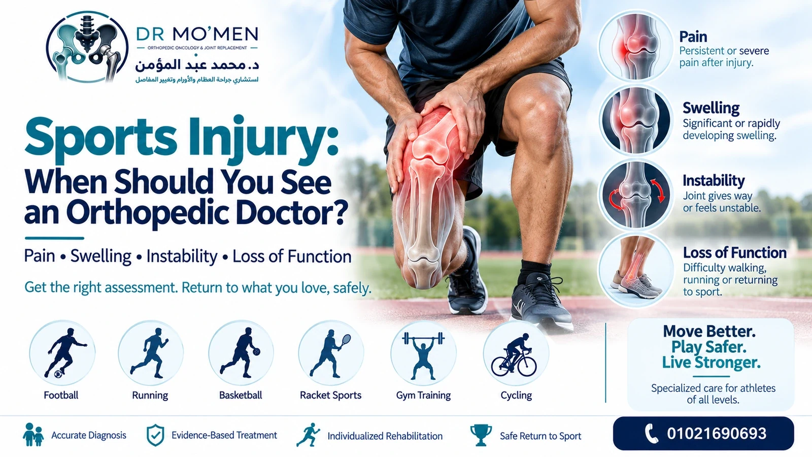
A painful ankle after football, a swollen knee after changing direction, or shoulder pain after training does not automatically mean a serious injury has occurred. Many mild sprains, muscle strains and overuse problems improve with appropriate activity modification and progressive rehabilitation.
However, sports injuries can also involve a fracture, significant ligament injury, tendon rupture, meniscal injury, joint instability, cartilage damage, dislocation or neurovascular injury. The practical question is therefore which injuries can be monitored initially and which should be assessed promptly.
What Is a Sports Injury?
A sports injury is a musculoskeletal injury occurring during competitive sport, recreational exercise, gym training, running, football, basketball, racket sports, cycling, weight training or other physical activity. It can affect bone, muscle, tendon, ligament, cartilage or a joint, and may occur suddenly or develop gradually through repeated loading.
Acute injuries
Acute injuries follow a specific event such as twisting the knee, rolling the ankle, falling onto the shoulder, being tackled, landing awkwardly from a jump, or sprinting and suddenly feeling muscle pain. Typical early features include pain, swelling, bruising, instability or immediate loss of function.
Overuse injuries
Overuse injuries develop when tissue is repeatedly stressed without sufficient recovery. Examples include tendinopathy, bone stress injury, throwing-related shoulder pain, running-related knee pain and repetitive muscle or tendon overload. Persistent or progressively worsening pain deserves assessment because continuing to load the injured tissue may delay recovery or allow a stress injury to progress.
Which Sports Injuries Can Often Be Monitored Initially?
A minor sports injury can sometimes be monitored initially when the overall pattern is reassuring: pain is mild, swelling is limited, there is no deformity, the joint feels stable, normal weight bearing remains possible, useful movement is preserved and symptoms are steadily improving. Activity modification should still be guided by symptoms rather than by an arbitrary number of rest days.
- Pain is mild and improving
- Swelling is limited
- There is no deformity
- The joint feels stable
- Normal weight bearing remains possible
- Movement is reasonably preserved
- Symptoms progressively improve rather than worsen
What Are the Warning Signs After a Sports Injury?
The most important warning signs are loss of normal function, major swelling, deformity, instability, mechanical locking and neurovascular symptoms. These findings do not identify one diagnosis by themselves, but they increase the need for timely examination and appropriate imaging rather than continued self-treatment.
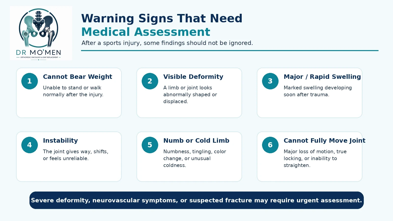
- You cannot bear weight Being unable to stand or walk normally after an injury may indicate a fracture, significant ligament injury, severe sprain or major joint injury. The ability to walk does not completely exclude a fracture.
- The joint or limb looks deformed Visible deformity after trauma can indicate fracture, dislocation or severe ligament injury. A visibly deformed joint should not be repeatedly manipulated outside an appropriate medical setting.
- There is major or rapid swelling Some swelling is common, but marked swelling developing rapidly after trauma can accompany substantial intra-articular or soft-tissue injury.
- The joint feels unstable or gives way Buckling, shifting, giving way or inability to trust a joint can occur after ligament injury and deserves assessment when persistent or recurrent.
- You cannot fully move the joint A knee that cannot straighten, a shoulder that cannot elevate after trauma or a mechanically blocked joint deserves evaluation.
- The knee is truly locked A true mechanical lock can occur with a displaced meniscal tear, loose body, osteochondral fragment or another intra-articular problem.
- You heard or felt a pop A pop is not diagnostic by itself. It becomes more concerning when combined with rapid swelling, instability, inability to continue sport or loss of function.
- There is numbness, tingling or a cold limb Neurological or circulation symptoms after trauma—including numbness, weakness, color change or unusual coldness—require urgent assessment.
- There is an open wound or bone is visible An open fracture is an emergency because contamination and soft-tissue injury materially affect treatment. Urgent hospital evaluation is required.
- Pain is severe or getting worse Progressively worsening pain, persistent severe pain or pain preventing normal function is a reason for medical review.
When Does a Sports Injury Require Emergency Care?
Emergency or urgent hospital assessment is appropriate when the injury may threaten limb function, involve an open fracture or dislocation, produce a new neurological or circulation deficit, or cause severe rapidly worsening symptoms. A second opinion or routine clinic appointment should never delay emergency care.
- Obvious fracture or major deformity
- Suspected dislocation
- Open wound with exposed bone or suspected open fracture
- Loss of circulation or an unusually cold/pale limb
- New neurological deficit
- Severe uncontrolled or rapidly worsening pain
- Rapidly increasing swelling with concern for compartment syndrome
- Significant head injury, concussion symptoms or loss of consciousness
When Should a Knee Injury Be Evaluated?
A knee injury should be evaluated when rapid swelling, instability, inability to bear weight, true locking, inability to fully straighten the knee, significant trauma or recurrent giving way develops. Sports involving pivoting, cutting, jumping, direct contact and sudden deceleration commonly expose the knee to ACL, meniscal, collateral-ligament, patellar and cartilage injuries.
- Rapid swelling
- Instability or giving way
- Inability to bear weight
- Locking
- Inability to fully straighten the knee
- Significant trauma
- Recurrent giving way
- Persistent pain despite appropriate initial rest
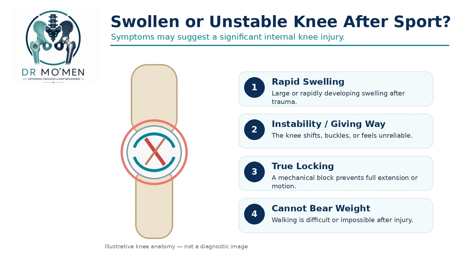
Possible injuries include ACL tear, meniscal tear, patellar dislocation, collateral ligament injury, cartilage injury and fracture. See the detailed ACL injury guide when instability or pivoting symptoms are prominent.
When Should an Ankle or Shoulder Injury Be Evaluated?
Ankle injuries
Most ankle injuries are ligament sprains, but fractures can look similar. Assessment is appropriate when weight bearing is difficult, swelling is substantial, pain is directly over bone, deformity is present, symptoms are worsening or recurrent instability develops. Being able to walk does not completely exclude an ankle fracture. See ankle sprain: cast or physiotherapy? for the rehabilitation pathway.
Shoulder injuries
Shoulder injuries may follow a direct fall, tackle, throwing episode, weightlifting or a fall onto an outstretched arm. Visible deformity, suspected dislocation, inability to lift the arm after trauma, marked weakness, persistent pain or recurrent instability should prompt assessment. The rotator cuff tear guide explains one important cause of persistent weakness and pain.
What About Muscle and Tendon Injuries?
Muscle injuries
Muscle strains are common in the hamstrings, quadriceps, calf and adductors. Mild strains may improve with appropriate rehabilitation, but a sudden severe tearing sensation, palpable defect, extensive bruising, difficulty walking, marked loss of strength or repeated recurrence deserves assessment.
Tendon injuries
A significant tendon rupture can be mistaken for a simple strain. Achilles, patellar, quadriceps, distal biceps and rotator cuff injuries are examples. A sudden snap, immediate loss of strength, inability to perform the tendon’s normal function, a visible or palpable gap, or major weakness should raise concern for a clinically important tear.
Do You Always Need an X-Ray or MRI?
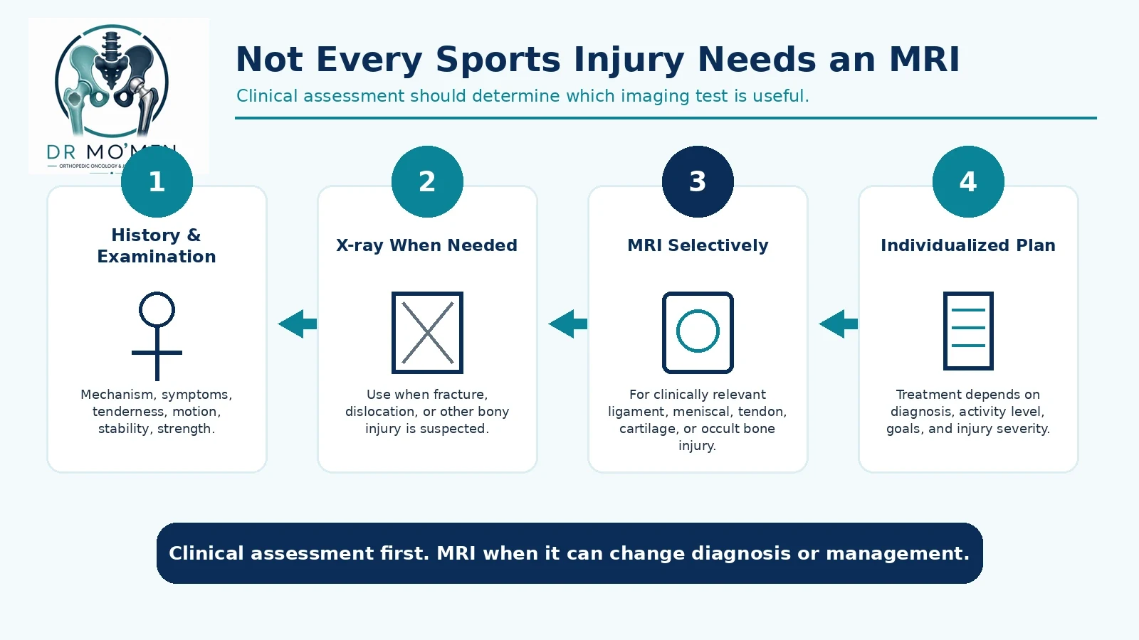
No. Imaging should answer a clinical question rather than be ordered automatically. X-rays are most useful when fracture, dislocation, alignment abnormality or another bony problem is suspected. MRI is useful selectively for clinically important ligament, meniscal, cartilage, tendon, muscle or bone-stress injury, but should be interpreted alongside the examination and mechanism of injury.
When is MRI useful?
MRI is particularly useful when the diagnosis remains uncertain after examination, significant internal soft-tissue injury is suspected, symptoms persist, surgery is being planned, or initial imaging does not answer the clinical question. An MRI abnormality can also be incidental, so imaging should not replace examination.
Does every sports injury need MRI?
No. Many sports injuries can be diagnosed and treated without MRI. The correct sequence is usually history and examination → selective X-ray when bony injury is suspected → MRI when it is likely to change diagnosis or management → individualized treatment plan.
Do Sports Injuries Always Need Surgery?
No. Many sports injuries can be treated non-operatively with activity modification, physiotherapy, progressive strengthening, neuromuscular rehabilitation, bracing when appropriate and gradual return to activity. Surgery is considered when the specific structural injury and the patient’s functional requirements justify it—not merely because an MRI uses the word “tear.”
An ACL tear does not automatically require reconstruction, and a rotator cuff tear does not automatically require immediate surgery. The decision should account for instability, strength, functional goals, associated injuries, response to rehabilitation and the risks of continued non-operative care.
When Is Physiotherapy Appropriate?
Physiotherapy is central to recovery from many sports injuries. Rehabilitation may restore movement, reduce swelling, rebuild strength, improve balance and proprioception, correct movement patterns and progress toward sport-specific activity. It should follow an appropriate diagnosis: a suspected fracture, locked joint, major tendon rupture or severely unstable joint should not simply be sent for exercises without assessment.
A structured program is usually progressed according to the injured tissue, pain response, swelling, range of motion, strength and the demands of the athlete’s sport. If symptoms worsen during rehabilitation, instability persists, or expected function is not returning, the diagnosis and treatment plan should be reviewed rather than simply increasing exercise intensity.
When Can You Return to Sport?
Return to sport should generally depend on function rather than on a fixed calendar date. The relevant criteria vary by injury, but commonly include minimal or absent pain, controlled swelling, appropriate range of motion, restored strength, joint stability, neuromuscular control, sport-specific movement and confidence.
- Minimal or absent pain
- Controlled swelling
- Appropriate range of motion
- Restored strength
- Stability
- Neuromuscular control
- Sport-specific movement
- Confidence and readiness
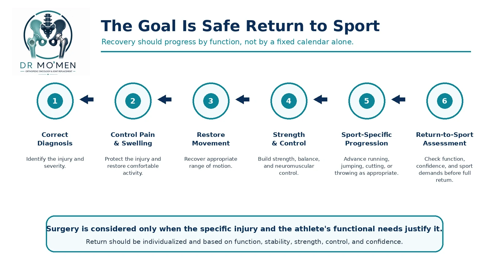
Returning too early can contribute to persistent symptoms or recurrent injury. The goal is not simply to become pain-free at rest; it is to regain the function required for the athlete’s actual sport and position.
What About Recurrent or Overuse Sports Injuries?
Recurrent injuries
An injury that repeatedly returns deserves reassessment. Common contributors include incomplete rehabilitation, residual instability, inadequate strength, altered biomechanics, an incorrect initial diagnosis, premature return to sport and untreated structural injury. Repeated ankle sprains, recurrent shoulder dislocations and repeated knee giving-way episodes should not be accepted as inevitable consequences of sport.
Overuse pain
More concerning overuse patterns include pain that progressively worsens during training, begins earlier in each session, persists after activity, occurs at night, produces focal bone tenderness, reduces performance or causes limping. These findings may indicate that loading is exceeding tissue tolerance; focal bone pain deserves particular caution when a stress injury is possible.
Should Children and Adolescents Be Assessed Differently?
Yes. A growing skeleton is not simply a smaller adult skeleton. Children and adolescents have growth plates, age-specific fracture patterns, apophyseal injuries and unique overuse problems. Persistent pain, limping, focal bone tenderness, swelling or loss of function in a young athlete deserves appropriate assessment rather than advice to simply “play through” the symptoms.
Growth-plate tenderness, persistent limping, inability to return to normal play, or pain that continues despite reduced activity should lower the threshold for assessment. Imaging choices also need to account for the child’s age, skeletal maturity and the suspected injury rather than copying an adult pathway.
Can You Play Through Pain?
Not every episode of exercise discomfort means tissue damage, but continuing activity is inappropriate when pain is accompanied by limping, instability, loss of strength, major swelling, restricted movement, progressive worsening or altered technique used to compensate. Those changes indicate that the athlete is no longer moving normally and that further loading may be unsafe.
Pain that causes an athlete to change running mechanics, avoid loading one limb or lose normal joint control is more clinically important than a mild transient ache that settles quickly. The decision to continue should be based on function and the suspected diagnosis, not on motivation alone.
What Should You Do Immediately After a Minor Sports Injury?
For an apparently minor soft-tissue injury, early management often includes protecting the injured area, stopping painful activity, appropriate compression, elevation when useful and gradual return to movement as tolerated. Cold application may temporarily help pain and swelling in selected acute injuries. The objective is not prolonged immobilization; rehabilitation should progress according to the diagnosis and symptoms.
If the injury is not clearly minor, or if weight bearing, circulation, sensation or joint position is abnormal, self-care is not the correct pathway. In that setting the priority is timely examination and, when indicated, imaging or emergency assessment.
When Should You See an Orthopedic Surgeon Rather Than Continue Self-Care?
Orthopedic review is appropriate when normal function has not returned, the joint is unstable or mechanically blocked, a fracture or major soft-tissue tear is possible, or symptoms persist despite reasonable initial care. It is also useful when imaging shows an injury but the recommended treatment does not clearly match the athlete’s symptoms and goals.
- You cannot bear weight
- A joint is unstable or repeatedly gives way
- Major swelling developed
- Movement remains restricted
- A joint locks
- Deformity occurred
- A fracture is possible
- A ligament or tendon rupture is suspected
- Symptoms are not improving
- Pain repeatedly returns with sport
- The same injury keeps happening
- Return to sport is uncertain
- Imaging shows an injury but the treatment recommendation is unclear
- Surgery has been recommended and you want to understand alternatives
Do You Need a Sports Orthopedic Second Opinion?
A second opinion can be useful when ACL reconstruction has been recommended, recurrent shoulder instability is being treated surgically, arthroscopy has been recommended mainly on MRI findings, symptoms do not match the imaging report, rehabilitation has failed, previous surgery did not solve the problem, or several different procedures have been proposed.
The purpose is not necessarily to avoid surgery. It is to determine whether the diagnosis, timing and proposed procedure are appropriate. See when a second opinion before orthopedic surgery may be useful or use the orthopedic second-opinion pathway.
Sports Injury Assessment in Heliopolis, Cairo
Patients who need an in-person examination can attend the main MoemenOrtho clinic at 50 El Hegaz Street, Heliopolis, Cairo, beside Heliopolis Metro Station. Sports injury assessment can include clinical examination, review of existing X-rays or MRI scans, stability testing and discussion of rehabilitation versus surgery when appropriate. See the Heliopolis orthopedic clinic page for directions, clinic hours and visit preparation.
For the visit, bring any original X-rays or MRI images—not only the written report—plus details of how the injury happened, when swelling started, whether the joint gives way or locks, and what sport or activity you want to return to. This makes the assessment more useful and reduces unnecessary repeat imaging when prior studies are adequate.
The Role of Dr. Mo’men in Sports Injury Assessment
Dr. Mohammed Abdelmoemen Abuelhadid evaluates acute and persistent orthopedic sports injuries with attention to the injury mechanism, clinical examination, joint stability, fracture exclusion, ligament and tendon integrity, meniscal and cartilage pathology, correlation of MRI findings with symptoms, rehabilitation options, whether surgery is actually required and safe progression back to activity.
Cases may include ACL injuries, meniscal injuries, recurrent shoulder instability, rotator cuff injuries, ankle injuries, selected tendon injuries, sports-related fractures and persistent symptoms after previous treatment. Patients outside Egypt may send available X-rays or MRI images and reports for an initial specialist review when appropriate before planning travel.
- Sports Injuries & Joint Arthroscopy service
- Fracture assessment
- Orthopedic Second Opinion
- International Patients
- Appointments or imaging review: 01021690693
Selected Evidence Sources
This article uses general orthopedic and sports-medicine principles and is intended for patient education. The following sources support the red-flag and imaging framework used above:
- NHS — Sprains and strains: urgent and emergency warning signs
- American College of Radiology — Appropriateness Criteria: Acute Trauma to the Knee
- American Academy of Orthopaedic Surgeons — ACL injury plain-language summary