
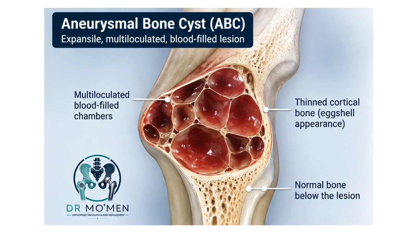
An aneurysmal bone cyst (ABC) is a benign but potentially locally aggressive bone lesion. Although it does not behave like a conventional metastatic bone cancer, it can enlarge, thin the cortex, damage nearby structures and sometimes cause a pathological fracture.
Key Takeaways
- ABC is benign, but it can be locally destructive and mechanically weaken bone.
- It is most common in children, adolescents and young adults, although it can occur at other ages.
- Many conventional ABCs harbor a USP6 rearrangement/fusion, supporting classification as a true benign neoplasm.
- MRI fluid-fluid levels are an important clue, but they are not diagnostic by themselves.
- A planned biopsy is commonly needed when the diagnosis is uncertain or before definitive treatment of an indeterminate lesion.
- Treatment is individualized and can include observation in selected cases, image-guided sclerotherapy, extended curettage, selective arterial embolization, stabilization/reconstruction, or selected off-label systemic therapy for difficult lesions.
What Is an Aneurysmal Bone Cyst?
Despite its name, an aneurysmal bone cyst is not a true arterial aneurysm and is more than a simple fluid cavity. Histologically, it contains blood-filled spaces that are not true endothelial-lined vascular channels. These spaces are separated by fibrous septa containing spindle cells, osteoclast-like giant cells and reactive woven bone.
Modern molecular pathology has changed how ABC is understood. Recurrent USP6 gene rearrangements are found in roughly two-thirds of conventional ABCs in major series and reviews. This supports the concept that many ABCs are true benign neoplasms rather than simply reactive vascular lesions. 2023 pathology review.
ABC Versus ABC-Like Changes in Another Tumor
Older literature often separates “primary” and “secondary” ABC. Current terminology increasingly distinguishes a conventional ABC from ABC-like changes developing within another bone tumor. These changes can occur in lesions such as giant cell tumor of bone, chondroblastoma, osteoblastoma, fibrous dysplasia and occasionally malignant tumors. The underlying lesion—not just the blood-filled cystic component—must therefore be identified before treatment.
Where Does ABC Usually Occur?
ABCs can occur in many bones. Common sites include the femur, tibia, humerus, pelvis and posterior elements of the spine. In long bones, the lesion is often eccentric and expansile in the metaphyseal or meta-diaphyseal region. Location matters because lesions near a growth plate, major joint, neurovascular bundle or spinal canal require more cautious planning.
What Symptoms Can an ABC Cause?
- Persistent localized bone pain or tenderness.
- Progressive swelling or a palpable bony prominence.
- Reduced motion when the lesion is close to a joint.
- A pathological fracture after relatively minor trauma if the cortex becomes severely thinned.
- Back or neck pain, deformity, radicular symptoms, numbness or weakness when the spine is involved.
How Is an Aneurysmal Bone Cyst Diagnosed?
1. Plain X-rays
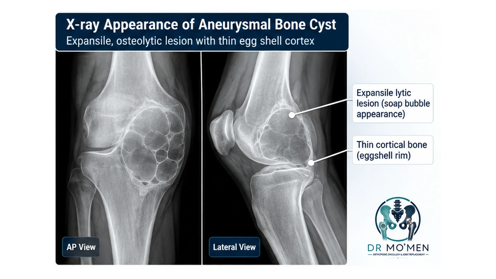
X-rays commonly show an eccentric, expansile radiolucent lesion with remodeling and cortical thinning. A “blow-out” or soap-bubble appearance may be seen, but these descriptions are not unique to ABC. The X-ray also helps assess fracture risk and mechanical stability.
2. MRI
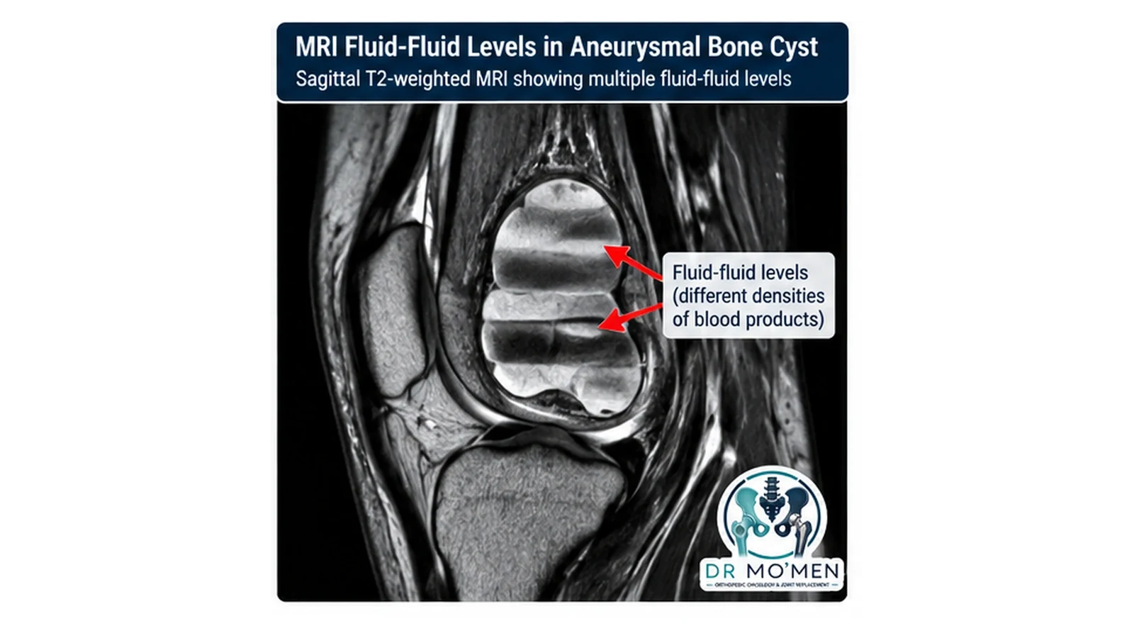
MRI defines the cystic architecture, surrounding soft tissues, growth plate or joint relationship, and possible solid components. Multiple fluid-fluid levels are a classic feature because blood products of different densities settle within cystic spaces. However, fluid-fluid levels can also occur in telangiectatic osteosarcoma and several other lesions, so they are not pathognomonic.
3. CT
CT is useful for showing cortical integrity, internal bony septa, subtle fracture and complex anatomy, especially in the pelvis or spine. It can also assist with image-guided biopsy or percutaneous treatment planning.
4. Planned Biopsy
When an ABC is suspected—particularly before curettage, injection or another definitive procedure—a biopsy is commonly recommended to confirm the diagnosis and exclude malignant mimics when imaging is not unequivocal. The biopsy tract should be planned with the orthopedic-oncology team so it does not compromise a later definitive operation. See planned bone-tumor biopsy principles.
ABC vs Important Clinical Mimics
The distinction between ABC, telangiectatic osteosarcoma and a simple/unicameral bone cyst can change the entire treatment pathway. The table below summarizes typical patterns rather than absolute diagnostic rules.
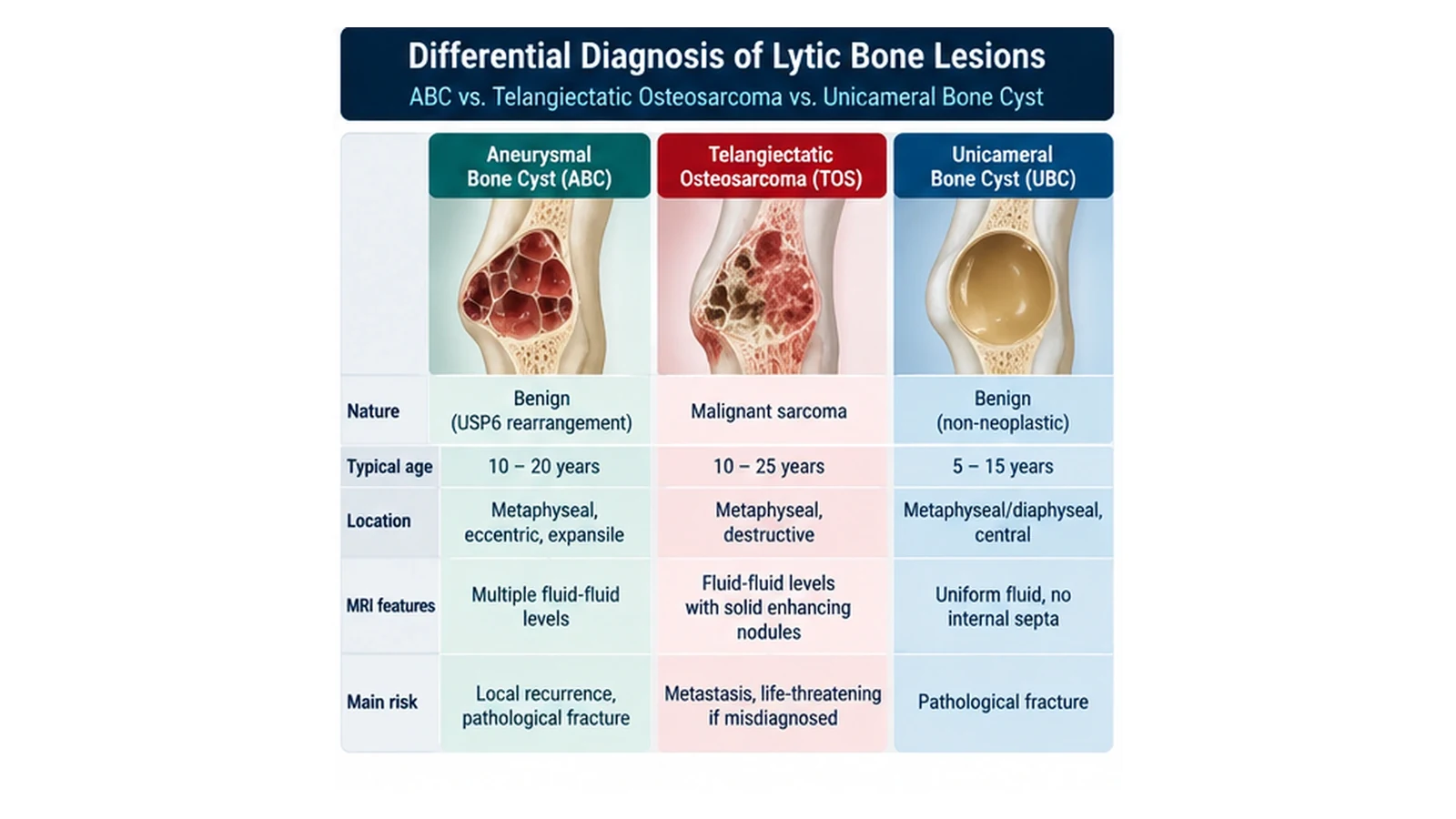
| Feature | Aneurysmal Bone Cyst (ABC) | Telangiectatic Osteosarcoma | Unicameral Bone Cyst |
|---|---|---|---|
| Biology | Benign, locally aggressive; many are USP6-rearranged | High-grade malignant bone sarcoma | Benign non-neoplastic cystic lesion |
| Typical setting | Usually children/adolescents; other ages possible | Usually children, adolescents or young adults | Usually skeletally immature patients |
| Radiographic pattern | Often eccentric and expansile with thin remodeled cortex | More aggressive destruction and/or soft-tissue component may be present | Usually central, often metaphyseal in long bones |
| MRI | Multiple fluid-fluid levels are common | Fluid-fluid levels may occur, often with more suspicious solid enhancing tissue | Usually more uniform fluid signal; fracture-related changes can occur |
| Why distinction matters | Recurrence, fracture and local structural damage | Requires oncologic staging and sarcoma treatment | Management centers on symptoms, activity and fracture risk |
Modern Treatment Options
There is no single treatment that is best for every aneurysmal bone cyst. The decision depends on age, site, size, symptoms, cortical integrity, fracture risk, proximity to the growth plate or joint, spinal or neurovascular involvement, previous treatment and local expertise.
1. Observation in Selected Cases
Short-interval surveillance may be considered for selected small, minimally symptomatic, structurally safe lesions that appear biologically quiet. Progressive pain, enlargement, fracture risk or a sensitive location usually favors active treatment.
2. Percutaneous Image-Guided Sclerotherapy
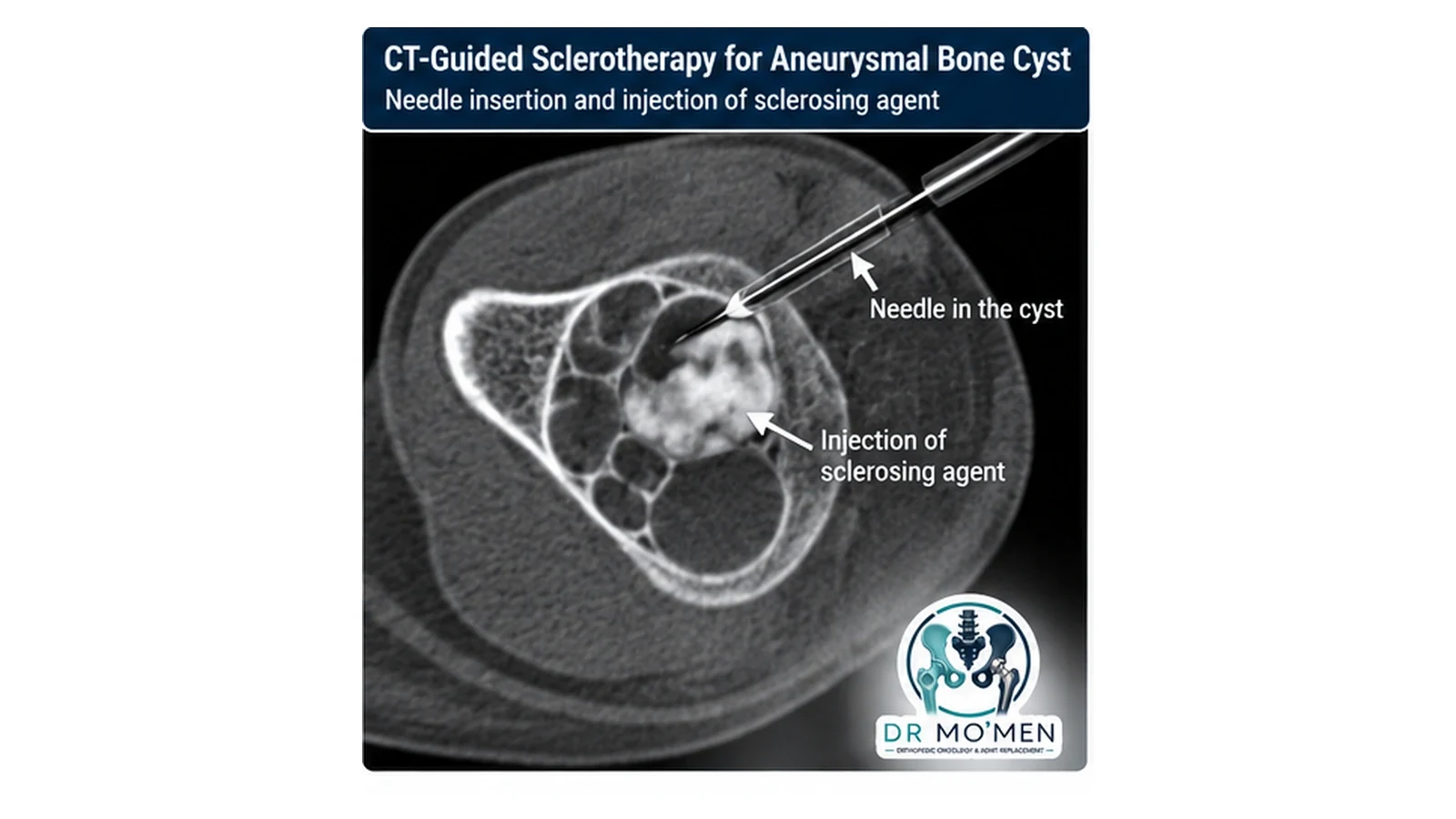
Percutaneous sclerotherapy is an accepted minimally invasive option for many accessible ABCs. Agents such as polidocanol or doxycycline are used in published protocols. Repeated sessions may be required; the number depends on response, agent, lesion size and location. A 2025 systematic review supports polidocanol as an effective option while emphasizing that the evidence remains heterogeneous and no single strategy is best for every ABC. 2025 systematic review.
3. Extended Curettage and Reconstruction
Surgery remains important when the lesion requires immediate mechanical stabilization, decompression, tissue diagnosis or direct local control. Extended intralesional curettage may include meticulous removal of the lesion and use of a high-speed burr where anatomically safe. The cavity may be reconstructed with graft, bone substitute, cement and/or fixation according to site and structural needs.
4. Selective Arterial Embolization
Selective arterial embolization can be used as a definitive or adjunctive treatment in selected highly vascular or difficult lesions, particularly in the pelvis and spine. It may also be used before surgery to reduce blood flow when a safe arterial target is identified.
5. Denosumab in Selected Difficult Lesions
Denosumab has been used off-label for selected unresectable, pelvic, spinal, recurrent or otherwise high-morbidity ABCs. It is not routine first-line therapy for most ABCs. Specialist monitoring is important because recurrence after discontinuation and metabolic complications—including rebound hypercalcemia, particularly in younger patients—have been reported. 2024 scoping review.
How Is the Best Treatment Chosen?
| Clinical situation | Potential approach | Important limitation |
|---|---|---|
| Accessible lesion without major instability | Image-guided sclerotherapy may be considered | May require repeated procedures and imaging follow-up |
| Large lesion with structural weakness, fracture risk or reconstruction need | Extended curettage ± graft/substitute ± fixation | Open surgery; recurrence remains possible |
| Pelvic/spinal or highly vascular lesion | Selective arterial embolization, surgery or combined strategy | Requires experienced multidisciplinary planning |
| Neurologic compromise or mechanical spinal instability | Urgent specialist assessment; decompression/stabilization may be necessary | Delay can risk permanent neurologic deficit |
| Difficult unresectable/high-morbidity lesion | Selected off-label systemic therapy such as denosumab may be considered | Monitoring, uncertain optimal duration and metabolic/recurrence risks |
Recovery, Recurrence, and Follow-Up
Recovery varies substantially. A small percutaneous procedure may permit relatively rapid return to daily activities, whereas a large curettage with grafting, fixation or treatment of a pathological fracture may require protected weight-bearing and months of radiographic healing. Return to sport should be based on symptoms, mechanical stability and serial imaging rather than a fixed calendar date.
Local recurrence remains possible after any treatment. Published rates vary because studies differ in age, site, technique and follow-up. Follow-up frequency should therefore be individualized, with clinical assessment and serial imaging focused on symptoms, re-expansion, cortical healing and stability.
When Is Specialist Orthopedic Oncology Assessment Important?
- An enlarging bone lesion in a child, adolescent or young adult.
- Persistent focal bone pain or swelling that does not fit a simple injury.
- A pathological fracture or marked cortical thinning.
- A report describing fluid-fluid levels, an expansile lytic lesion or uncertainty between ABC and another tumor.
- A lesion in the pelvis, spine or close to a growth plate or major joint.
- Recurrence after previous curettage or injection.
- A proposed operation before the diagnosis has been confirmed or the biopsy route planned.
Emergency Red Flags
Urgent assessment is appropriate for new limb weakness or numbness, loss of bladder or bowel control, rapidly progressive spinal symptoms, severe acute deformity, or inability to bear weight after a suspected pathological fracture.
Evidence & Sources
- Nasri E, Reith JD. Aneurysmal bone cyst: a review. J Pathol Transl Med. 2023. PMID 36950810.
- van Geloven TPG, van de Sande MAJ, van der Heijden L. The treatment of aneurysmal bone cysts. Curr Opin Pediatr. 2023.
- Bakarman KA. Diagnosis and Current Treatment of Aneurysmal Bone Cysts. Cureus. 2024. PMID 38449944.
- Prabowo Y, et al. Sclerotherapy with polidocanol as a treatment for aneurysmal bone cyst: a systematic review. J Clin Orthop Trauma. 2025. PMID 40292395.
- El Masry AM, Azmy SI, Mustafa MAR, Abuelhadid MA. Using Denosumab as a Nonsurgical Management of Aneurysmal Bone Cysts in the Pelvis. Clin Orthop Surg. 2024. PMID 38304208.