
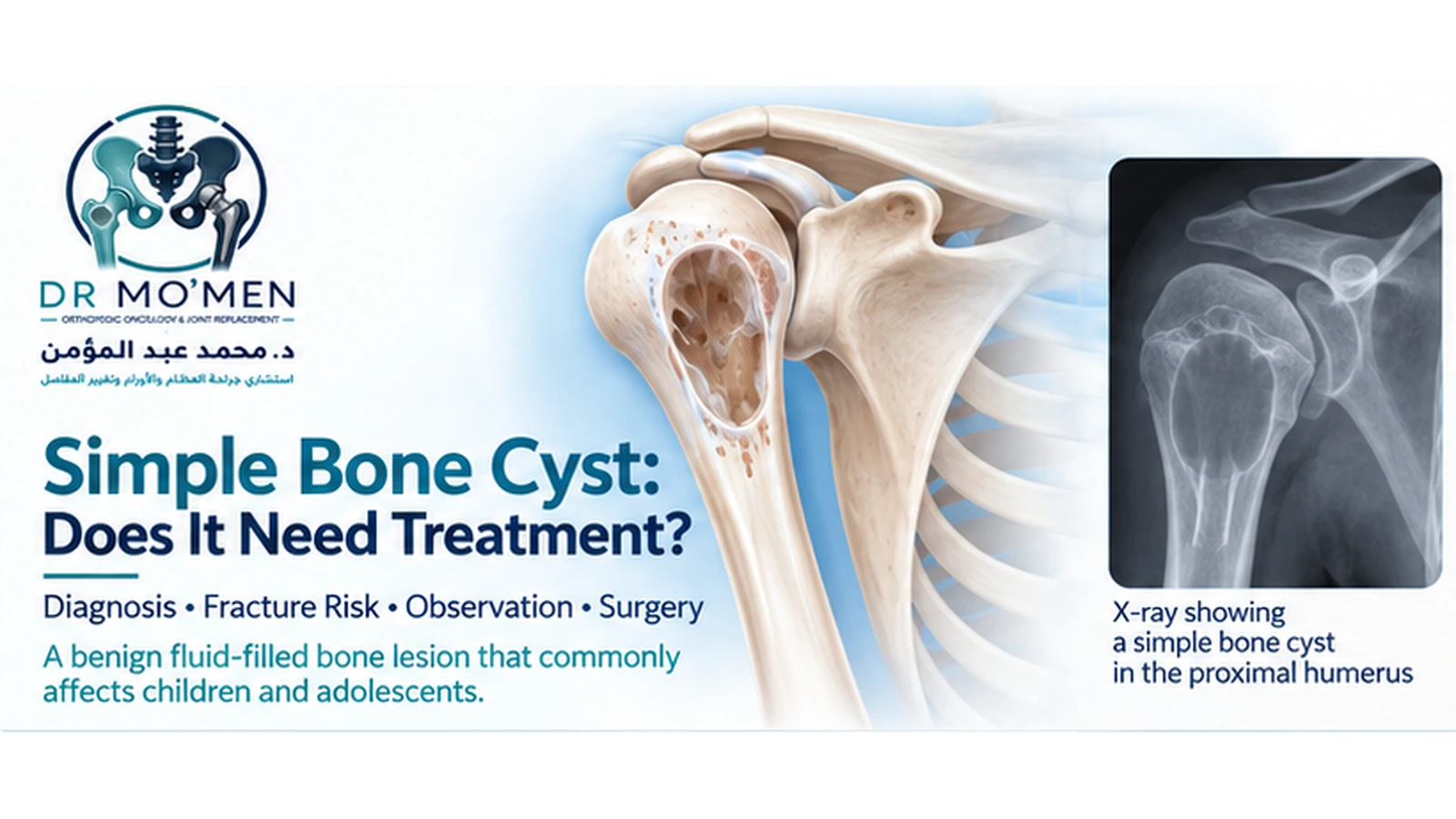
A simple bone cyst, also called a unicameral bone cyst (UBC), is a benign fluid-filled cavity that develops inside bone.
It occurs mainly in children and adolescents and is particularly common in the proximal humerus and proximal femur. Many cysts are discovered only after an X-ray is taken for an injury, and some first present because a fracture occurs through the weakened bone.
Despite the word “cyst,” this is not a cyst that can simply be removed like a skin cyst.
The important questions are:
Is the diagnosis secure?
How weak is the bone?
Has a fracture occurred?
Where is the cyst located?
Is observation safe, or is treatment needed to reduce mechanical risk?
What Is a Simple Bone Cyst?
A simple bone cyst is a benign, non-neoplastic intramedullary bone lesion containing fluid.
It usually develops in the metaphysis of a long bone in a growing child.
The exact cause remains uncertain. Several theories have been proposed, including disturbance of local venous drainage and altered bone remodeling, but no single mechanism has been definitively established.
Who Usually Develops a Unicameral Bone Cyst?
Simple bone cysts occur predominantly in children and adolescents.
Most occur during the first two decades of life, with proximal humeral and proximal femoral involvement being particularly common.
The lesion may gradually move farther away from the growth plate as the child grows.
Where Do Simple Bone Cysts Usually Occur?
The most common sites are:
- proximal humerus
- proximal femur
Other possible locations include:
- tibia
- fibula
- calcaneus
- pelvis
- other long bones
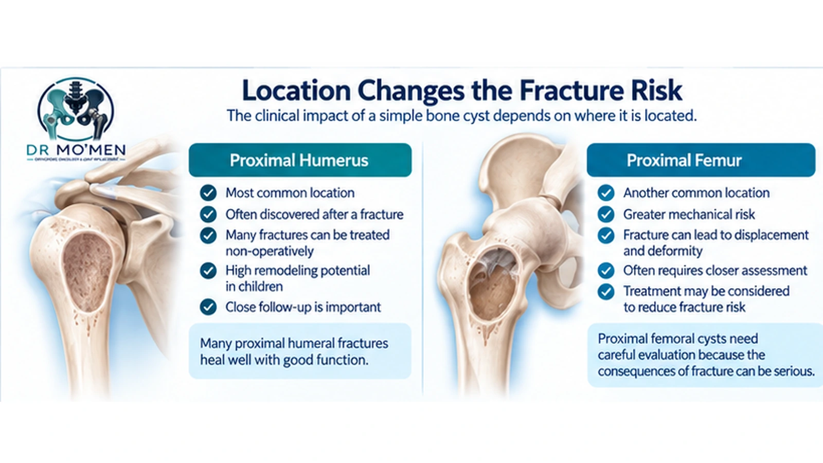
The location matters greatly because the consequences of fracture differ.
A cyst in the proximal humerus may often be managed quite differently from a large cyst in the proximal femur, where fracture may lead to more serious mechanical problems.
What Symptoms Does a Simple Bone Cyst Cause?
Many UBCs cause no symptoms at all.
They may be discovered incidentally.
However, a substantial number present after a pathological fracture through the cyst.
Symptoms may include:
- sudden pain after minor trauma
- swelling
- inability to use the limb normally
- tenderness
- deformity when fracture displacement occurs
Pain without fracture can occur but should prompt review of the diagnosis and mechanical situation rather than automatically being attributed to the cyst.
Is a Simple Bone Cyst a Bone Tumor?
A UBC is considered a benign cystic bone lesion, not a malignant tumor.
However, not every cystic-looking bone lesion is a simple bone cyst.
Other lesions may enter the differential diagnosis, including:
- aneurysmal bone cyst
- fibrous dysplasia
- non-ossifying fibroma
- chondroblastoma
- infection
- other benign lesions
- occasionally more serious pathology
Therefore, the diagnosis should be based on the complete radiographic and clinical picture.
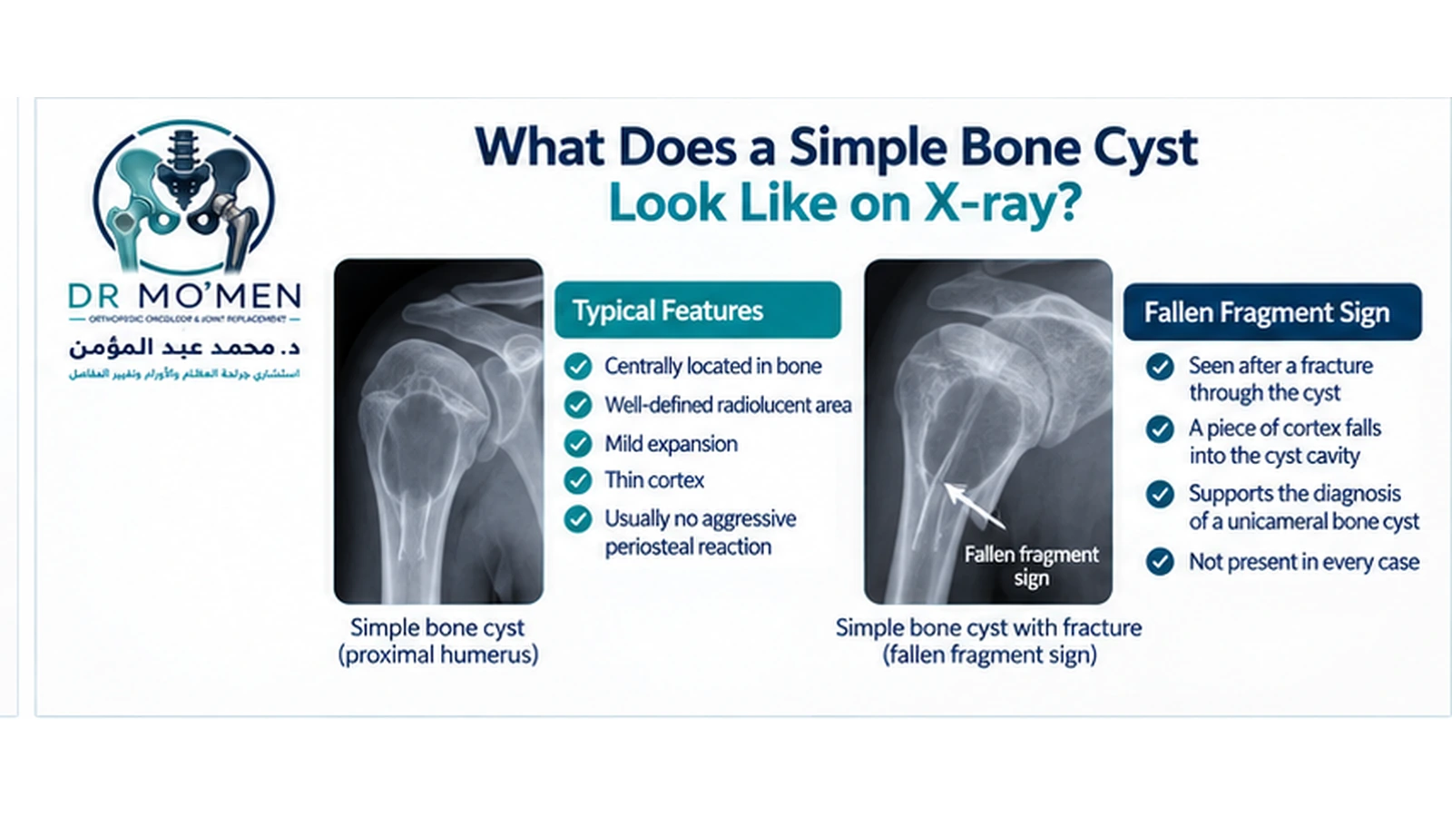
What Does a Simple Bone Cyst Look Like on X-Ray?
A typical UBC is:
- centrally located within the bone
- radiolucent
- well defined
- usually mildly expansile
- associated with cortical thinning
- usually without an aggressive periosteal reaction unless a fracture has occurred
The typical lesion is a centrally located metaphyseal radiolucency with a thin cortical rim.
What Is the Fallen Fragment Sign?
When a fracture occurs through a simple bone cyst, a small piece of cortex may fall into the fluid-filled cavity.
This produces the classic fallen fragment, or fallen leaf, sign.
When present, it strongly supports the diagnosis of a unicameral bone cyst.
However, it is not seen in every case.
Do You Need MRI?
Not always.
For a characteristic lesion on plain radiographs, X-rays may provide enough information.
MRI can be useful when:
- the diagnosis is uncertain
- the lesion is atypical
- another cystic or tumor-like lesion is being considered
- soft-tissue findings need clarification
- the extent is difficult to define
MRI typically shows a fluid-containing lesion, but the imaging appearance after fracture can become more complex.
When Is CT Useful?
CT is not routinely required for most straightforward long-bone UBCs.
It may help in:
- complex anatomy
- unusual locations
- cortical assessment
- selected calcaneal lesions
- preoperative planning
Plain radiographs remain the fundamental investigation in most cases.
Does Every Simple Bone Cyst Need a Biopsy?
No.
A typical UBC in the correct age group and anatomical location may often be diagnosed radiographically.
Biopsy becomes more relevant when:
- imaging is atypical
- the lesion behaves unusually
- another diagnosis remains possible
- surgery is planned and tissue confirmation is needed
As with other bone lesions, an uncertain diagnosis should be reviewed before an unplanned curettage is performed.
What Is an Active Bone Cyst?
An active cyst is generally located close to the growth plate.
These lesions are more common in younger children and may behave more persistently.
As the child grows, some cysts become more separated from the physis and are often termed latent cysts.
This distinction can matter when considering recurrence risk, growth and treatment strategy.
Why Do Pathological Fractures Occur?
As the cyst enlarges, the cortex may become thin.
The bone can therefore become mechanically weaker.
A relatively minor injury that would not normally fracture healthy bone may cause a pathological fracture through the cyst.
This is one of the main reasons treatment decisions focus on fracture risk rather than simply cyst size.
Does a Fracture Make the Cyst Heal?
Sometimes—but not reliably.
After fracture, some cysts show partial or substantial healing.
Others remain visible or recur.
Therefore, a fracture is not automatically considered a definitive treatment for the cyst.
Follow-up X-rays are important after fracture healing.
Simple Bone Cyst in the Proximal Humerus
The proximal humerus is one of the most common locations.
When a pathological fracture occurs here, many children can initially be treated non-operatively with immobilization because the proximal humerus has substantial remodeling potential and many fractures heal satisfactorily.
After fracture healing, the cyst should be reassessed.
Persistent cysts with significant structural weakness may require further treatment depending on age, size, activity and recurrence risk.
Simple Bone Cyst in the Proximal Femur
The proximal femur deserves greater caution.
A fracture in this area can potentially lead to:
- displacement
- deformity
- varus malalignment
- limb-length problems
- more complex reconstruction
Because the mechanical consequences are greater, proximal femoral cysts may justify more proactive treatment when fracture risk is significant.
How Is Fracture Risk Assessed?
There is no single measurement that perfectly predicts fracture.
Assessment considers:
- site of the cyst
- cortical thickness
- extent of the lesion
- age
- activity
- previous fracture
- remaining bone stock
- proximity to the physis
- whether the cyst occupies most of the bone diameter
The mechanical consequences of a fracture are also important.
A lesion with moderate fracture risk in the humerus may be treated differently from a similar-looking lesion in the proximal femur.
When Can a Simple Bone Cyst Be Observed?
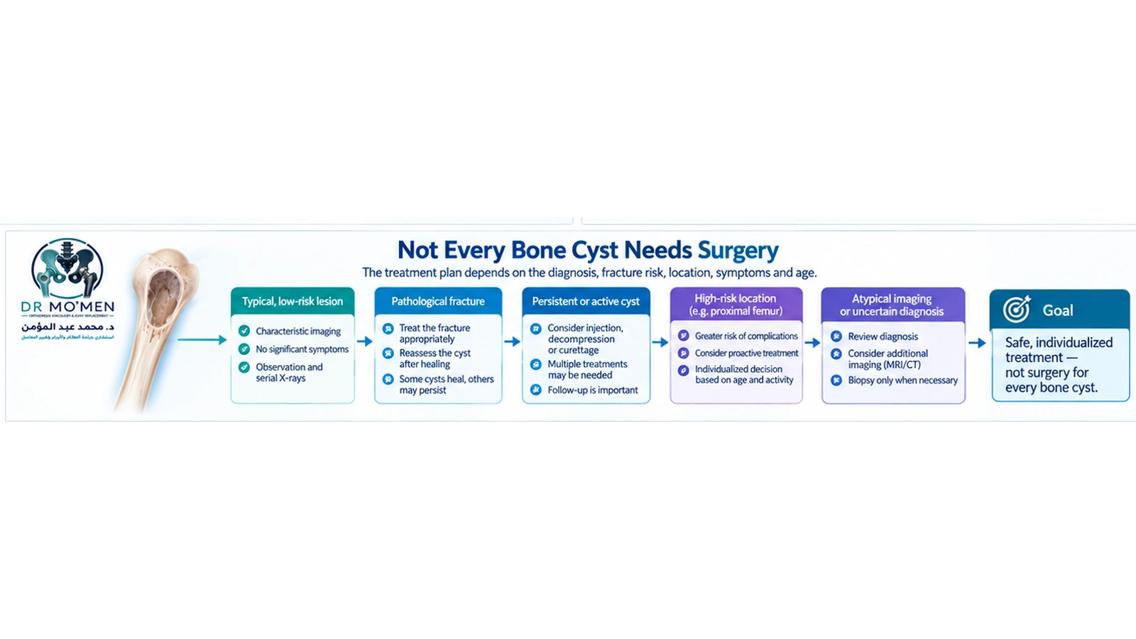
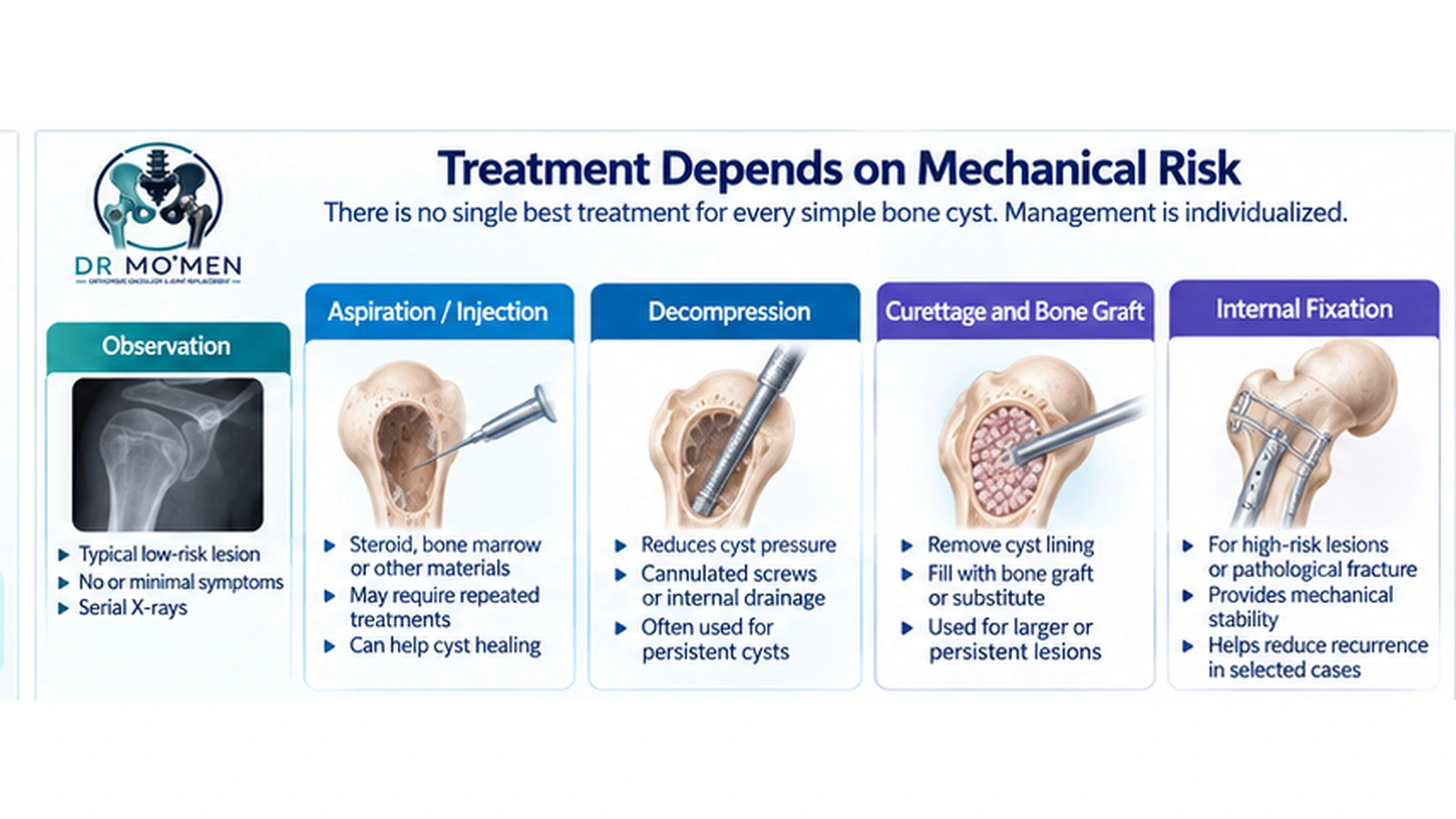
Observation can be appropriate when:
- imaging is typical
- the child has minimal or no symptoms
- fracture risk is low
- the lesion is in a mechanically forgiving location
- the cortex remains sufficiently strong
- there is no concerning progression
Observation with serial radiographs is appropriate for many upper-extremity UBCs and lower-extremity lesions with low pathological-fracture risk.
How Is a Pathological Fracture Treated?
Treatment depends on the location and fracture pattern.
Upper extremity
Many proximal humeral fractures through UBCs can initially be treated with:
- sling or immobilization
- pain control
- radiographic follow-up
Once the fracture heals, the residual cyst can be reassessed.
Lower extremity
A fracture through a proximal femoral cyst may require a more aggressive stabilization strategy because of the consequences of displacement and deformity.
Treatment should be individualized.
What Treatments Are Available for Persistent or High-Risk Cysts?
There is no single universally accepted treatment that is superior in every patient.
Options may include:
Percutaneous aspiration and injection
The cyst can be entered through a needle and treated with substances such as:
- corticosteroid
- bone marrow aspirate
- bone graft substitutes
- combinations of biologic materials
Multiple procedures may sometimes be required.
Decompression
Because increased cyst pressure may contribute to persistence, treatment strategies often attempt to create ongoing decompression.
Techniques may include:
- cannulated screws
- flexible intramedullary nails
- other internal drainage strategies
Curettage
Curettage removes the cyst lining and tissue through an opening in the bone.
The resulting cavity may be filled with:
- autograft
- allograft
- bone substitute
However, curettage alone does not guarantee permanent healing, and recurrence or persistence remains possible.
Internal fixation
Internal fixation may be appropriate when:
- the bone is significantly weakened
- a fracture is present
- the proximal femur is involved
- recurrent fracture is likely
- deformity risk is substantial
Fixation may simultaneously provide mechanical protection and, depending on the implant, cyst decompression.
Is Steroid Injection Still Used?
Yes, in selected situations.
Steroid injection has historically been widely used for UBCs.
However, repeated injection treatments may be required, and more recent approaches increasingly emphasize decompression and mechanical management in appropriate cases.
Is Bone Marrow Injection Better Than Steroid?
Not clearly.
Multiple comparative studies have produced conflicting results.
The evidence does not support presenting one injection material as universally superior.
This is why the treatment decision should focus on:
- patient age
- cyst location
- fracture risk
- mechanical stability
- cyst activity
- prior treatment
rather than simply choosing an injectable substance.
Why Is Decompression Important?
The biology of UBC remains incompletely understood, but cyst pressure and local fluid dynamics are thought to contribute to persistence.
Creating a sustained pathway for decompression may therefore promote bone healing.
Can a Simple Bone Cyst Recur?
Yes.
Persistence or recurrence is one of the main challenges of UBC treatment.
Recurrence risk varies according to:
- patient age
- cyst activity
- proximity to the growth plate
- treatment method
- residual cyst size
- follow-up duration
Patients therefore need appropriate radiographic follow-up even after treatment.
Does Recurrence Mean the Lesion Is Malignant?
No.
Recurrence or persistence is common in UBCs and does not automatically indicate cancer.
However, if the lesion behaves unexpectedly or develops atypical imaging features, the diagnosis should be reassessed.
What About a Simple Bone Cyst in the Calcaneus?
Calcaneal cysts behave differently from typical proximal humeral lesions.
They may be discovered incidentally or present with heel pain.
Treatment depends on:
- symptoms
- cyst size
- exact location
- cortical strength
- fracture risk
Can the Cyst Affect Growth?
Most patients do not develop major growth disturbance.
However, lesions located near the growth plate require thoughtful treatment planning, particularly in younger children.
Both the lesion itself and overly aggressive surgery near the physis can potentially create problems.
This is another reason treatment should be individualized.
When Should the Diagnosis Be Reconsidered?
Reassessment is appropriate when:
- the lesion is not centrally located
- marked cortical destruction is present
- there is a soft-tissue mass
- aggressive periosteal reaction occurs without fracture
- symptoms are disproportionate
- the lesion progresses unexpectedly
- imaging does not fit the typical age or site
- recurrent “bone cyst” behaves unusually
The safest approach is to confirm the diagnosis rather than repeatedly treat an atypical lesion under the assumption that it is a UBC.
When Should You Seek Specialist Orthopedic Oncology Assessment?
Specialist review may be particularly useful when:
- imaging is atypical
- diagnosis remains uncertain
- a pathological fracture has occurred
- the proximal femur is involved
- multiple fractures have occurred
- the cyst is large and mechanically significant
- surgery is being considered
- a previous treatment has failed
- recurrence occurs repeatedly
- biopsy is being considered
The Role of Dr. Mo’men in Simple Bone Cyst Cases
Dr. Mohammed Abdelmoemen Abuelhadid evaluates cystic bone lesions with attention to:
- confirming whether imaging is typical of a UBC
- differentiating simple bone cyst from other benign or aggressive lesions
- reviewing fracture risk
- assessing proximal femoral lesions carefully
- determining whether observation is appropriate
- deciding whether biopsy is genuinely necessary
- reviewing pathological fractures
- choosing between injection, decompression, curettage and fixation when treatment is required
- evaluating recurrent or previously treated cysts
The aim is not to operate on every bone cyst.
The treatment plan depends primarily on:
diagnostic certainty + fracture risk + anatomical location + symptoms + mechanical stability + age.
Patients outside Egypt may send their X-rays, CT/MRI studies and previous treatment records for an initial review before deciding whether in-person assessment is required.
Orthopedic Oncology: /en/services/orthopedic-oncology/
Bone Biopsy: /en/services/bone-biopsy/
Fracture Service: /en/services/fractures/
Second Opinion: /en/second-opinion/
International Patients: /en/international-patients/
For appointments or imaging review: 01021690693