
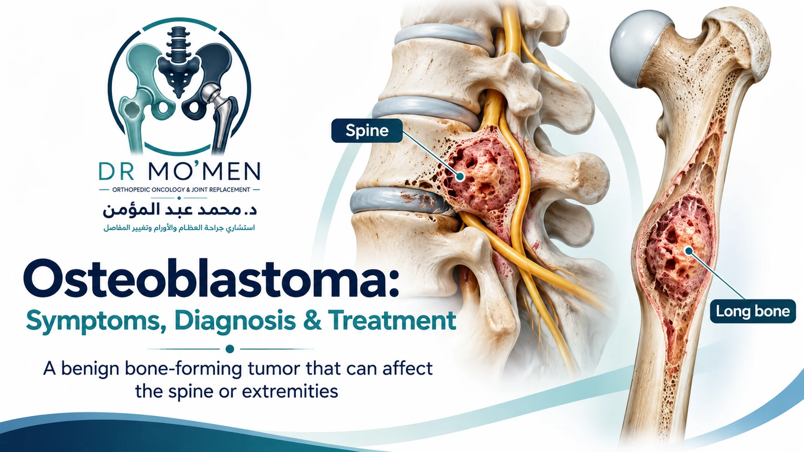
Osteoblastoma is a rare benign bone-forming tumor that usually affects adolescents and young adults. It can arise in the spine, long bones, pelvis, and other skeletal sites. Although benign, some osteoblastomas are expansile or locally aggressive and can weaken bone, extend toward nearby soft tissues, or compress neural structures depending on their location.
Key Takeaways
- Osteoblastoma is benign, but “benign” does not mean biologically inactive; selected lesions can be locally aggressive.
- It usually affects younger patients. In the large Lucas series, the mean age was about 20 years and males were affected more often than females.
- The spine is an important site but not the only site. The vertebral column including sacrum accounted for 32% of cases in that large series; long bones and other skeletal locations also occur.
- Osteoblastoma is typically larger than osteoid osteoma, but size alone does not establish the diagnosis.
- CT is particularly useful for mineralization and cortical anatomy; MRI is important for marrow, soft-tissue, joint and neural relationships.
- Biopsy is not an isolated test. When histology is required, it should be planned after appropriate imaging and with the definitive treatment pathway in mind.
- Selected smaller, non-aggressive lesions may be candidates for image-guided thermal ablation in experienced centers; larger, aggressive, structurally threatening or diagnostically uncertain lesions more often require surgery.
- Follow-up matters because local recurrence can occur, particularly after incomplete treatment or in biologically aggressive lesions.
What Is Osteoblastoma?
Osteoblastoma is a benign osteogenic neoplasm composed of osteoid and woven bone produced by osteoblasts in a vascular stroma. It shares important histologic features with osteoid osteoma, but osteoblastoma is usually larger, more expansile and less associated with the classic “night pain relieved dramatically by NSAIDs” pattern.
A size threshold around 2 cm is commonly used as a practical distinction from osteoid osteoma, but it is not an absolute rule. The final diagnosis depends on the combined clinical, radiologic and histopathologic picture.
Where Can Osteoblastoma Occur?
Osteoblastoma can occur in both the axial and appendicular skeleton. The vertebral column — especially posterior elements such as the pedicle or lamina — is a frequent site, but the tumor also occurs in long bones of the extremities, the pelvis, foot and other bones. Therefore, evaluation should be based on the actual lesion location rather than assuming that osteoblastoma is primarily a spinal disease.
What Symptoms Can Osteoblastoma Cause?
Symptoms vary according to location, tumor size and biological behavior. Pain is the most common complaint, but the exact pattern is less stereotyped than in osteoid osteoma.
- Persistent localized bone pain, which may be dull or progressive.
- Pain that may occur at night but often does not show the dramatic NSAID response typical of osteoid osteoma.
- Localized swelling, tenderness or a palpable bony prominence when the lesion is superficial.
- Limping or reduced function when a weight-bearing bone is involved.
- Joint stiffness, effusion or restricted movement when the lesion lies close to a joint.
- Painful scoliosis or focal muscle spasm when the lesion involves the spine.
- Radiating pain, numbness, weakness or other neurological symptoms if a spinal lesion narrows the canal or neural foramen.
Osteoblastoma in the Spine
Spinal osteoblastoma deserves specific attention because a posterior-element lesion can cause persistent back or neck pain, painful scoliosis, and — in selected cases — nerve-root or spinal-cord compression. CT defines the bony anatomy well, while MRI helps show the epidural, marrow and soft-tissue relationships. Surgical planning may also need to address stability if a large part of the posterior elements or facet complex must be removed.
Osteoblastoma in Long Bones and the Extremities
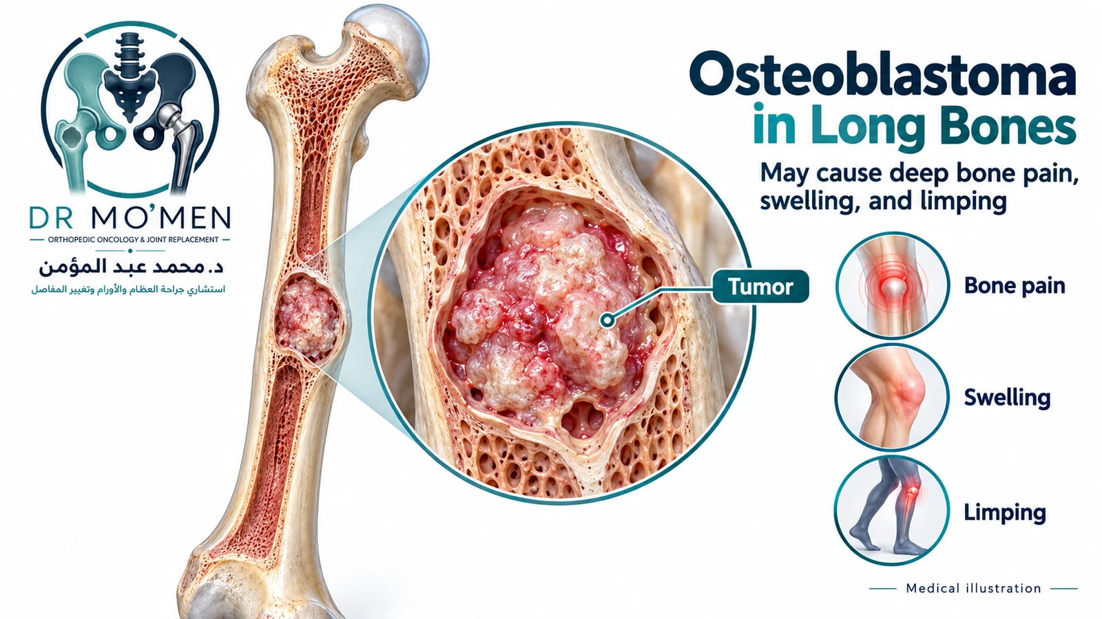
In the extremities, patients may present with focal pain, swelling, limping, or loss of motion near a joint. Imaging can show an expansile lytic or mixed lesion, cortical thinning or expansion, and variable internal mineralization. Structural risk is important in a weight-bearing bone because a large lesion can weaken the cortex even though the tumor is benign.
How Is Osteoblastoma Diagnosed?
A secure diagnosis requires correlation between symptoms, imaging and, when indicated, pathology. The diagnostic sequence should be deliberate because treatment differs substantially between osteoblastoma, osteoid osteoma, aneurysmal bone cyst, infection and osteosarcoma.
1. Plain Radiographs
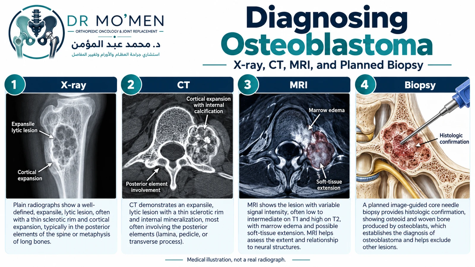
X-rays may show a well-defined lytic or mixed lesion with variable sclerosis, internal mineralization, cortical expansion or thinning. Some osteoblastomas look relatively circumscribed, while aggressive lesions can appear more destructive.
2. CT
CT is especially useful for defining mineralized osteoid matrix, cortical anatomy and complex sites such as the spine or pelvis. It can also help plan a percutaneous procedure or biopsy route when those are appropriate.
3. MRI
MRI defines intramedullary extent, soft-tissue extension, joint involvement and proximity to nerves and vessels. Reactive marrow and soft-tissue edema can be extensive, so MRI should be interpreted together with radiographs and CT rather than in isolation.
4. Planned Biopsy When Needed
Histologic confirmation is commonly required when the imaging is atypical, an aggressive lesion is possible, the diagnosis will change the surgical plan, or major reconstruction is being considered. A classic benign-appearing lesion may sometimes be managed without a separate biopsy, particularly if an image-guided ablation strategy includes planned tissue sampling. The decision should be individualized.
Can Osteoblastoma Be Confused With Osteosarcoma?
Yes. Some bone-forming tumors can overlap on imaging and in small biopsy samples. If an osteoblastoma looks unusually aggressive, the pathology does not fit the imaging, or the lesion behaves unexpectedly after treatment, the diagnosis should be reviewed by a specialist bone-tumor team rather than relying on one feature alone.
In difficult cases, specialist pathology may use ancillary molecular tests such as FOS/FOSB studies, but these are supporting tools rather than stand-alone proof. A 2025 pathology review emphasizes integrated differential diagnosis for benign bone-forming tumors, and 2026 molecular evidence shows that even FOS rearrangements are not perfectly specific for osteoblastoma. The safest approach is correlation of the clinical picture, imaging and pathology before definitive treatment. 2025 benign bone-forming tumor review · 2026 FOS-fusion study.
Osteoblastoma vs Osteoid Osteoma
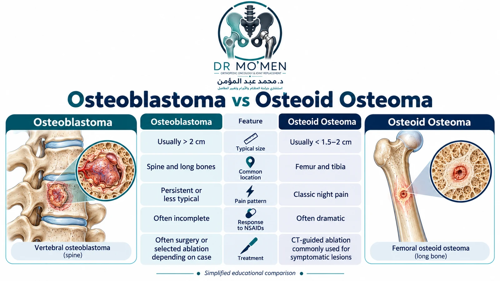
The two tumors are related osteogenic lesions, but their clinical behavior and treatment are not identical. The comparison below uses typical patterns rather than absolute rules.
| Feature | Osteoblastoma | Osteoid Osteoma |
|---|---|---|
| Typical size | Usually larger; commonly > about 2 cm | Usually smaller; often <1.5–2 cm |
| Common sites | Spine and long bones; other sites also occur | Femur and tibia are especially common |
| Pain | Persistent or less stereotyped; NSAID response may be incomplete | Classically worse at night with marked NSAID response |
| Imaging | Expansile lytic/mixed lesion with variable mineralization and sclerosis | Small nidus often surrounded by reactive sclerosis |
| Behavior | May be active or locally aggressive | Usually limited and self-limiting |
| Treatment | Ablation or surgery depending on location, behavior and anatomy | CT-guided ablation commonly used for symptomatic lesions |
How Is Osteoblastoma Treated?
There is no single operation or procedure that is correct for every osteoblastoma. Treatment should consider biological behavior, location, size, cortical integrity, neurological or joint involvement, diagnostic certainty, previous procedures, and the morbidity of the proposed treatment.
1. Image-Guided Thermal Ablation
CT-guided radiofrequency ablation or other thermal techniques can be effective in selected osteoblastomas, particularly smaller or non-aggressive lesions that can be safely accessed and adequately covered by the ablation zone. Published series include both spinal and extremity lesions. These techniques require experienced interventional teams, careful thermal protection near nerves or joints, and multidisciplinary case selection.
Thermal ablation is not automatically suitable for a large, aggressive, structurally unstable or diagnostically uncertain lesion. In those settings, surgery may provide better local control, tissue diagnosis and reconstruction.
2. Extended Intralesional Curettage
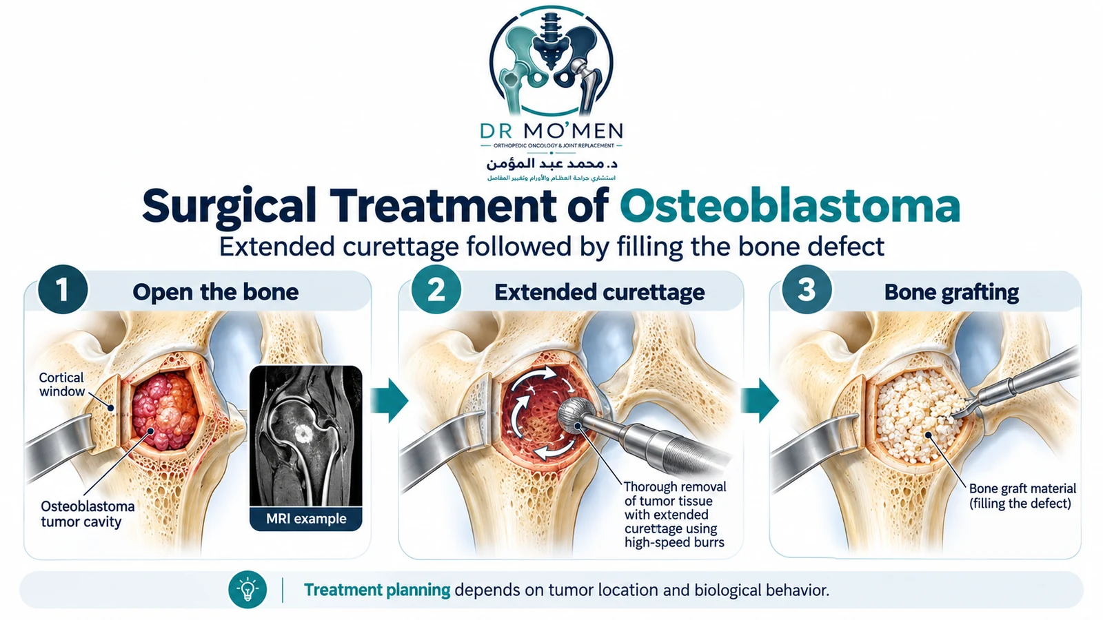
For many active but resectable lesions, extended intralesional curettage is an established surgical strategy. The surgeon removes the visible tumor and may use a high-speed burr to extend the margin within the cavity. Depending on location and bone strength, the defect can be left to heal, filled with bone graft or bone substitute, or reconstructed with cement or fixation. Local adjuvants may be considered in selected cases; they are not mandatory in every osteoblastoma.
3. En Bloc or Wider Resection
For selected aggressive lesions, recurrent tumors, lesions with major cortical/soft-tissue extension, or tumors in which intralesional treatment is unlikely to provide adequate local control, en bloc resection may be considered when anatomically feasible. In the spine, this can be complex and may require decompression, reconstruction and instrumentation to preserve stability.
4. Reconstruction and Stabilization
When tumor removal creates a significant structural defect, reconstruction may involve bone grafting, internal fixation, spinal instrumentation, or other site-specific methods. The reconstruction plan is part of tumor surgery, not an afterthought.
What About Enneking Stage and “Aggressive Osteoblastoma”?
Osteoblastoma can behave as an active benign lesion or as a more aggressive benign tumor. In specialist practice, Enneking staging and the radiologic border of the lesion may help frame the treatment strategy, particularly in the spine. Historical series support intralesional excision for many active stage-2 lesions and consideration of wider/en bloc surgery for selected stage-3 lesions, but anatomy, morbidity and modern minimally invasive options must also be weighed. The stage should guide — not mechanically dictate — treatment.
Can Osteoblastoma Recur?
Yes. Local recurrence can occur, and the risk is not represented well by one universal percentage. It depends on tumor biology, location, completeness of treatment, treatment method and whether the lesion has already been disturbed by previous surgery. The 306-case Lucas series reported recurrence in both institutional and consultation cohorts, and spinal series show higher recurrence in biologically aggressive or previously treated lesions. This is why planned first treatment and structured follow-up are important.
Recovery and Follow-Up
Recovery varies widely. A patient treated with percutaneous ablation may resume daily activity relatively quickly, whereas extended curettage, long-bone fixation, spinal decompression or fusion may require protected loading, bracing or longer rehabilitation. Follow-up typically includes clinical assessment and interval imaging tailored to the treated site and recurrence risk.
When Do You Need Specialist Assessment?
- Persistent focal bone pain with an unexplained lesion on X-ray, CT or MRI.
- A painful or enlarging bony swelling.
- Back or neck pain associated with painful scoliosis or a known posterior-element lesion.
- Numbness, weakness, radiating pain or other neurological symptoms.
- A lesion that has been called “osteoblastoma” but the imaging and pathology do not clearly match.
- A planned biopsy, curettage or ablation where the route or definitive treatment strategy has not yet been discussed with a tumor specialist.
- Recurrence after previous treatment.
Urgent Red Flags
Urgent assessment is appropriate for new or progressive limb weakness, loss of bladder or bowel control, rapidly worsening spinal neurological symptoms, a pathological fracture, severe systemic illness, or a rapidly enlarging painful mass. These features require broader evaluation and are not explained simply by the label “benign.”
Dr. Mo’men’s Role in Complex Osteoblastoma Cases
Dr. Mohammed Abdelmoemen Abuelhadid evaluates bone and soft-tissue tumors, complex biopsy planning, limb-salvage questions and reconstruction problems. For osteoblastoma, the priority is to confirm that imaging and pathology fit the diagnosis, determine whether image-guided treatment or surgery is more appropriate, protect nearby joints and neurovascular structures, and plan reconstruction when tumor removal could weaken the skeleton.
Patients outside Egypt can send available X-rays, CT/MRI studies, pathology reports and previous operative notes for a preliminary second-opinion review before travel. Remote review does not replace physical examination when one is required.
Evidence & Sources
- WHO Classification of Tumours Editorial Board. Soft Tissue and Bone Tumours. 5th ed., Volume 3. IARC; 2020.
- Lucas DR, Unni KK, McLeod RA, O'Connor MI, Sim FH. Osteoblastoma: clinicopathologic study of 306 cases. Hum Pathol. 1994;25(2):117–134. doi:10.1016/0046-8177(94)90267-4.
- Boriani S, et al. Staging and treatment of osteoblastoma in the mobile spine: a review of 51 cases. Eur Spine J. 2012;21(10):2003–2010. PMCID: PMC3463681.
- Rehnitz C, et al. CT-guided radiofrequency ablation of osteoid osteoma and osteoblastoma: clinical success and long-term follow up in 77 patients. Eur J Radiol. 2012;81(11):3426–3434. doi:10.1016/j.ejrad.2012.04.037.
- Weber MA, et al. Clinical long-term outcome, technical success, and cost analysis of radiofrequency ablation for the treatment of osteoblastomas and spinal osteoid osteomas in comparison to open surgical resection. Skeletal Radiol. 2015;44(7):981–993. doi:10.1007/s00256-015-2139-z.
- Osteoblastoma: When the Treatment Is Not Minimally Invasive, an Overview. PubMed PMID: 34682768.
- MoemenOrtho: Does Every Bone Tumor Need a Biopsy?
- McHugh KE, Reith JD, Kilpatrick SE. Benign Bone Forming (Osteoblastic) Tumors. Surg Pathol Clin. 2025;18(3):471–488. PMID: 40716916. doi:10.1016/j.path.2025.01.006.
- Bontoux C, et al. FOS gene fusions in osteosarcoma raise the hypothesis of malignant transformation of osteoblastoma. Virchows Arch. 2026;488(3):607–616. PMID: 41081889. doi:10.1007/s00428-025-04202-6.