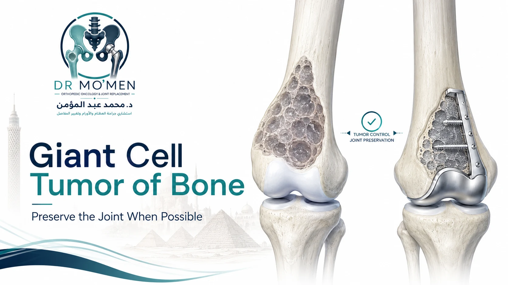
A diagnosis of giant cell tumor of bone, often abbreviated as GCTB, can be confusing. Patients may hear that it is not a typical bone cancer while also hearing terms such as aggressive, recurrence, bone destruction, surgery or pathological fracture.
One of the central goals of treatment is to achieve local tumor control while preserving the patient’s own joint whenever this can be done safely. For many lesions, this can be achieved using extended curettage. Other tumors are too extensive for reliable joint-preserving surgery and may require wide resection and reconstruction.
The decision depends on imaging, pathology, bone destruction, joint involvement, soft-tissue extension, fracture pattern and previous treatment.
What Is a Giant Cell Tumor of Bone?
Giant cell tumor of bone is a primary bone tumor characterized histologically by numerous giant cells within a background of other tumor cells. It most often occurs after skeletal maturity.
Although it is generally not classified in the same way as conventional high-grade bone sarcomas such as osteosarcoma, GCTB can behave aggressively at the site where it develops.
- Expand through bone
- Thin or destroy the cortex
- Extend into surrounding soft tissue
- Weaken joint-supporting bone
- Cause pathological fracture
- Recur after treatment
Rarely, more unusual biological behavior can occur. This is why giant cell tumor should be treated as a genuine orthopedic oncology problem rather than simply as a benign cyst.
Is Giant Cell Tumor of Bone Cancer?
Most giant cell tumors of bone are not conventional malignant bone cancers. However, they are also not completely harmless. They are generally regarded as tumors with intermediate or locally aggressive behavior, meaning they can destroy substantial amounts of bone, recur locally and extend into soft tissue.
Rare malignant forms exist but are a distinct clinical situation. “Not typical cancer” should not be interpreted as meaning that no treatment or follow-up is required.
Where Does Giant Cell Tumor Usually Occur?
GCTB commonly develops near the end of a long bone, usually close to a joint.
- Distal femur
- Proximal tibia
- Distal radius
- Proximal humerus
- Other long-bone ends
- Selected pelvic and sacral locations
The area around the knee is particularly important because the tumor can extend close to the subchondral bone supporting the articular surface. The challenge is often how to treat the tumor adequately while preserving the natural knee joint.
What Symptoms Can Giant Cell Tumor Cause?
- Gradually increasing localized pain
- Swelling or tenderness
- Reduced joint movement
- Difficulty walking or activity-related discomfort
- Increasing deformity in advanced cases
- Sudden severe pain if pathological fracture occurs
Symptoms may develop gradually over weeks or months. Persistent localized bone pain and swelling deserve appropriate assessment rather than repeated symptomatic treatment without imaging.
What Does Giant Cell Tumor Look Like on Imaging?
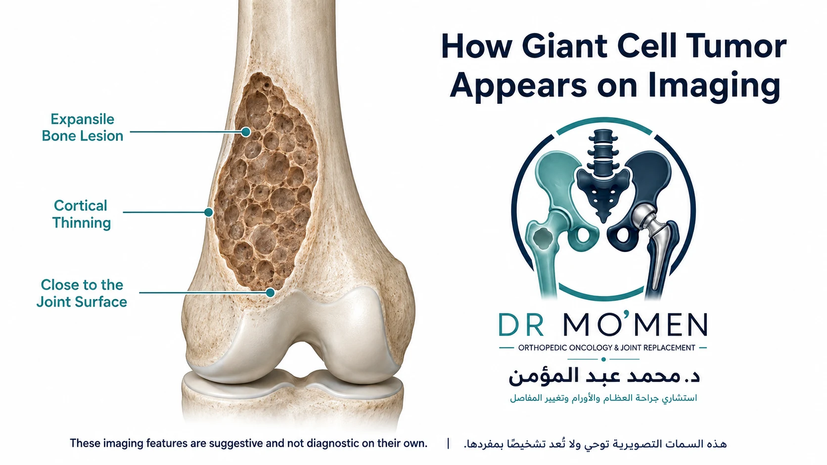
Plain X-rays are very useful in the initial assessment. A typical lesion may appear as an expansile lytic bone lesion close to the joint surface, with bone destruction, cortical thinning or breakthrough, and extension toward the subchondral bone.
MRI helps define intraosseous extent, relation to the joint, remaining subchondral bone, cortical breakthrough, soft-tissue extension, surrounding structures, neurovascular relationships where relevant and pathological fracture. CT can add information about cortical bone, subtle fractures and complex anatomy.
Why Must the Diagnosis Be Confirmed Before Surgery?
A giant-cell-rich lesion on imaging is not always a giant cell tumor of bone. Other conditions can appear similar clinically, radiologically or histologically. Definitive treatment should therefore be based on accurate diagnosis.
Biopsy planning matters
A biopsy should be performed through a route that does not unnecessarily compromise definitive surgery. If a primary bone tumor is suspected, biopsy planning should be coordinated with the team responsible for definitive tumor treatment.
Read more about bone tumor biopsy planning.
Does Every Giant Cell Tumor Need Biopsy?
In most cases where the diagnosis is not already securely established, tissue confirmation is important before definitive tumor surgery. The exact diagnostic pathway depends on imaging, age, location, clinical context, previous treatment and the differential diagnosis.
The key principle is that biopsy should be planned, not simply performed through the most convenient route.
How Is Giant Cell Tumor Treated?
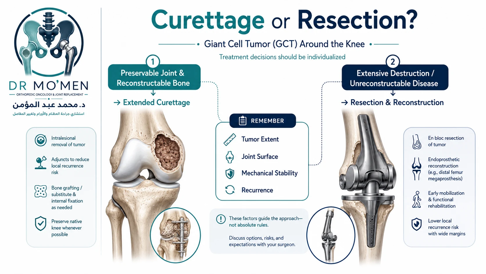
The major surgical options are broadly joint-preserving extended curettage or wide resection with reconstruction. The choice is individualized.
1. Joint-Preserving Extended Curettage
The tumor is removed from inside the bone while the surrounding joint and much of the bone structure are preserved.
2. Wide Resection
The entire tumor-bearing segment is removed together with an appropriate margin. The resulting defect then requires reconstruction.
What Is Extended Curettage?
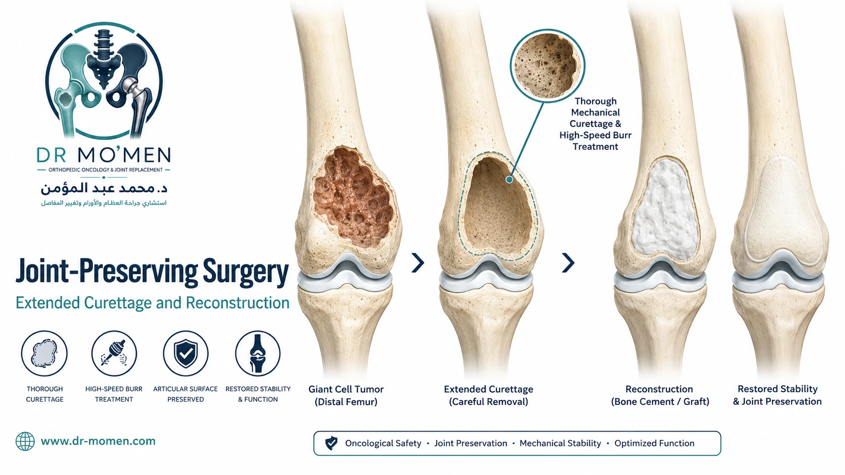
Extended curettage is a joint-preserving strategy frequently used for appropriately selected giant cell tumors. The principle is to remove the visible tumor from within the bone and treat the cavity thoroughly in an attempt to reduce microscopic residual tumor.
- Creating an appropriate cortical window
- Thorough mechanical curettage
- Visual inspection of the cavity
- High-speed burr in selected practice
- Local adjuvant treatment depending on surgeon and institutional protocol
- Reconstruction of the remaining defect
The exact technique varies. No single adjuvant should be described as universally necessary or superior for every lesion.
Why Is a High-Speed Burr Used?
After gross tumor has been removed with curettes, a high-speed burr may be used to mechanically extend treatment of the cavity by removing additional potentially involved bone from the internal cavity walls.
It must be used carefully because the lesion may be close to articular cartilage, subchondral bone, thin cortex or neurovascular structures.
What Local Adjuvants May Be Used?
Various local adjuvants have historically been used, including thermal techniques, chemical agents, cryotherapy and other local methods. Choice varies according to location, cavity anatomy, remaining bone, available resources and institutional practice.
The central principle remains meticulous tumor removal and appropriate reconstruction rather than reliance on one adjuvant alone.
How Is the Bone Defect Filled?
After curettage, the resulting cavity may be reconstructed using bone cement, bone graft, a combination of materials, or fixation when required. The best option depends on defect size, proximity to the joint, quality of surrounding bone, fracture risk and the reconstruction strategy.
Why Is Bone Cement Used?
Bone cement may provide relatively immediate structural filling, mechanical support and radiographic contrast with surrounding bone. It should not be described as a guarantee against recurrence.
When Is Bone Graft Used?
Bone graft may be considered when biological reconstruction is desirable. Options can include autograft, allograft or combinations with other materials.
When Is Internal Fixation Needed?
Internal fixation may be considered when there is a large cortical defect, pathological fracture, very thin remaining cortex, concern about postoperative mechanical failure, or a need to support the reconstructed cavity.
Giant Cell Tumor Around the Knee
The distal femur and proximal tibia are among the clinically important sites for GCTB. The treatment decision considers the amount of subchondral bone remaining, articular surface integrity, lesion size, cortical destruction, soft-tissue extension, pathological fracture and mechanical stability.
Whenever feasible, preserving the native joint may offer an important functional advantage. Joint preservation should not be attempted if the remaining anatomy cannot provide a safe or durable reconstruction.
Can the Knee Joint Usually Be Preserved?
In many appropriately selected cases, yes. Extended curettage can often preserve the native knee joint, provided adequate tumor removal is feasible, enough joint-supporting bone remains, reconstruction can be mechanically stabilized and the anatomy is suitable for curettage.
When Is Wide Resection Considered?
Wide resection may be considered when joint-preserving surgery is unlikely to provide adequate local control or reliable reconstruction.
- Extensive joint destruction
- Massive cortical loss
- Major soft-tissue extension
- Highly destructive recurrent disease
- Multiply recurrent tumors
- Unreconstructable pathological fracture
- Anatomy unsuitable for safe curettage
- Selected unusual locations
Wide resection is a substantially larger operation and may require megaprosthetic reconstruction or a biological reconstruction.
Curettage or Resection: Which Is Better?
There is no universal answer. The aim is to balance local tumor control, joint preservation, mechanical stability and long-term function. Extended curettage may preserve the patient’s own joint; wide resection removes more bone and potentially the joint, but may be necessary in selected destructive disease.
Why Can Giant Cell Tumor Recur?
Local recurrence can occur after treatment. This does not automatically mean the original treatment was inappropriate. Factors include biological aggressiveness, lesion extent, soft-tissue extension, previous surgery, completeness of local tumor removal and anatomical limitations.
What Are the Signs of Recurrence?
- New or increasing pain
- Recurrent swelling
- Change around the previous surgical site
- New radiographic lucency
- Progressive bone destruction
- New soft-tissue mass
Pain after surgery does not automatically mean recurrence. Mechanical weakness, fracture, infection, scar tissue or joint problems may also cause symptoms.
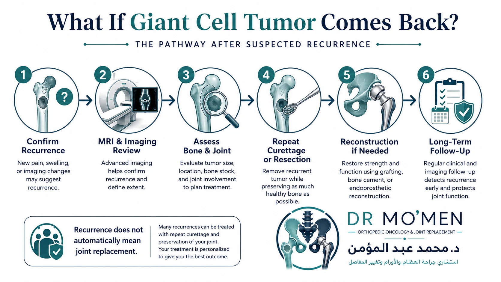
What Happens If Giant Cell Tumor Comes Back?
Recurrence does not automatically mean the joint must be removed. Some recurrent GCTBs may still be treated with repeat extended curettage, while others require wider resection. The decision depends on recurrence location, remaining bone, joint condition, soft-tissue extension, prior reconstruction and the number of previous recurrences.
Can Repeat Curettage Be Performed?
Yes, in selected patients when recurrence is localized, joint-supporting bone remains salvageable, anatomy permits thorough tumor removal and reconstruction remains mechanically feasible.
When Does Recurrence Require Resection?
Resection may be considered when recurrent disease has caused extensive bone destruction, major soft-tissue extension, loss of joint-supporting bone, repeated local failure, an unreconstructable pathological fracture or anatomy unsuitable for another curettage procedure.
For a broader recurrence pathway, see what to do if a bone tumor comes back.
What Is the Role of Denosumab?
Denosumab is a targeted medication that can have a role in selected patients with GCTB, including tumors that are difficult to resect, cases where surgery would cause major morbidity, selected unresectable disease, or other specialist circumstances.
It should not be viewed as an automatic replacement for surgery in every patient. Timing, duration and relationship to surgery require specialist consideration within an orthopedic oncology and multidisciplinary framework.
Can Denosumab Make Surgery Easier?
In selected cases, medical treatment can alter the appearance and structure of a GCTB and may affect surgical planning. The implications are more complex than simply “shrinking the tumor,” so use before or instead of surgery should be individualized.
Can Giant Cell Tumor Spread to the Lungs?
Pulmonary metastases are uncommon but recognized in GCTB. This does not make every giant cell tumor a conventional malignant bone cancer. The need for chest imaging and surveillance depends on tumor behavior, recurrence, treatment history and specialist assessment.
Is Malignant Giant Cell Tumor Different?
Yes. Malignant transformation or primary malignant forms are unusual and represent a different clinical problem from conventional GCTB. If pathology suggests malignant giant cell tumor, management should be discussed within a sarcoma multidisciplinary team.
What If There Is a Pathological Fracture?
A pathological fracture can occur when the tumor has weakened the bone substantially. It can complicate treatment but does not automatically require amputation, joint replacement or wide resection.
The decision depends on fracture pattern, joint surface, remaining bone, soft-tissue extension, tumor anatomy and mechanical reconstructability. Some pathological fractures can still be managed with tumor curettage and appropriate reconstruction; others require wider surgery.
When Does a Pathological Fracture Need Prompt Assessment?
Sudden severe pain, inability to bear weight, new deformity, rapidly increasing swelling, or new neurological or vascular symptoms require prompt local medical assessment. A fracture through a suspected bone tumor should not automatically be treated as a routine trauma fracture before the diagnosis and tumor extent are understood whenever clinically possible.
How Long Does Recovery Take?
Recovery depends on the procedure. After curettage, recovery is influenced by defect size, cement or graft reconstruction, fixation, pathological fracture, joint involvement and weight-bearing restrictions.
After wide resection and megaprosthetic reconstruction, recovery is usually more extensive and may involve early mobilization, structured physiotherapy, muscle strengthening, gait training and long-term implant monitoring. There is no universal recovery timeline.
When Can the Patient Walk?
Weight-bearing depends on location, defect size, reconstruction, fixation, remaining bone, fracture status and the surgeon’s protocol. Patients should follow an individualized postoperative plan rather than a fixed internet timetable.
Follow-Up After Giant Cell Tumor Treatment
Follow-up is important because recurrence may occur. Monitoring may include clinical examination, local X-rays, MRI or CT in selected circumstances, functional assessment and chest imaging where appropriate. The surveillance schedule should reflect treatment type, recurrence history, anatomical location and biological behavior.
When Should You Seek Specialist Orthopedic Oncology Assessment?
- Giant cell tumor has been diagnosed
- The tumor is close to a major joint
- Pathological fracture has occurred
- Joint replacement has been recommended
- The lesion has recurred
- Multiple previous curettage procedures have failed
- Wide resection has been proposed
- Denosumab treatment is being considered
- Imaging and pathology do not appear to match
- You want another opinion before irreversible surgery
A second opinion does not necessarily mean the original recommendation is incorrect. It can clarify whether joint preservation remains feasible. Request an orthopedic oncology second opinion.
International Patients: What Should You Send Before Travelling?
- Plain X-rays
- MRI
- CT where available
- Biopsy and pathology reports
- Pathology slides or blocks if specialist review is required
- Previous operative report and reconstruction details
- Recent and original imaging for recurrent tumors
- Treatment and denosumab history if used
- Current symptoms
A preliminary review may help determine whether the diagnosis is secure, whether biopsy planning is needed, whether the joint appears preservable, whether extended curettage or fixation may be considered, and whether wider resection and reconstruction should be assessed.
Have You Been Diagnosed With a Giant Cell Tumor Near a Joint?
Specialist assessment may help clarify whether the diagnosis is secure, whether the native joint can be preserved, whether extended curettage or fixation is appropriate, and whether recurrence can still be treated with joint-preserving surgery.
International contact: +20 102 1690693
For the joint-preservation decision after recurrence following curettage, see Bone Tumor Recurrence After Curettage.
Frequently Asked Questions
Is giant cell tumor of bone cancer?
Most GCTBs are not conventional malignant bone cancers. They are generally considered locally aggressive tumors that can destroy bone and recur. Rare malignant forms are different clinical entities.
Can giant cell tumor be treated without replacing the joint?
Often, yes. Many appropriately selected tumors can be treated with extended curettage while preserving the native joint. Feasibility depends on bone destruction, joint involvement, fracture and soft-tissue extension.
Why is bone cement used after curettage?
Bone cement can provide structural filling of a large tumor cavity and can be useful in reconstruction. It does not guarantee that the tumor will not recur.
Does recurrence mean I need a megaprosthesis?
No. Some recurrent giant cell tumors can still be treated with repeat curettage. Wider resection is considered when recurrent disease has caused extensive destruction or cannot be reliably treated with another joint-preserving procedure.
What is denosumab used for?
Denosumab may have a role in selected giant cell tumor cases, particularly where surgery is difficult or would involve major morbidity. It is not automatically required for every patient.
Can giant cell tumor cause a pathological fracture?
Yes. A large lesion can weaken the bone enough to fracture. Treatment depends on the fracture pattern, remaining bone, joint surface and extent of the tumor.
Can I get a second opinion before travelling to Egypt?
Yes. Available X-rays, MRI, pathology reports, previous surgery records and current imaging can be reviewed preliminarily before travel. The final treatment decision requires clinical assessment and complete review of the case.
How this article was reviewed
This patient-education article was reviewed for clinical accuracy, patient safety and decision-making clarity by Dr. Mohammed Abdelmoemen Abuelhadid. It is intended to support, not replace, clinical examination, pathology review and review of the actual imaging and reports.

Author and medical reviewer
Dr. Mohammed Abdelmoemen Abuelhadid — Orthopedic Oncology & Joint Replacement Surgeon and Lecturer of Orthopedic Surgery at Ain Shams University.
Medical Disclaimer: This article is for patient education and does not replace clinical examination, review of imaging, pathology, investigations, or individualized multidisciplinary medical advice.