
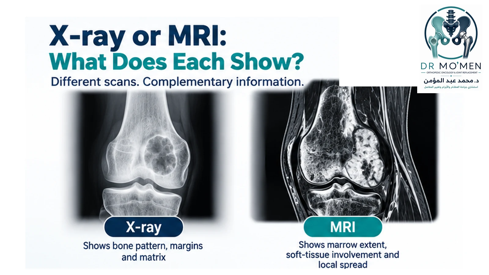
When a bone tumor or unexplained bone lesion is suspected, patients often assume that the newest or most expensive scan must be the most useful. That is not necessarily true.
X-rays and MRI answer different questions. A plain X-ray may reveal the lesion's biological pattern surprisingly well, while MRI provides more detailed information about how far the lesion extends through the bone and into surrounding tissues.
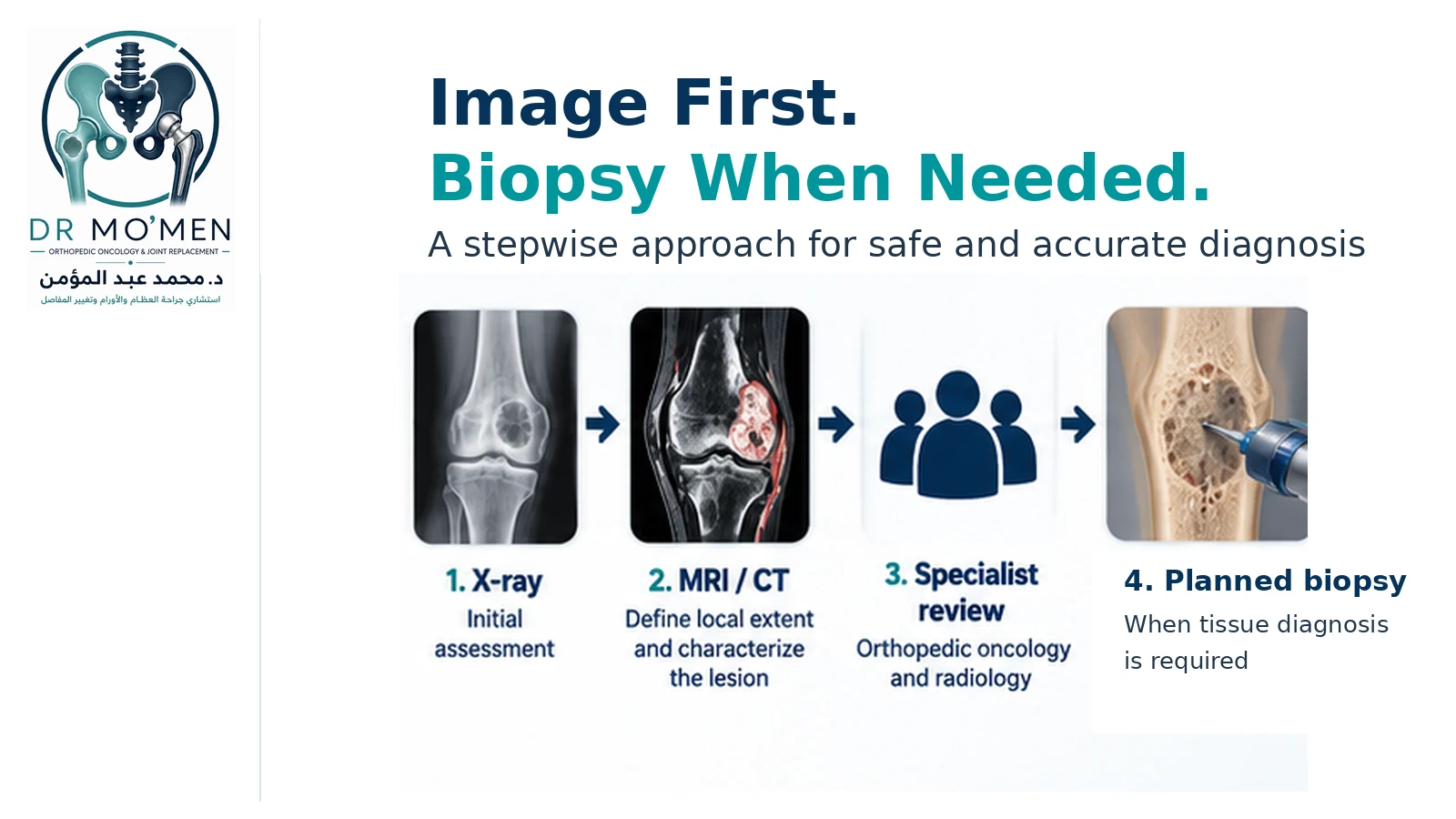
The practical sequence is often: X-ray → characterize the lesion → MRI when indicated → specialist review → planned biopsy if tissue diagnosis is required.
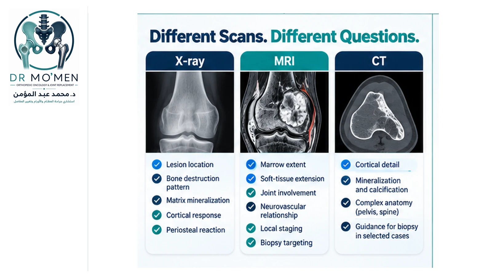
X-ray vs MRI vs CT for Bone Tumors — At a Glance
| Imaging test | Best at answering | Typical role |
|---|---|---|
| X-ray | Margins, matrix, pattern of bone destruction, cortex, periosteal reaction and pathological fracture | First-line assessment of a suspected primary bone lesion |
| MRI | Marrow extent, soft-tissue extension, joints, muscle compartments, nerves and vessels | Local extent/staging and biopsy targeting when the lesion is indeterminate or aggressive |
| CT | Cortical detail, mineralization/calcification and complex anatomy | Complementary problem-solving study and selected image-guided biopsy planning |
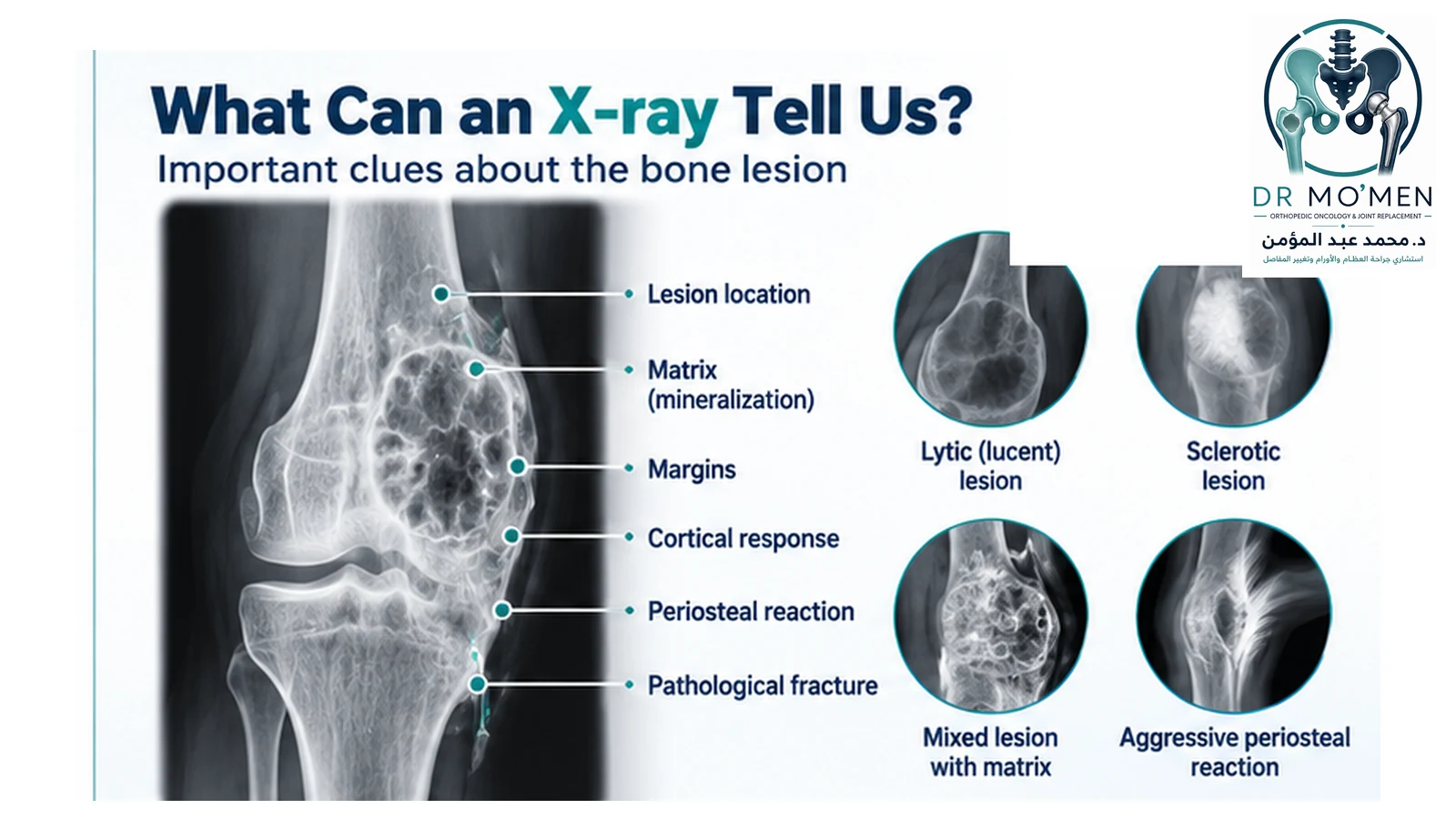
X-ray is particularly useful for:
- Lesion location.
- Pattern of bone destruction.
- Margins.
- Periosteal reaction.
- Mineralized matrix.
- Cortical response.
- Pathological fracture.
MRI is particularly useful for:
- Marrow extent.
- Soft-tissue extension.
- Relationship to nerves and vessels.
- Relationship to adjacent joints.
- Local staging.
- Biopsy targeting.
Why is X-ray usually the first test?
Plain radiographs remain highly informative in bone tumors because the relationship between the lesion and surrounding bone can provide clues about biological behavior.
A good X-ray can show whether the lesion is lytic, sclerotic or mixed; whether its margins are sharply or poorly defined; whether the cortex is preserved, expanded or destroyed; whether the periosteum has reacted; whether mineralized matrix is present; and whether a pathological fracture has occurred.
Can an X-ray show bone cancer?
An X-ray can show features that are concerning for an aggressive or malignant process, but it usually cannot establish the final diagnosis by itself.
Concerning findings may include aggressive bone destruction, poorly defined margins, cortical destruction, aggressive periosteal reaction, pathological fracture or a suspected soft-tissue mass.
An aggressive-looking X-ray does not automatically mean cancer. Infection and some benign but locally aggressive lesions can produce similar appearances.
Can a normal X-ray exclude a bone tumor?
No. A normal radiograph does not completely exclude a clinically important primary bone tumor. Persistent focal pain, an enlarging mass or other concerning symptoms can still justify urgent MRI or specialist referral.
A normal X-ray does not completely exclude an important bone tumor when symptoms remain clinically concerning.
What does “lytic bone lesion” mean?
A lytic or lucent lesion appears darker than surrounding bone because the affected area contains less mineralized bone. Lytic lesions can occur with benign tumors, cysts, giant cell-rich lesions, infection, metastases, myeloma and primary malignant tumors.
Lytic is an imaging description, not a diagnosis of cancer.
What does “sclerotic bone lesion” mean?
A sclerotic lesion appears denser or whiter than surrounding bone. Possible causes range from a benign bone island or reactive bone formation to osteoid-producing tumors and selected metastatic lesions.
What do lesion margins tell us?
The transition between abnormal and normal bone is one of the most useful radiographic features. A narrow transition and well-defined margin can suggest slower behavior, while a wide transition, poorly defined border or permeative pattern can indicate a more aggressive process.
These patterns guide risk assessment but do not replace final diagnosis.
What is periosteal reaction?
The periosteum is the tissue covering the outer surface of bone. When irritated, it can produce new bone. Rapidly progressive lesions may create interrupted or aggressive periosteal patterns, but fracture, infection and inflammation can also produce periosteal reaction.
Can X-ray suggest the type of tumor?
Sometimes. Age, anatomical site, lesion location within the bone, matrix, margins and periosteal reaction can strongly suggest a particular diagnosis. Radiologists may look for chondroid mineralization, osteoid production, fibrous matrix or cystic features.
However, imaging features can overlap and a likely radiological diagnosis must still fit the clinical picture.
Why do I need MRI after an abnormal X-ray?
X-ray often helps answer: “What kind of biological process might this be?” MRI helps answer: “Exactly how far does it extend and what structures does it involve?”
When radiographs show an indeterminate or aggressive lesion, MRI is generally the key next local imaging study.
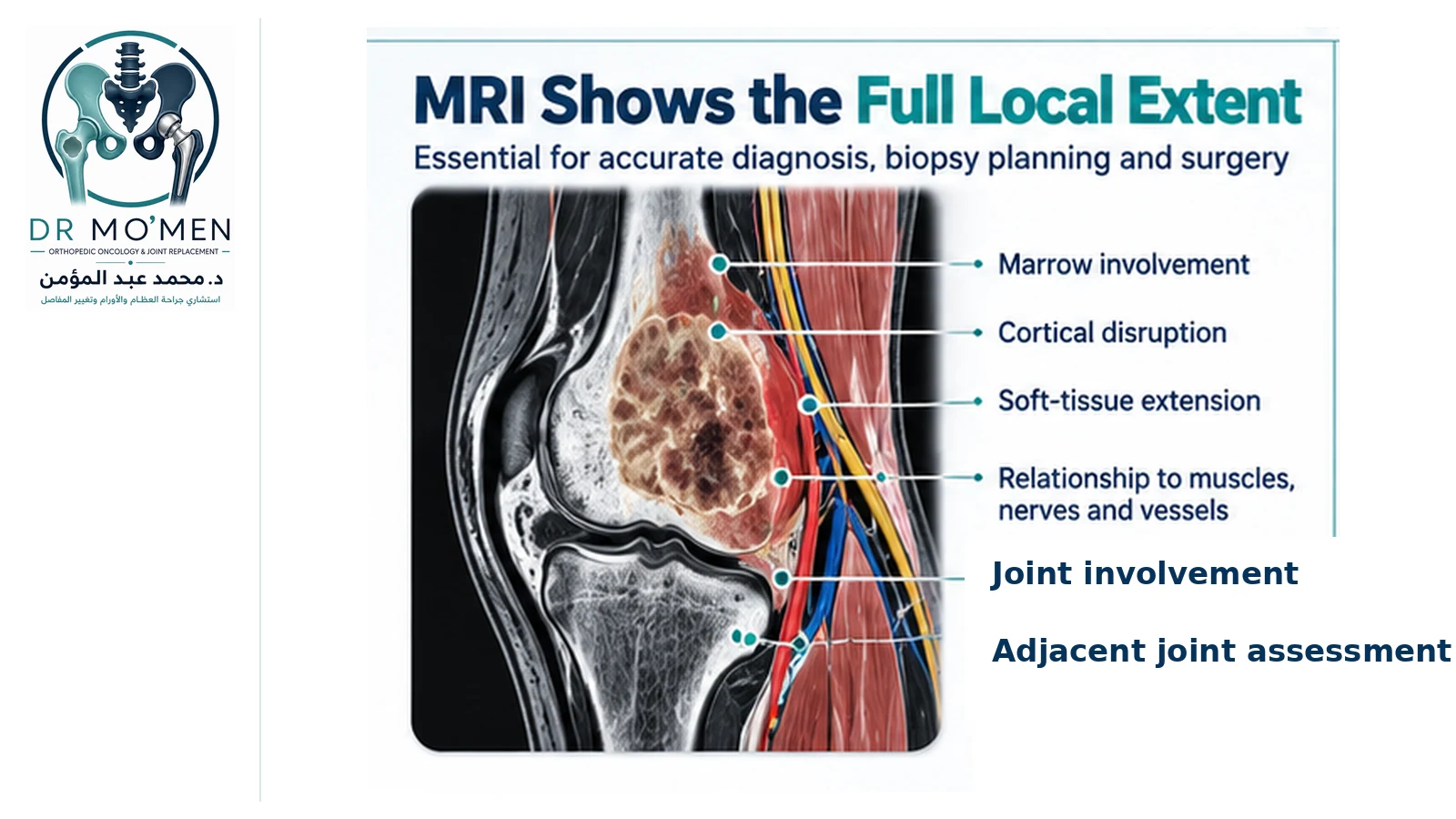
What does MRI show in a bone tumor?
- Intramedullary extent.
- Soft-tissue extension.
- Relationship to muscle compartments.
- Relationship to major nerves and vessels.
- Relationship to adjacent joints.
- Surrounding marrow changes.
- Hemorrhage or necrosis.
- Heterogeneous tumor components.
This information becomes especially important when biopsy or limb-salvage surgery is being considered.
Does MRI need to include the whole bone?
When a primary malignant bone tumor is suspected, MRI should assess the full local extent rather than only a tightly cropped area around the visible lesion.
Current specialist guidance recommends imaging the whole involved bone, the relevant local anatomical compartment and adjacent joints.
When a primary malignant bone tumor is suspected, MRI should evaluate the full local extent rather than only a small cropped area around the visible mass.
Does MRI need contrast?
The answer depends on the clinical question. MRI without contrast can already define marrow involvement, anatomical extent and soft-tissue extension. Contrast-enhanced MRI can add value in selected situations, including complex heterogeneous lesions, assessment of viable versus necrotic tissue and biopsy targeting.
Can MRI tell whether a bone tumor is benign or malignant?
MRI can show features that increase or reduce suspicion, but it usually cannot replace histology when a primary malignant bone tumor remains suspected.
Some benign but aggressive tumors can look alarming on MRI, while some low-grade malignant tumors may appear less dramatic. Diagnosis therefore depends on the complete clinical, radiological and pathological picture.
Why is X-ray still needed if I already have an MRI?
MRI is highly sensitive to marrow and soft-tissue changes, but X-ray may demonstrate matrix mineralization, periosteal bone formation, cortical remodeling and the pattern of bone destruction more clearly.
In orthopedic oncology, X-ray and MRI are usually complementary rather than competing tests.
When is CT useful?
CT provides excellent bone detail and is particularly useful for cortical destruction, subtle mineralization, calcification, periosteal bone formation and complex anatomy.
CT can also be valuable in pelvic lesions, when MRI is contraindicated or incomplete, and for image-guided biopsy in selected cases.
Is CT better than MRI?
No single modality is universally better. CT is stronger for cortical detail and mineralization; MRI is stronger for marrow extent, soft-tissue extension and local staging.
Should PET/CT or a bone scan replace MRI?
Usually not for local tumor assessment. MRI remains the major modality for defining local anatomical extent. PET/CT, whole-body MRI and bone scintigraphy answer different questions, particularly staging, evaluation of additional skeletal sites and treatment response in selected tumors.
Should imaging be done before biopsy?
Yes, when a primary malignant bone tumor is suspected. Cross-sectional imaging should define the lesion and help select the biopsy target and route before tissue sampling is performed.
Poorly planned biopsy can complicate later surgery, which is why biopsy should be integrated into the orthopedic-oncology treatment plan.
Why does MRI help plan the biopsy?
Bone tumors can be heterogeneous. MRI may show necrotic or cystic areas as well as viable solid regions or potentially higher-grade components. Sampling the wrong area can produce an inconclusive or misleading result.
Should a bone tumor be biopsied because MRI looks abnormal?
Not automatically. Some lesions have classic benign imaging patterns and do not require biopsy. Other lesions may need comparison with prior imaging, CT, follow-up or specialist review before deciding whether tissue diagnosis is necessary.
What if the MRI report says “cannot exclude malignancy”?
This phrase does not mean that cancer has been diagnosed. It means the radiologist cannot confidently classify the lesion as benign based on that study alone.
The next step can include specialist image review, correlation with X-rays, CT, additional MRI sequences, comparison with older studies or planned biopsy depending on the lesion.
Does the written MRI report tell the whole story?
Not always. The report is important, but orthopedic oncology decisions often require review of the actual images because the exact tumor boundaries, cortical relationship, joint involvement, muscle compartments, vessels, nerves, heterogeneous regions and possible biopsy trajectory can affect management.
Can MRI be wrong?
MRI is highly informative but not infallible. Infection, stress injury, healing fracture, inflammatory disease, bone infarction and postoperative change can mimic tumor. Benign and malignant tumors can also overlap in appearance.
What if X-ray and MRI do not seem to agree?
The studies should be reviewed together. X-ray may show matrix and biological pattern better, while MRI may show extent and surrounding tissue involvement more clearly. If uncertainty remains, CT, comparison with prior studies or biopsy may be needed.
What if pathology does not match the imaging?
The case requires reassessment. A diagnosis should make sense across the clinical presentation, radiology and pathology. See how to interpret imaging–pathology discordance.
Possible explanations include sampling error, tumor heterogeneity, incorrect targeting, pathology interpretation difficulty or imaging interpretation difficulty.
Does previous cancer change the imaging pathway?
Yes. In an older adult or a patient with a known malignancy, the differential diagnosis may include metastasis, myeloma, a primary bone tumor and benign incidental lesions. Previous cancer does not mean every new bone lesion is metastatic.
What imaging findings need more urgent assessment?
- Aggressive bone destruction.
- Cortical breakthrough.
- Pathological fracture.
- Large soft-tissue mass.
- Rapid progression.
- Neurovascular involvement.
- Spinal or pelvic lesion with neurological symptoms.
Practical bone tumor imaging pathway
- Step 1 — X-ray: assess location, margin, matrix, cortex, periosteal reaction and fracture.
- Step 2 — If confidently benign: observation or follow-up when appropriate.
- Step 3 — If indeterminate or aggressive: MRI to define local extent.
- Step 4 — CT when useful for cortex, mineralization or complex anatomy.
- Step 5 — Orthopedic oncology review.
- Step 6 — Planned biopsy when required.
- Step 7 — Correlate radiology + pathology + clinical findings before definitive treatment.
When should you see an orthopedic oncologist?
- An X-ray shows an indeterminate bone lesion.
- Imaging appears aggressive.
- MRI shows a soft-tissue component.
- The cortex is substantially weakened.
- Pathological fracture is possible.
- A biopsy is being considered.
- Imaging and pathology disagree.
- Surgery has been proposed before the diagnosis is secure.
- A second opinion is needed before major treatment.
Specialist review of bone tumor imaging
Dr. Mohammed Abdelmoemen Abuelhadid evaluates bone and soft-tissue tumors and complex bone lesions where X-ray, CT, MRI and pathology need to be interpreted together.
A specialist review may help determine whether the lesion has a convincing benign imaging pattern, whether MRI or CT is needed, whether bone strength is threatened, whether biopsy is required, which region should be targeted and whether pathology fits the imaging.
For patients outside Egypt, the actual imaging files, reports and previous investigations can also be reviewed as part of an online second medical opinion before biopsy or travel when appropriate. How to send DICOM imaging files.
For appointments or imaging review: 01021690693
Evidence Behind Bone Tumor Imaging
Current guidance supports plain radiography as the initial imaging study for suspected primary bone tumors, MRI for local extent when lesions are indeterminate or aggressive, CT as a complementary modality for cortical/mineral detail, and completion of appropriate cross-sectional imaging before planned biopsy.
- ACR Appropriateness Criteria: Suspected Primary Bone Tumors — revised 2024 — supports radiography first and MRI as usually appropriate when a radiographic lesion is indeterminate or aggressive.
- British Sarcoma Group: UK Guidelines for the Management of Bone Sarcomas — supports X-ray as the first investigation, MRI of the involved bone/local compartment and adjacent joints, complementary CT, and imaging before biopsy.