
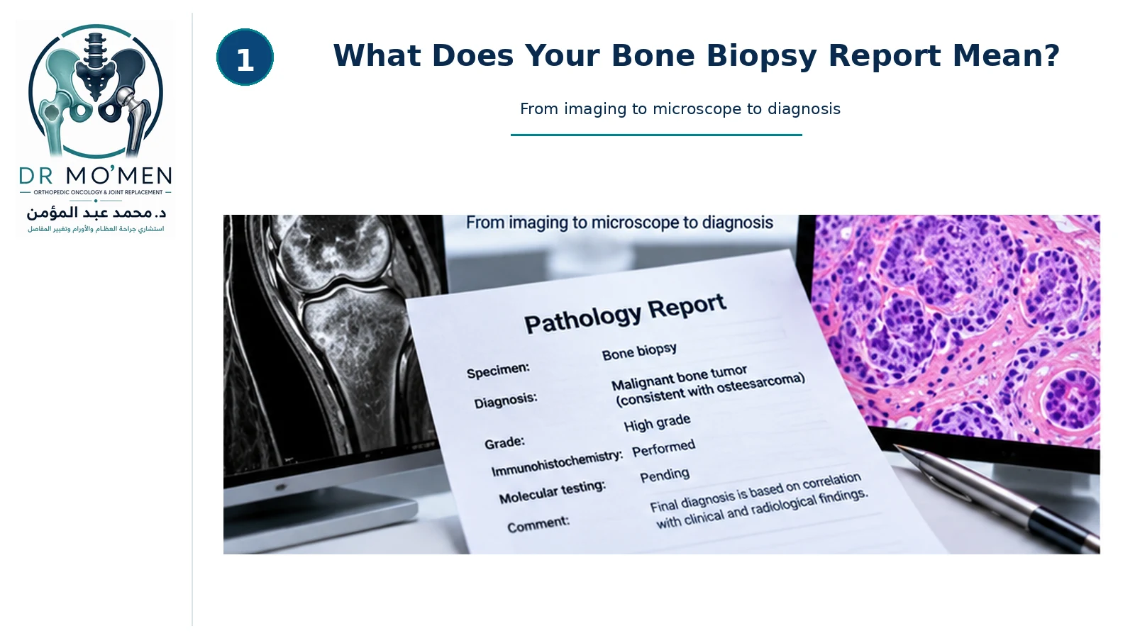
Waiting for the result of a bone-tumor biopsy can be stressful. When the pathology report arrives, it may contain unfamiliar terms such as benign, malignant, atypical, spindle cell, giant cell-rich, osteoid, chondroid, low grade, high grade, immunohistochemistry, molecular testing or margins.
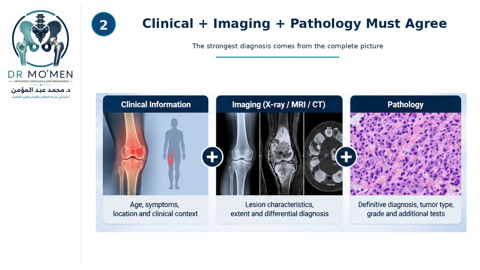
The most important principle is that a bone-tumor pathology report should not be interpreted in isolation. Modern bone-tumor diagnosis depends on correlation among the clinical history, X-ray/CT/MRI findings and pathology.
The key question is not simply “What does the microscope show?” but “Does the pathology diagnosis explain the lesion seen on the imaging and fit the clinical picture?”
Bone Tumor Pathology Report Terms at a Glance
| Report term | What it usually means | What it does not mean by itself |
|---|---|---|
| Benign | No malignant cancer identified in the sampled tissue. | It does not automatically mean no treatment or follow-up is needed. |
| Malignant | A cancerous tumor diagnosis that usually triggers staging and multidisciplinary treatment planning. | It does not define the complete stage from pathology alone. |
| Indeterminate / suspicious | The available tissue does not establish a completely secure diagnosis. | It is not the same as a benign result. |
| Nondiagnostic | The sample is insufficient or nonrepresentative for a reliable diagnosis. | It does not exclude malignancy. |
| Grade | A tumor-specific estimate of microscopic aggressiveness when applicable. | It is not interchangeable across all bone-tumor types. |
| IHC / molecular testing | Additional tests that can help classify selected tumors or identify defining alterations. | Not every bone tumor requires every available test. |
Who should review a bone tumor biopsy?
When a primary malignant bone tumor is suspected, the specimen should ideally be reviewed by a specialist bone-tumor pathologist within a multidisciplinary sarcoma team.
Current bone-sarcoma guidance recommends specialist pathology review of primary malignant bone-tumor diagnoses because these tumors are rare, heterogeneous and increasingly dependent on integrated molecular and radiological interpretation.
What information does the pathologist need?
- Patient age.
- Exact anatomical site and involved bone.
- Location within the bone.
- Relevant symptoms and clinical history.
- Previous cancer history or previous biopsy/surgery.
- The radiological differential diagnosis.
Bone-tumor pathology is more informative when the pathologist knows what the imaging shows and what diagnoses the radiologist and orthopedic oncology team are considering.
What does “benign” mean?
A benign bone tumor is not a malignant cancer. Examples include osteochondroma, enchondroma, non-ossifying fibroma, simple bone cyst and chondroblastoma.
Benign does not always mean that no treatment is needed. Some benign lesions can enlarge, weaken bone, cause pathological fracture, damage a nearby joint or recur. Management still depends on tumor type, location, symptoms and structural risk.
What does “malignant” mean?
A malignant bone tumor has the potential for destructive local growth and, depending on tumor type, spread to other parts of the body. Examples include osteosarcoma, Ewing sarcoma and chondrosarcoma.
A malignant diagnosis usually leads to formal staging and multidisciplinary treatment planning. The appropriate surgery, systemic therapy or radiotherapy depends on the specific tumor entity.
What does “atypical” mean?
Atypical means that some cells or tissue features are not completely normal. It does not automatically mean cancer. Depending on context, atypia can reflect reactive change, a borderline or intermediate process, a low-grade neoplasm or a malignant tumor.
Its significance therefore depends on morphology, imaging, clinical setting and any additional tests.
What does “indeterminate” or “suspicious” mean?
These terms usually mean that the available tissue does not allow a completely secure diagnosis. Reasons can include too little tissue, necrosis, crushed tissue, tumor heterogeneity or overlapping microscopic appearances.
The next step may include deeper sections, IHC, molecular testing, specialist pathology review or repeat biopsy.
What does “nondiagnostic biopsy” mean?
A nondiagnostic biopsy means that the sample does not provide enough reliable information to establish the diagnosis. This can occur if insufficient tissue was obtained, the biopsy sampled necrosis, the lesion was difficult to target or the sampled region was not representative.
A nondiagnostic biopsy is not equivalent to a benign biopsy. Read when a bone lesion actually needs biopsy.
Can a bone tumor be heterogeneous?
Yes. Some bone tumors contain different regions with different biological characteristics. One part may be necrotic or lower grade, while another area may be solid, viable or higher grade.
MRI performed before biopsy can help identify the most representative target. Tumor heterogeneity is also one reason a small biopsy may occasionally underestimate the lesion.
What is tumor grade?
Tumor grade describes how aggressive the tumor appears microscopically. Higher-grade tumors often show greater cellular atypia, more mitotic activity, necrosis and less resemblance to normal tissue.
Grading systems vary by tumor type. A low-grade or high-grade label must therefore be interpreted for that specific diagnosis rather than compared directly across different sarcomas.
Why does grade matter?
Grade can influence staging, prognosis, surgical planning, systemic-treatment decisions and surveillance. It is only one component of the overall diagnosis and must be interpreted together with tumor type and anatomical extent.
Can a biopsy underestimate tumor grade?
Yes. A heterogeneous tumor can be undergraded if the biopsy samples a less aggressive region. Cartilage tumors are a particularly important example because imaging and histological features can overlap and grade may vary within the same lesion.
If the MRI appears substantially more aggressive than the biopsy result, the case should be reviewed rather than assuming the small sample represents the entire tumor.
What is immunohistochemistry?
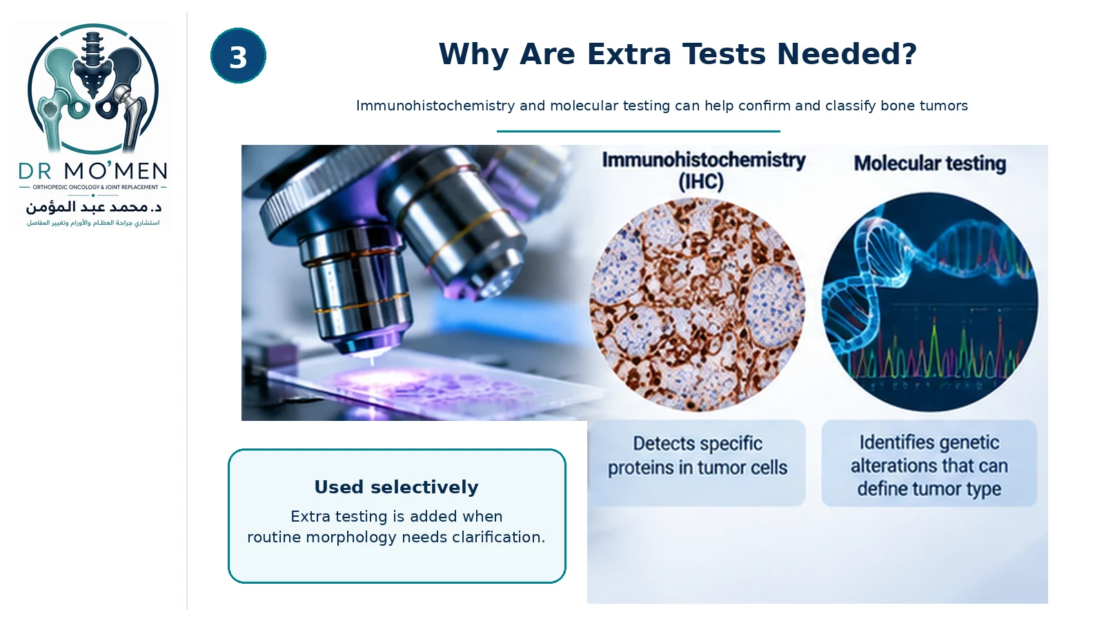
Immunohistochemistry, or IHC, uses antibodies to detect specific proteins in tumor cells. It can help identify cell lineage, support a particular diagnostic category and distinguish between tumors with similar microscopic appearances.
IHC is interpreted as a pattern of findings. One positive stain rarely establishes a bone-tumor diagnosis by itself.
Does every bone tumor need immunohistochemistry?
No. Some lesions can be diagnosed confidently from the clinical information, imaging and routine histology. IHC is used selectively when it helps distinguish among realistic competing diagnoses.
What are molecular tests?
Molecular tests look for selected genetic abnormalities in tumor cells. Depending on the diagnostic question, these methods can include FISH, PCR, DNA or RNA sequencing, next-generation sequencing, mutation analysis and gene-fusion testing.
Modern classification of bone and soft-tissue tumors increasingly incorporates molecular features, particularly for entities with overlapping morphology.
Why are molecular tests important?
Molecular testing can confirm or refine a diagnosis, distinguish tumors that look similar under the microscope, identify specific sarcoma subtypes and occasionally identify clinically relevant molecular targets.
Not every bone tumor needs diagnostic molecular testing. The need depends on the suspected tumor type, morphology, IHC results and level of diagnostic uncertainty.
What does “gene fusion” mean?
A gene fusion occurs when parts of two genes become joined together. Some sarcomas have characteristic fusions or rearrangements that serve as important diagnostic signatures.
Examples include Ewing sarcoma and other molecularly defined round-cell sarcomas such as CIC-rearranged and BCOR-altered sarcomas.
What does positive or negative IHC mean?
A positive marker means the tested protein is detected in the cells; a negative marker means it is not detected. The pathologist interprets the whole pattern together with morphology, patient age, tumor location, imaging and molecular findings.
What does “margins” mean?
Margins are mainly relevant to a definitive resection specimen, not to a small diagnostic core biopsy. A margin is the edge of the tissue removed during tumor surgery.
After resection, pathology may describe margins as clear/negative or involved/positive and may report the distance between the tumor and the nearest surgical margin.
Why doesn't a needle-biopsy report usually discuss margins?
A core needle biopsy is intended to diagnose the lesion, not remove it completely. It therefore cannot normally determine whether the entire tumor has been removed with an adequate surgical margin.
What does “necrosis” mean?
Necrosis means dead tissue. It may occur because a rapidly growing tumor outgrows its blood supply, because of spontaneous degeneration, or because chemotherapy has killed tumor cells.
Sampling only necrotic tissue can make a biopsy nondiagnostic.
What does treatment response mean after chemotherapy?
For selected tumors such as osteosarcoma and Ewing sarcoma, the definitive resection specimen after preoperative treatment may be assessed for histological response, including the amount of viable and necrotic tumor. This is different from the initial diagnostic biopsy.
What if the biopsy says benign but MRI looks aggressive?
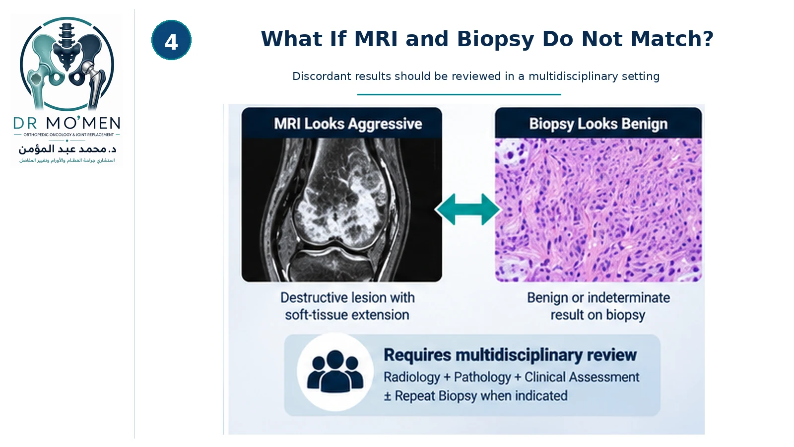
This is an important orthopedic-oncology safety situation. A benign biopsy should not automatically overrule an aggressive MRI when the two do not fit together.
For the imaging side of this decision, see what X-ray, MRI and CT each show in a bone tumor.
Possible explanations include sampling error, tumor heterogeneity, biopsy of a nonrepresentative region, insufficient tissue, pathology interpretation difficulty or an imaging diagnosis that also needs reconsideration.
The appropriate next step is usually specialist radiology review, pathology review and multidisciplinary discussion, with repeat biopsy when indicated.
What if the MRI looks benign but pathology looks malignant?
The same principle applies. An unexpectedly malignant pathology result should be reconciled with the imaging, exact anatomical site and clinical history. Discordant findings require review rather than automatic acceptance of one source in isolation.
Can the diagnosis change after expert review?
Yes. Rare bone tumors can be diagnostically difficult. Specialist review may refine the diagnosis, change the subtype or grade, or recommend additional molecular testing.
For suspected primary malignant bone tumors, specialist sarcoma pathology review is part of the recommended multidisciplinary pathway.
Should pathology slides be reviewed before major bone tumor treatment?
Specialist review is particularly important when the diagnosis was made outside a sarcoma center, when a primary malignant bone tumor is suspected, when imaging and pathology do not match, when the diagnosis is rare or indeterminate, or when major limb-salvage surgery or systemic treatment depends on the histological diagnosis.
What is radiology-pathology correlation?
Radiology-pathology correlation asks whether the tissue diagnosis explains what is visible on the imaging. For example, a destructive lesion with cortical breakthrough and a large soft-tissue mass would be difficult to reconcile with a tiny sample showing only bland fibrous tissue unless sampling error or another explanation is considered.
Why do patient age and tumor location matter?
Bone-tumor differential diagnosis changes substantially with age and anatomical location. The same microscopic pattern may have different significance in a child, a young adult and an older patient, or when it occurs in the epiphysis, metaphysis, diaphysis, bone surface or axial skeleton.
What if infection is still possible?
Bone infection can mimic tumor clinically and radiologically. In selected cases, biopsy material may also be sent for microbiology, bacterial culture or other infectious testing when infection remains in the differential diagnosis.
What should the final diagnosis section tell me?
A well-structured pathology report may include:
- Specimen type and anatomical site.
- Microscopic description.
- Final diagnosis.
- Tumor type and subtype.
- Grade when applicable.
- Immunohistochemistry results.
- Molecular results or tests still pending.
- Comments or differential diagnosis.
A definitive resection report may additionally include tumor size, local extent, margins and treatment response.
A practical patient reading guide
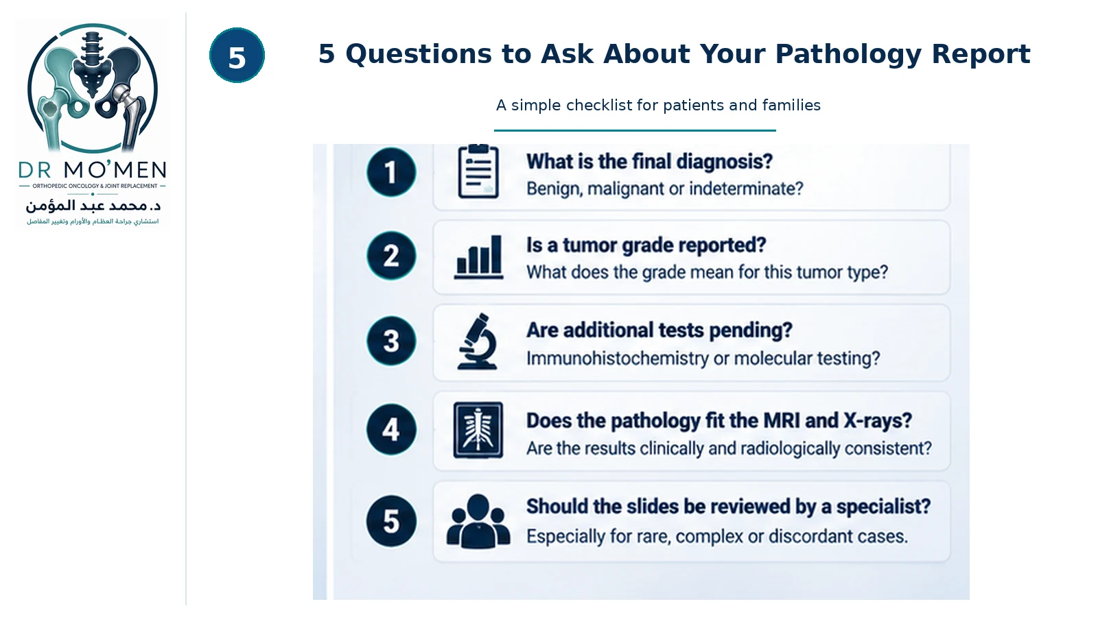
When you receive a pathology report, ask five questions:
- What is the final diagnosis: benign, intermediate/locally aggressive, malignant or indeterminate?
- Is the diagnosis complete, or does the report say suspicious, compatible with, favors, or pending additional tests?
- Is a tumor grade reported, and what does that grade mean for this specific tumor type?
- Are IHC, molecular or other tests still pending?
- Does the pathology result fit the MRI and X-rays?
When should you ask for specialist review?
- The diagnosis is unusual or rare.
- The biopsy is indeterminate or nondiagnostic.
- Pathology and imaging disagree.
- A primary malignant bone tumor is suspected.
- Tumor grade remains uncertain.
- Major limb-salvage surgery is planned.
- Systemic treatment depends on the precise histological diagnosis.
- Different centers have provided different diagnoses or treatment recommendations.
Specialist review of bone tumor pathology
Dr. Mohammed Abdelmoemen Abuelhadid evaluates bone and soft-tissue tumor cases in which the pathology result needs to be interpreted together with the original imaging and clinical findings.
The objective is not simply to read the biopsy report. The key question is whether the pathology diagnosis explains the lesion seen on X-ray, CT and MRI.
When necessary, the diagnostic pathway may include original-image review, specialist pathology-slide review, multidisciplinary discussion, additional IHC or molecular testing, and repeat biopsy when the existing specimen is inadequate or discordant.
For patients outside Egypt, existing imaging, pathology reports and medical records can also be reviewed as part of an online second medical opinion before treatment or travel when appropriate.
For appointments or imaging review: 01021690693
Evidence Behind Bone Tumor Pathology Interpretation
The central safety principle is integration: suspected primary malignant bone tumors should be interpreted by specialist teams using clinical information, imaging and pathology together, with additional immunohistochemical or molecular testing when the diagnosis requires it.
- British Sarcoma Group: UK Guidelines for the Management of Bone Sarcomas — recommends specialist pathology review and multidisciplinary correlation for suspected primary malignant bone tumors.
- Kovacs et al., Histopathology — reviews current immunohistochemical and molecular tools used to classify difficult bone tumors.
- Berkeley, Andrei & Saifuddin, Skeletal Radiology — emphasizes imaging–pathology correlation in rare primary bone sarcomas.
- Systematic review of expert sarcoma pathology second opinion — supports specialist review for rare or potentially discordant bone and soft-tissue sarcoma diagnoses.