
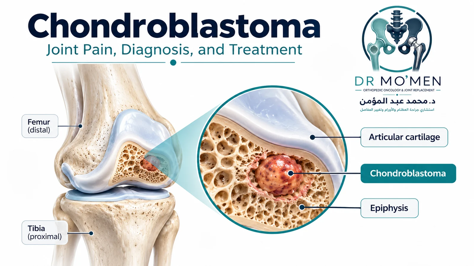
Persistent knee, shoulder, or hip pain in a teenager or young adult is usually caused by a much more common problem than a bone tumor. However, when pain, swelling, stiffness, or a limp persists despite appropriate treatment, an epiphyseal bone lesion such as chondroblastoma should be considered in the differential diagnosis.
Key Takeaways
- Chondroblastoma is rare and benign, but it can be locally aggressive and recur after treatment.
- It most often affects adolescents and young adults and commonly arises near the knee, shoulder, or hip in the epiphysis or apophysis.
- Joint pain, swelling, restricted motion and a limp may mimic a sports injury or inflammatory joint problem.
- X-ray, CT and MRI provide different information: location and margins, mineralization/cortical detail, and edema/joint/soft-tissue extent respectively.
- Secondary aneurysmal bone cyst (ABC)-like change can occur and may make the lesion look more expansile or aggressive.
- Curettage with careful reconstruction remains the conventional treatment for many lesions; selected appendicular lesions may be suitable for image-guided RFA.
- Because the tumor often lies directly beneath joint cartilage, treatment planning must balance local control with preservation of the subchondral bone and native joint.
What Is Chondroblastoma?
Chondroblastoma is a rare benign bone tumor composed of chondroblastic cells and characteristic matrix. Current AAOS patient guidance describes it as accounting for fewer than 1% of bone tumors. It occurs predominantly in adolescents and young adults, with a male predominance.
The classic lesion is centered in the epiphysis at the end of a long bone or in an apophysis, an area where tendons or ligaments attach. Common sites include the proximal tibia, distal femur, proximal humerus, and proximal femur. Less common locations include the pelvis, patella, foot, and other bones.
Although the tumor is histologically benign, it may expand, erode surrounding bone, damage the subchondral support beneath cartilage, and recur locally. This is why “benign” does not mean it should simply be ignored.
Why Can It Feel Like a Sports Injury?
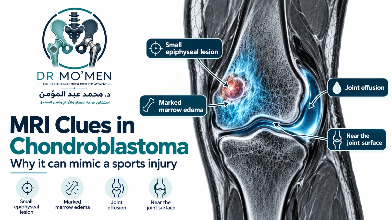
The lesion sits close to the joint and often produces substantial local inflammation. That can generate symptoms that overlap with meniscal, ligament, or overuse problems, particularly in a young active patient. See also when a sports injury needs orthopedic assessment.
- Persistent deep pain close to a joint.
- Joint swelling or effusion.
- Stiffness or reduced range of motion.
- Limping or reduced use of the limb.
- Occasional local tenderness or muscle wasting from prolonged underuse.
A lack of improvement after an appropriate period of conservative treatment should prompt reassessment rather than simply repeating physiotherapy or analgesics indefinitely.
How Is Chondroblastoma Diagnosed?
1. Plain X-rays
Radiographs often show a small, well-defined lytic lesion near the joint, commonly with a thin sclerotic rim. Internal mineralization can be visible in some cases. X-rays are useful for defining the lesion’s location, margins, relationship to the growth plate, and degree of cortical expansion.
2. CT
CT is particularly useful for showing cortical detail and internal matrix mineralization that may be subtle on plain radiographs. It can also help define how close the lesion is to the subchondral bone and can assist procedural planning.
3. MRI
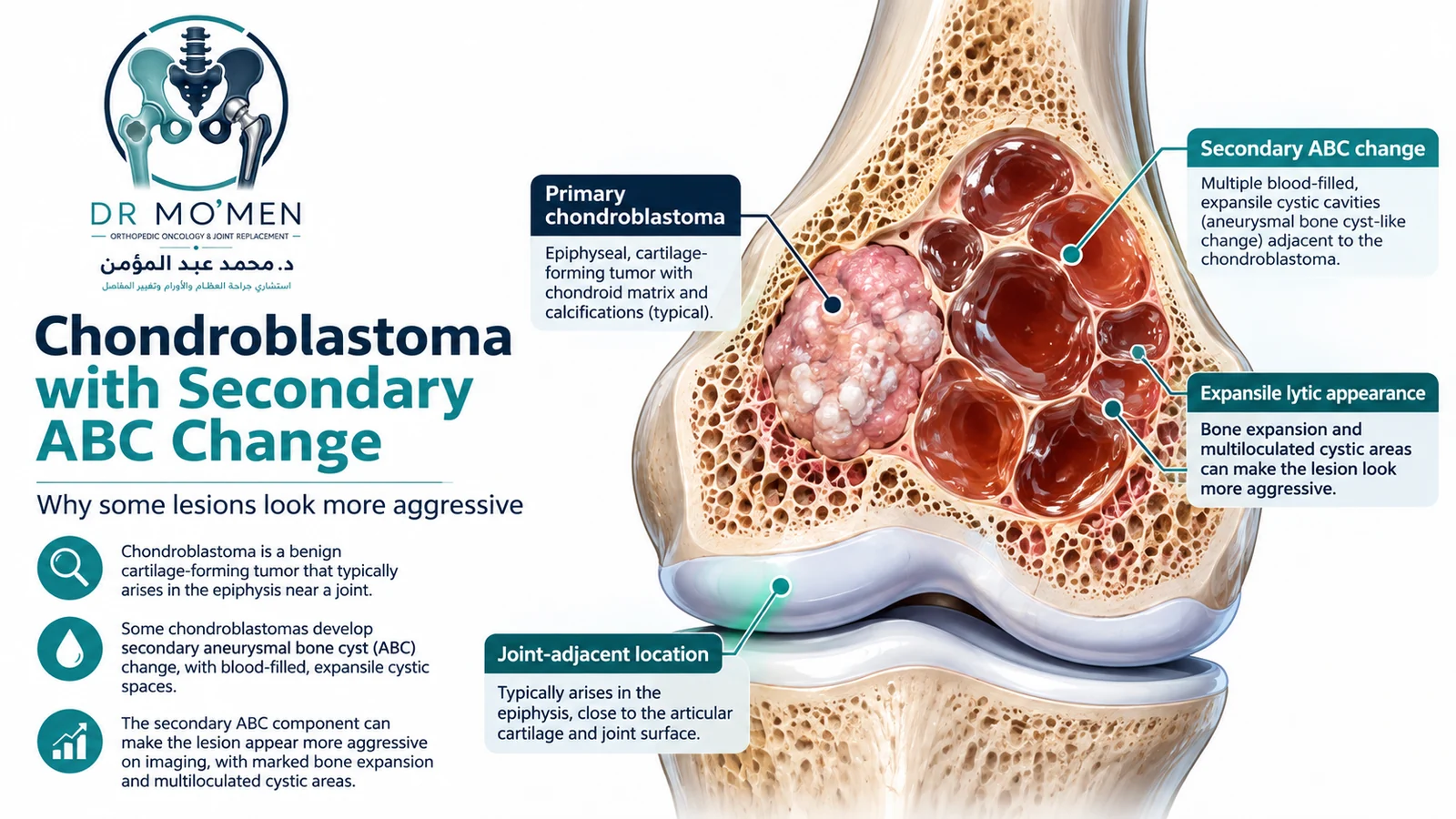
MRI is valuable for defining the lesion, the surrounding marrow and soft tissues, joint effusion, and any extension toward the articular surface. A relatively small chondroblastoma may be surrounded by disproportionately extensive bone-marrow edema. Secondary ABC-like change may produce cystic areas or fluid-fluid levels.
Does Every Chondroblastoma Need a Biopsy?
Histologic confirmation is generally obtained before definitive treatment. The biopsy should be planned after complete imaging review, particularly when the appearance is atypical, the patient is older than expected, an ABC-like component obscures the solid lesion, or the differential diagnosis includes infection or a malignant tumor.
Typical pathology includes chondroblasts, multinucleated giant cells, chondroid matrix and characteristic pericellular “chicken-wire” calcification. This term refers to the microscopic histologic pattern—not to the appearance of calcification on CT.
Differential Diagnosis: What Else Can Look Similar?
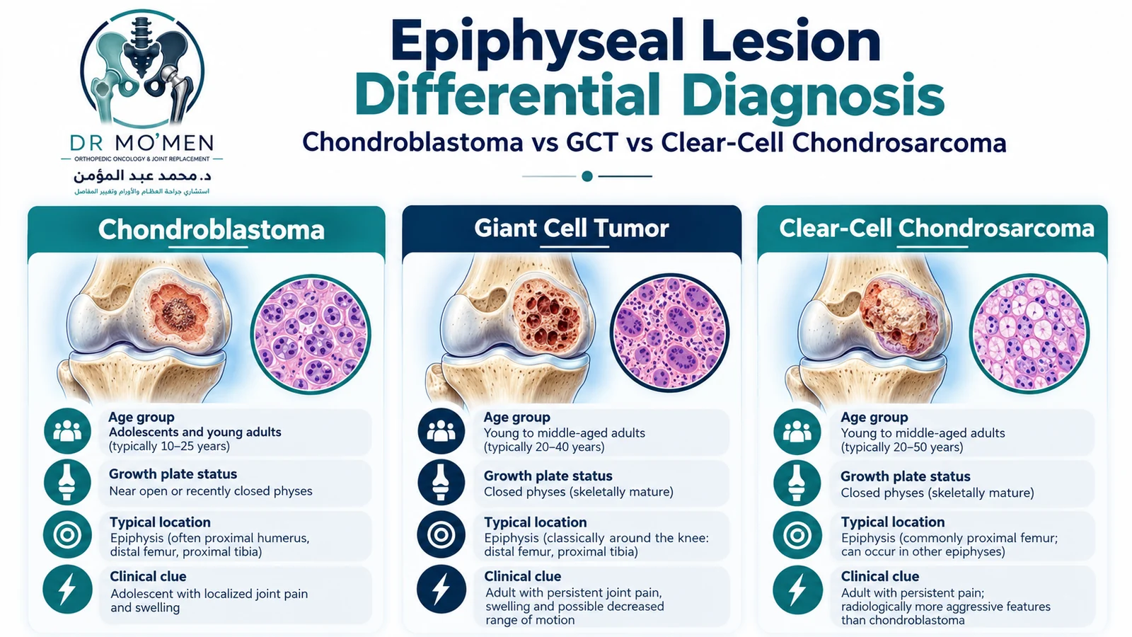
| Condition | Typical context | Important distinction |
|---|---|---|
| Chondroblastoma | Usually adolescents/young adults; epiphysis or apophysis | Well-defined lytic lesion, possible mineralization, often marked edema and joint symptoms. |
| Giant cell tumor of bone | Usually skeletally mature young adults | Typically reaches the subchondral bone after physeal closure; matrix mineralization is not typical. |
| Brodie abscess / subacute osteomyelitis | Any age; often children or young adults | Clinical and imaging context of infection; inflammatory markers may be normal, so normal ESR/CRP does not exclude infection. |
| Clear-cell chondrosarcoma | Usually older adults | Low-grade malignant cartilage tumor that can also involve the epiphysis; imaging and pathology correlation is essential. |
| Secondary ABC change | Can occur within chondroblastoma | Cystic/hemorrhagic spaces can dominate imaging; the underlying solid tumor still needs recognition and sampling. |
What Does Secondary ABC Change Mean?
A chondroblastoma may contain secondary aneurysmal bone cyst-like areas: blood-filled cystic spaces that can make the lesion appear more expansile and may create fluid-fluid levels on MRI. The presence of secondary ABC change does not transform the lesion into a different malignant tumor. For the primary lesion itself, see the aneurysmal bone cyst guide.
Some studies have associated secondary ABC change with recurrence, while others—including Ramappa et al.—did not find it to be an independent recurrence predictor. It should therefore be treated as an imaging/pathology feature rather than a guaranteed marker of poor outcome.
Treatment: How Is the Joint Preserved?
Treatment depends on the tumor’s size, location, subchondral support, growth-plate status, symptoms, recurrence history, and diagnostic certainty. There is no single operation that is optimal for every chondroblastoma.
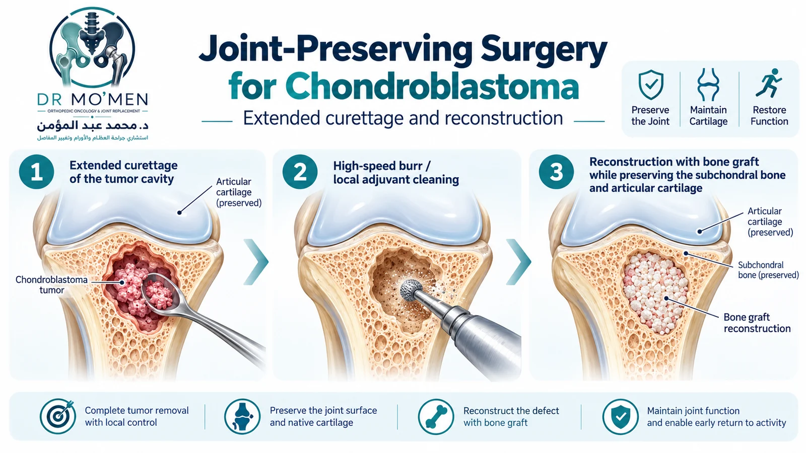
1. Extended Intralesional Curettage
Curettage remains the conventional treatment for many chondroblastomas. The surgeon creates a planned cortical window, removes the tumor under direct visualization, and may use a high-speed burr to extend the cavity and reduce residual tumor in bony ridges.
2. Local Adjuvants
Depending on location and surgeon preference, selected local adjuvants may be used after curettage. Examples described in clinical practice include phenol, liquid nitrogen, hydrogen peroxide, alcohol, or thermal methods. No single adjuvant is mandatory for every lesion, and tissue close to articular cartilage or the growth plate requires particular caution.
3. Reconstruction of the Defect
The cavity may be reconstructed with autograft, allograft, bone substitutes, bone cement, or combinations. The choice depends on defect size, location, age, remaining subchondral bone, and the need for structural support. Near a joint in a young patient, preserving the articular surface and supporting subchondral bone are central goals.
4. Image-Guided Radiofrequency Ablation in Selected Lesions
RFA is not appropriate for every chondroblastoma, but published series and a 2022 systematic review support it as a minimally invasive alternative in selected appendicular lesions. Suitability depends on lesion size, diagnostic confidence, safe probe access, distance from cartilage and neurovascular structures, and the ability to monitor for residual or recurrent disease.
Large lesions immediately beneath a weight-bearing surface require particular caution because thermal treatment or structural weakening may threaten the articular surface.
5. Resection in Selected Complex Cases
Segmental or wider resection is uncommon but may be necessary for selected destructive, recurrent, anatomically difficult, or unreconstructable lesions. The surgical plan should be individualized rather than dictated by the tumor name alone.
Recovery, Recurrence, and Follow-up
Recovery varies substantially. A small lesion treated through a limited cortical window may require a different rehabilitation plan from a large subchondral lesion or a lesion in the proximal femur. Weight-bearing restrictions and return to sport should therefore be individualized according to structural stability, reconstruction, pain, and follow-up imaging.
Local recurrence is a recognized risk. AAOS describes recurrence after curettage in roughly 10–20% of cases, while the Ramappa series reported 15%. Rates vary across cohorts and may be influenced by site, treatment technique, lesion complexity and follow-up duration. Recurrence usually appears within the first few years, so regular clinical and radiographic surveillance is important.
Can Chondroblastoma Spread to the Lungs?
Chondroblastoma is classified as a benign tumor, but pulmonary metastases have been reported very rarely. A 2020 multi-institutional pediatric series found no lung metastases among 130 children, while other published cohorts and case series confirm that distant spread can occur. Because reported rates vary, chest imaging and surveillance should be individualized according to age, tumor behavior, recurrence history and specialist judgment rather than applied identically to every patient.
When Should You Seek Specialist Orthopedic Oncology Assessment?
- Persistent joint pain or swelling without a clear injury or without expected improvement.
- A lytic epiphyseal lesion reported on X-ray, CT, or MRI.
- A proposed biopsy or curettage before the diagnosis has been securely correlated with imaging.
- Marked edema, an ABC-like component, cortical thinning, or concern about the joint surface.
- Recurrent pain or a new lesion after previous chondroblastoma treatment.
- Conflicting radiology and pathology reports, or concern about GCT, infection, clear-cell chondrosarcoma, or another tumor.
Urgent Red Flags
Urgent assessment is appropriate if joint or bone pain is accompanied by high fever and systemic illness, rapidly progressive swelling, inability to use the limb, a pathological fracture, or new neurological or vascular symptoms. These features are not specific to chondroblastoma and require broader evaluation.
Dr. Mo’men’s Role in Complex Cases
Dr. Mohammed Abdelmoemen Abuelhadid evaluates bone and soft-tissue tumors, limb-salvage problems, and complex reconstruction. For a suspected chondroblastoma, the key questions are whether the imaging pattern is truly compatible with the diagnosis, whether tissue confirmation is needed, how to preserve the native joint, and whether curettage, ablation, or another strategy best matches the lesion.
For patients outside Egypt, available X-rays, CT/MRI images, pathology reports, and previous operative notes can be reviewed as part of a preliminary second-opinion pathway before travel. Remote review does not replace examination when physical findings are required.
Evidence & Sources
- AAOS OrthoInfo — Chondroblastoma.
- Ramappa AJ et al. Chondroblastoma of bone. JBJS Am. 2000;82:1140–1145.
- Chen W, DiFrancesco LM. Chondroblastoma: An Update. Arch Pathol Lab Med. 2017;141:867–871.
- Dahlin DC, Ivins JC. Benign chondroblastoma: a study of 125 cases. Cancer. 1972;30:401–413.
- Sulaiman SRK et al. Radiofrequency ablation for appendicular chondroblastoma: systematic review and meta-analysis. Indian J Radiol Imaging. 2022;32:523–530.
- Arkader A et al. Pediatric Chondroblastoma and the Need for Lung Staging at Presentation. J Pediatr Orthop. 2020;40:e894–e897.
- WHO/IARC. WHO Classification of Tumours: Soft Tissue and Bone Tumours. 5th ed. 2020.
- Karbhari A et al. Retrospective Study of Multimodality Imaging Features of Chondroblastoma. Indian J Orthop. 2024.