
Pigmented Villonodular Synovitis (PVNS): Arthroscopic vs Open Surgery
Persistent knee swelling, repeated joint fluid, stiffness, or pain that keeps returning can occasionally be caused by pigmented villonodular synovitis (PVNS). The name can sound alarming, but PVNS is usually not a malignant cancer. It is a locally active synovial condition that can damage a joint if it becomes extensive or repeatedly recurs.
PVNS is an older term that patients and doctors still commonly use. In current terminology, it belongs to the spectrum of tenosynovial giant cell tumor (TGCT), and the classic intra-articular PVNS pattern generally corresponds to diffuse-type TGCT. The practical question after diagnosis is not simply “keyhole or open surgery?”—it is which approach gives safe access to the full extent of disease while preserving joint function.
What Is PVNS / Tenosynovial Giant Cell Tumor?
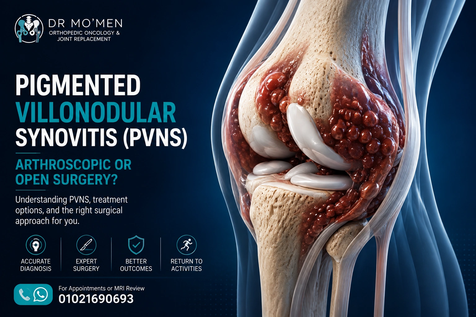
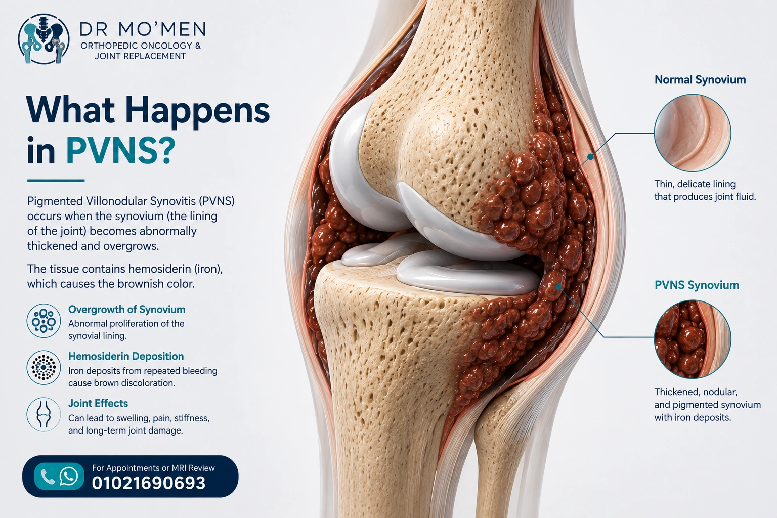
The inside of a joint is lined by synovium, a thin tissue that helps produce joint fluid. In TGCT, this synovial tissue proliferates abnormally and may form nodules or more extensive thickened tissue.
The tissue often contains hemosiderin, an iron-containing pigment associated with repeated microscopic bleeding. This helps explain the characteristic brown-red appearance during surgery and the dark “blooming” appearance that can be seen on certain MRI sequences.
TGCT is described as localized-type or diffuse-type. Localized disease is generally a discrete, well-defined lesion. Diffuse-type disease involves a broader area of synovium, may extend through several joint recesses, and can occasionally grow outside the joint. The knee is the most commonly involved large joint.
What Symptoms Can PVNS Cause?
Symptoms are often gradual and can resemble more common knee problems. This is one reason the diagnosis may be delayed.
- Recurrent or persistent knee swelling
- Aching pain or discomfort
- Stiffness or reduced range of motion
- Repeated joint effusion
- A feeling of fullness inside the knee
- Clicking, catching or occasional locking
- Difficulty with sport, stairs or prolonged walking
- Sometimes a bloody or brown-tinged joint aspirate
A swollen knee does not automatically mean PVNS. Meniscal injuries, cartilage problems, inflammatory arthritis, infection and other synovial disorders can produce overlapping symptoms, so the diagnosis should be based on the full clinical and imaging picture.
Is PVNS a Cancer?
TGCT/PVNS is generally considered a benign or non-metastasizing tumor-like neoplasm, but diffuse disease can behave in a locally aggressive way. It does not usually spread through the body like a conventional malignant cancer.
However, “benign” does not mean harmless. Persistent diffuse disease can erode bone, damage cartilage, restrict movement and contribute to secondary osteoarthritis. The clinical importance therefore depends on symptoms, location, disease extent, joint damage and previous treatment.
How Is PVNS Diagnosed?
Clinical assessment
Assessment starts with the history and examination. The surgeon looks for the pattern of swelling, tenderness, range-of-motion loss, mechanical symptoms, instability and alternative causes of recurrent joint effusion.
X-rays
Plain X-rays may be normal, especially early in the disease. In more advanced cases they may show bone erosions, joint-space loss or secondary degenerative change.
MRI
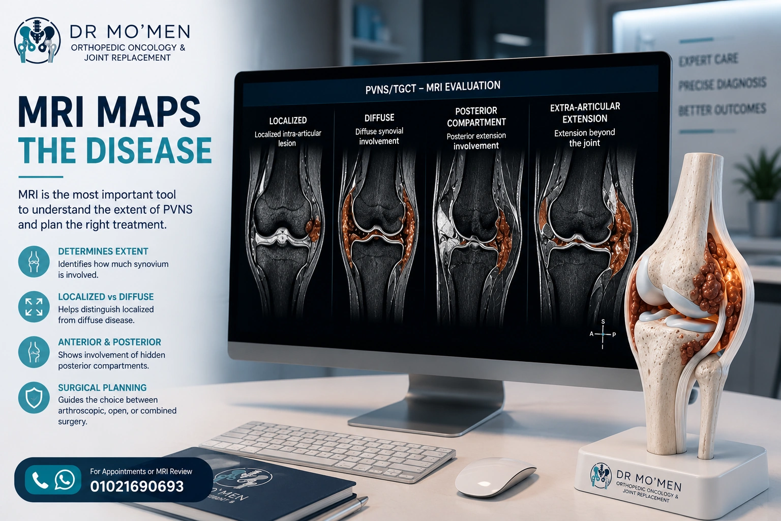
MRI is the key imaging study for suspected knee TGCT. It helps show whether the process is localized or diffuse and maps the anterior, posterior and extra-articular compartments that may need to be addressed during treatment.
Hemosiderin often produces low-signal areas and susceptibility or “blooming” on MRI. Just as importantly, MRI helps the surgeon identify areas that may be difficult to reach with arthroscopy alone and assess whether cartilage or bone has already been affected.
Is a Biopsy Always Necessary?
Not every patient follows the same diagnostic pathway. When imaging is classic and a clearly resectable lesion is being treated, the diagnosis is confirmed by histopathology of the removed tissue. When the appearance is atypical, the diagnosis is uncertain, or another tumor is possible, a planned biopsy may be appropriate before definitive surgery.
Important: a suspicious or atypical tumor-like lesion should not undergo an unplanned excision. If biopsy is needed, its route should be coordinated with the team responsible for definitive treatment.
Related reading: Bone Tumor Biopsy: Why Planning Matters
What Are the Treatment Options?
Treatment is individualized. Not every MRI abnormality requires immediate surgery; selected patients with limited symptoms may be observed with specialist follow-up. For symptomatic, progressive or function-limiting disease that can be resected with acceptable morbidity, surgery remains a central treatment option.
The surgical procedure is usually an excision or synovectomy—removing the abnormal tissue while preserving the surrounding joint structures whenever possible. The choice between arthroscopic, open or combined surgery depends on disease distribution rather than incision size alone.
Arthroscopic Excision / Synovectomy
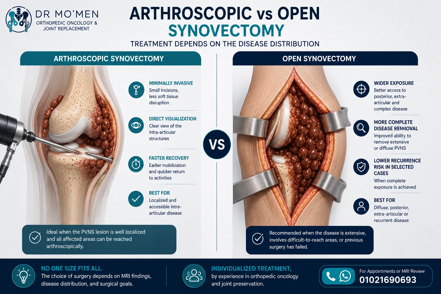
Arthroscopy uses a camera and small instruments passed through portals around the knee. It can provide excellent visualization of accessible intra-articular compartments and is particularly attractive for a localized lesion that can be completely reached.
Potential advantages
- Smaller skin incisions
- Less superficial soft-tissue disruption
- Direct visualization of intra-articular structures
- Ability to inspect several knee compartments through multiple portals
- Often faster early rehabilitation after limited procedures
Arthroscopy can also be used in selected diffuse cases, especially in experienced hands. The key limitation is access: if important disease lies in a blind or technically difficult area, leaving residual tissue simply to keep the procedure “minimally invasive” may not be the best strategy.
When Can Arthroscopy Be Insufficient?
Diffuse TGCT may extend into the posterior knee, extra-articular tissues or multiple recesses. Some of these areas are technically challenging to reach completely through arthroscopy.
For that reason, MRI should be reviewed as a surgical map. The objective is not the smallest incision; it is adequate control of the disease with acceptable risk to cartilage, ligaments, nerves, vessels and other structures.
Open Synovectomy
Open surgery provides direct exposure through a larger incision. It may be considered when the disease is extensive, involves difficult posterior compartments, extends outside the joint, has recurred after previous surgery, or cannot be safely and adequately addressed arthroscopically.
Potential advantages
- Wider exposure of extensive or diffuse disease
- Direct access to posterior or extra-articular areas when required
- Ability to protect important structures under direct visualization
- Useful for selected recurrent or previously operated cases
The trade-off is greater soft-tissue exposure and potentially more postoperative stiffness, wound morbidity or rehabilitation demand. These disadvantages must be balanced against the risk of inadequate access to the disease.
Arthroscopic vs Open Synovectomy: Which Is Better?
There is no universal winner. Published studies are heterogeneous, and recurrence is influenced by the biology and extent of disease, previous treatment, anatomical access and how completely the abnormal tissue can be removed.
For a small, well-defined and accessible lesion, arthroscopic excision may be appropriate. For extensive diffuse disease—particularly when important posterior or extra-articular areas are involved—an open approach may provide safer or more complete access. Some patients benefit from a combined arthroscopic and open strategy.
This is why an MRI-based treatment plan is more useful than a rule such as “arthroscopy is always better” or “open surgery is always safer.”
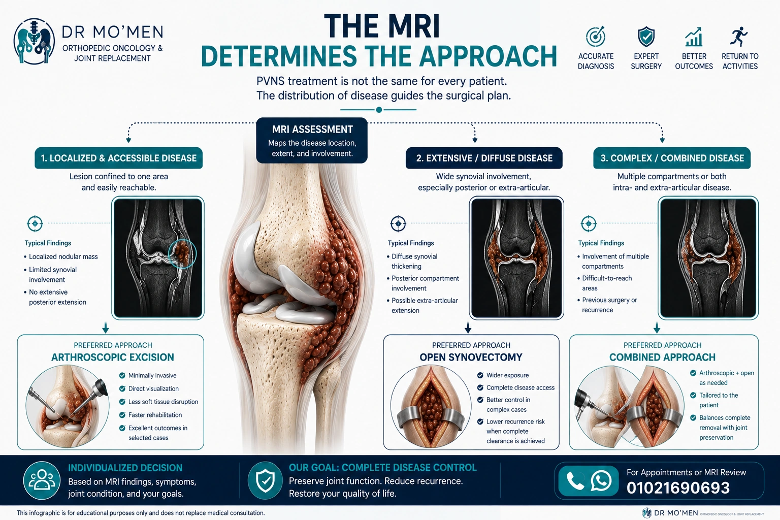
How the MRI Helps Choose the Surgical Approach
Before surgery, the MRI should answer practical questions: Is the disease localized or diffuse? Is the posterior compartment involved? Is there extra-articular extension? Is cartilage or bone damaged? Are there areas that cannot be reached safely through the planned approach?
A localized, accessible lesion may favor arthroscopic excision. Extensive diffuse or posterior disease may favor open exposure. Complex disease involving several compartments may require a combined strategy. These are treatment-planning principles, not rigid rules.
Can PVNS Come Back After Surgery?
Yes. Recurrence is an important issue, especially in diffuse-type TGCT. The risk is not determined by the incision alone; it is influenced by disease extent, anatomical accessibility, previous surgery, residual disease and the underlying biology of the tumor.
Follow-up is therefore individualized. Patients with diffuse or recurrent disease may need longer clinical and MRI surveillance than patients with a completely excised localized lesion.
What If the Joint Is Already Damaged?
Long-standing diffuse disease can occasionally produce cartilage loss, bone erosion and secondary osteoarthritis. In a relatively preserved joint, the goal is usually disease control while preserving the native joint whenever appropriate.
If severe end-stage joint destruction has already developed, treatment planning may need to address both the TGCT and the damaged joint. Joint replacement is considered only in selected cases after specialist evaluation.
Related service: Hip & Knee Joint Replacement
Are There Non-Surgical or Systemic Treatments?
Surgery is not the only possible option in every complex case. For recurrent, unresectable or diffuse TGCT in which another operation may cause major functional loss or morbidity, a specialist multidisciplinary team may discuss systemic treatment directed at the CSF1/CSF1R pathway.
These treatments are not routine substitutes for surgery in every patient. Suitability, drug approval, availability, monitoring and potential adverse effects vary by country and individual circumstances, so this part of management should be handled through an experienced tumor team.
Recovery After PVNS Surgery
Recovery depends on the amount of tissue removed, the surgical approach, associated cartilage or bone procedures, and the condition of the knee before surgery. A limited arthroscopic excision may recover more quickly than an extensive open synovectomy, but rehabilitation should be tailored to the actual procedure.
- Swelling and pain control
- Progressive range-of-motion exercises
- Quadriceps and lower-limb strengthening
- Physiotherapy when indicated
- Gradual return to work, walking and sport
- Planned clinical and imaging follow-up
When Should You Seek Specialist Assessment?
- Persistent or unexplained recurrent knee swelling
- Repeated bloody joint effusions
- An MRI report suggesting PVNS or TGCT
- Diffuse disease or posterior-compartment involvement
- Bone erosion or extra-articular extension
- Recurrence after previous arthroscopy or open surgery
- Uncertainty about arthroscopic versus open treatment
Fever, a hot red joint, rapidly worsening severe pain, or inability to bear weight should be assessed urgently because infection and other acute conditions must be excluded.
The Role of Dr. Mo’men in Complex PVNS / TGCT Cases
PVNS/TGCT sits at the intersection of joint surgery and orthopedic tumor surgery. Complex cases require review of the original MRI, the exact distribution of disease, previous procedures, joint damage and the functional goals of the patient.
Dr. Mohammed Abdelmoemen Abuelhadid evaluates complex and recurrent cases to determine whether arthroscopic, open, combined or non-surgical strategies should be discussed. The treatment decision depends on examination, imaging and final diagnosis—not on a single MRI sentence or a preference for one surgical technique.
For appointments or MRI review: 01021690693
Patients outside Egypt: an online second opinion can be used to review available MRI images, reports, pathology and previous treatment before deciding whether an in-person visit or travel is appropriate.
Second Opinion Service
Frequently Asked Questions
Is PVNS the same as TGCT?
PVNS is an older term that is still widely used. In current classification, classic PVNS generally corresponds to diffuse-type tenosynovial giant cell tumor (D-TGCT), while TGCT also includes a localized type.
Is PVNS cancer?
PVNS/TGCT is usually not a malignant cancer and does not typically spread through the body. Diffuse disease can nevertheless be locally aggressive and may damage cartilage and bone.
Can PVNS be treated completely with arthroscopy?
Some localized and appropriately accessible lesions can be removed arthroscopically. Extensive diffuse, posterior or extra-articular disease may require open or combined surgery.
Can PVNS return after surgery?
Yes, particularly in diffuse-type disease. Recurrence depends on disease extent, accessibility, previous treatment, residual disease and tumor biology, so follow-up is individualized.
Does every patient with PVNS need surgery?
No. The decision depends on symptoms, progression, disease distribution, joint damage and the expected morbidity of treatment. Selected patients may be observed, while complex unresectable or recurrent cases may need multidisciplinary discussion of other options.
Medical Disclaimer
For persistent knee swelling or synovial disease requiring procedural assessment, see sports injuries and arthroscopy care.