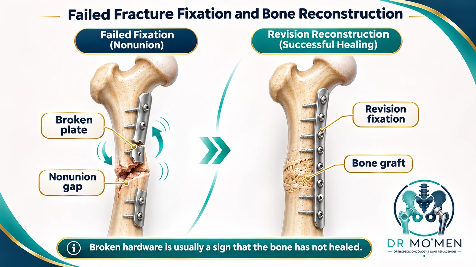
Failed fracture fixation means that the fracture or its fixation construct is no longer progressing toward a stable, functional union. Plates, screws or intramedullary nails may loosen, bend, cut out or break when stability is inadequate, bone-healing biology is compromised, infection is present, or several factors coexist. Revision planning should identify the cause first, then restore alignment, stability, biology and infection control when needed.
Key Takeaways
- Hardware failure is a signal, not a diagnosis. The revision plan should identify why union or fixation failed.
- Mechanical instability, biological compromise and fracture-related infection may coexist; alignment and bone stock also matter.
- CRP and ESR can support the infection work-up but cannot reliably exclude low-grade FRI. Deep intraoperative sampling is more important when revision is undertaken.
- Revision options include exchange nailing, augmentative plating, revision compression/locking constructs, external fixation, autologous grafting, induced-membrane reconstruction and, in selected periarticular failures, arthroplasty.
- An atypical destructive lesion or low-energy pathological fracture should trigger tumor-aware evaluation and biopsy planning before routine revision fixation.
Why Do Plates, Screws and Intramedullary Nails Fail?
Internal fixation is designed to stabilize a fracture while the patient’s own bone heals. If union is delayed or does not occur, more of the repeated bending, torsional and axial load continues to pass through the implant. Over time, this may lead to screw loosening, plate bending, nail or plate fatigue failure, loss of reduction, or cut-out into an adjacent joint.
Hardware failure should therefore be treated as a clue to the underlying problem rather than as the diagnosis itself. A revision operation that only replaces a broken implant without correcting the reason for failure can reproduce the same mechanical or biological environment.
The main contributors usually fall into four interacting groups:
- Mechanical / construct factors: instability, poor alignment, inadequate rotational control, an unfavorable plate working length or screw distribution, insufficient nail fit, an unaddressed gap or loss of medial support, or a construct that does not match the intended healing strategy.
- Biological / local factors: severe soft-tissue injury, open fracture, devascularized bone, bone loss, repeated surgical stripping or a poor soft-tissue envelope.
- Fracture-related infection: biofilm around implants can interfere with healing and may be present even without fever or obvious pus.
- Host factors: nicotine exposure, diabetes, malnutrition, anemia, severe osteoporosis and other metabolic or systemic problems may reduce healing potential or fixation purchase.
What Are the Main Failure Patterns?
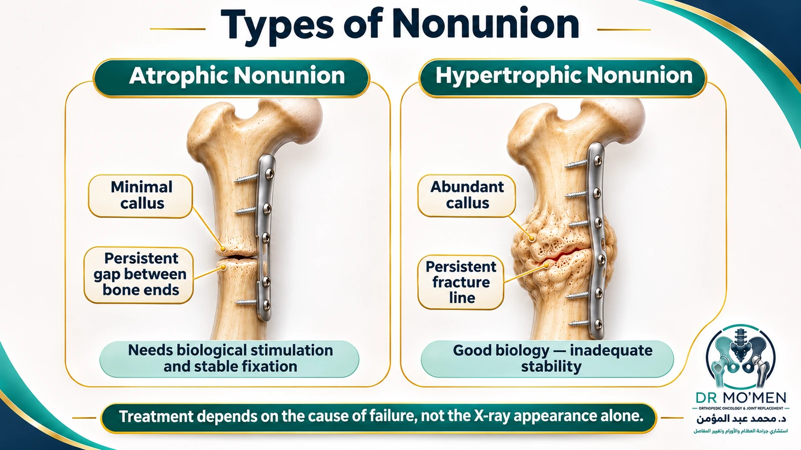
The pattern matters because treatment for early loss of fixation is not the same as treatment for an established nonunion or fracture-related infection.
Early mechanical failure may present with loss of reduction, screw pull-out or cut-out, plate bending, or nail-related instability before a mature nonunion has developed. This may follow poor bone quality, an unstable construct, an early fall, or a fracture pattern that was difficult to control.
Hypertrophic nonunion usually shows abundant callus on radiographs, indicating that the fracture is attempting to heal but remains mechanically unstable. Atrophic or oligotrophic nonunion shows little radiographic callus and may require attention to both stability and the biological environment. Importantly, these radiographic labels do not directly measure blood supply or cellular viability; they are patterns that must be interpreted with the clinical history, imaging and operative findings.
Infected nonunion combines impaired healing with fracture-related infection (FRI). It may be obvious, with pus or a communicating sinus, or subtle, with pain, implant loosening or failure of progression of healing and no fever.
Quick Comparison
| Pattern | Typical clue | Main revision objective |
|---|---|---|
| Hypertrophic nonunion | Abundant callus with persistent fracture line; often points to inadequate stability | Improve mechanics/alignment; biological augmentation only when indicated |
| Oligotrophic / atrophic pattern | Little callus; may reflect poor mechanics, biology, gap, bone loss or prior damage | Restore stability and address the biological/defect problem as needed |
| Fracture-related infection | Sinus/pus may be obvious; otherwise pain, loosening, nonunion or suggestive imaging/labs | Debridement + stable fracture strategy + deep sampling + targeted antimicrobial plan; retention/exchange/staging individualized |
| Early mechanical failure | Loss of reduction, cut-out, screw pull-out or implant deformation before established nonunion | Reassess reduction, bone quality, construct and joint damage; revise when needed |
What Symptoms Suggest Failed Fixation or Nonunion?
Persistent or recurrent pain at the fracture site, especially with loading or use of the limb, is one of the most common presentations. A sudden increase in pain after a period of improvement may occur when an implant bends or breaks, but pain alone does not establish the diagnosis.
Other warning features include a feeling of movement at the fracture, clicking, progressive deformity or shortening, a newly prominent screw or plate edge, reduced function, and stiffness or muscle wasting around adjacent joints.
Which Red Flags Need Urgent Orthopedic Assessment?
Urgent assessment is appropriate when failed fixation threatens the skin, joint, nerves, blood supply or overall health.
A fistula or sinus that communicates with the fracture or implant, or visible pus, is a confirmatory sign of fracture-related infection under international consensus criteria. Persistent or new wound drainage without an alternative explanation is concerning and should prompt investigation, but drainage alone is not automatically a confirmatory criterion in every case.
Other red flags include sudden collapse or inability to bear weight, new deformity, hardware tenting or penetrating the skin, new foot drop or wrist drop, a cold or pale limb, progressive numbness or weakness, systemic illness, or grinding pain suggesting that a screw or blade has entered a joint.
How Is the Cause of Failure Diagnosed?
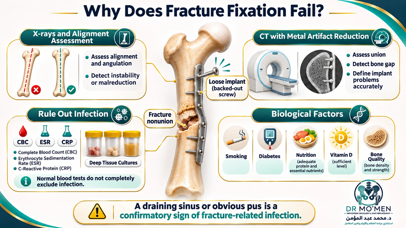
The central question is not only whether the metal is broken, but why the reconstruction failed. A structured work-up reviews the original fracture pattern, reduction, implant choice, alignment, serial healing, bone quality, soft tissues, host factors and the possibility of infection.
Radiographs are usually the first step. Orthogonal views and, when relevant, full-length alignment films can show loss of reduction, deformity, hardware breakage, screw loosening, fracture-gap persistence and progression—or absence—of callus. CT may be useful when plain films do not adequately show bridging, bone defects, articular damage or complex anatomy. Metal-artifact-reduction (MAR) reconstruction can improve CT assessment where available.
CBC, CRP and ESR can support the infection work-up, but they are suggestive tests rather than reliable rule-in or rule-out tests for FRI. Normal markers do not exclude a low-grade infection.
If revision surgery is performed for possible FRI, a structured sampling protocol is important. International recommendations favor multiple deep tissue or bone samples taken early in the procedure with separate sterile instruments; five samples are commonly preferred when feasible. Superficial wound or sinus swabs are not substitutes for deep sampling. Microbiology and histopathology should be interpreted together with the clinical and operative findings.
Selected patients also need assessment of modifiable host factors such as nicotine use, glycemic control, nutrition, anemia, vitamin-D status and osteoporosis. These tests should be individualized rather than ordered as a fixed panel for every patient.
Could a Tumor or Pathological Fracture Be the Hidden Cause?
Occasionally, unexpectedly rapid fixation failure, an unusual lytic lesion, a fracture after minimal trauma, or progressive bone destruction raises concern for a pathological fracture caused by a primary bone tumor or metastasis.
In that situation, revision fixation or curettage should not proceed as a routine nonunion operation until the lesion has been appropriately characterized. Cross-sectional imaging and oncological work-up may be required, and if biopsy is needed it should be planned so that the biopsy tract does not compromise definitive tumor surgery. Unplanned surgery through a bone tumor can complicate later limb-salvage treatment.
Can Failed Fixation Heal Without Major Revision Surgery?
Not every delayed fracture requires a large revision operation. If the hardware is intact, alignment is acceptable, the construct remains stable and infection is not suspected, selected delayed unions may continue to heal with protected loading, smoking or nicotine cessation, correction of relevant metabolic problems and close radiographic follow-up.
Dynamization of an intramedullary nail can help selected femoral or tibial shaft delayed unions when controlled axial compression is desirable and the fracture pattern, alignment and implant configuration are suitable. It is not appropriate for every nonunion and can worsen shortening or instability in the wrong setting.
A physically broken implant across an ununited, unstable or progressively deformed fracture usually shifts the discussion toward revision reconstruction. The decision is based on mechanics, biology, infection and patient factors rather than on a single time threshold. Although a 9-month/no-progress-for-3-months definition is often cited for nonunion, clinically important failure may require action earlier in selected fracture patterns.
How Is Bone Reconstructed During Revision Surgery?
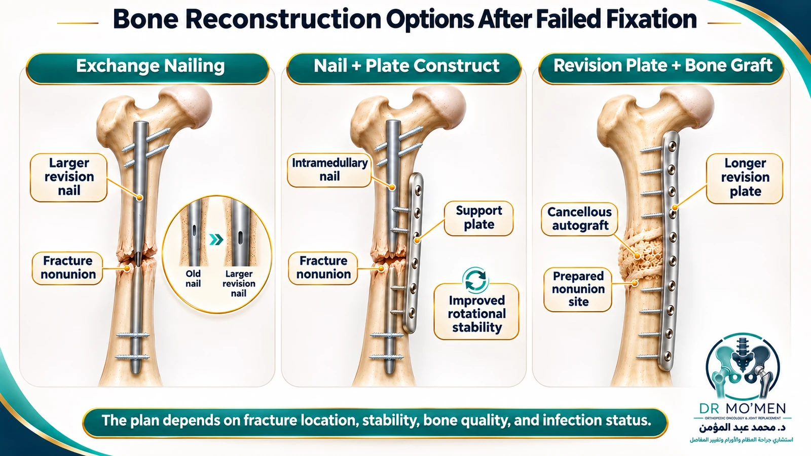
Revision fracture surgery is planned around the mechanical and biological deficiencies that caused the first reconstruction to fail. The 'Diamond Concept' is a useful framework: stable mechanics, osteogenic cells, an osteoconductive scaffold, biological signals and a viable vascular environment all contribute to healing.
Failed-hardware removal may require broken-screw extractors, trephines, reverse-thread devices, burrs or nail-extraction tools. The goal is to remove what must be removed while preserving as much host bone as possible.
For selected aseptic femoral or tibial shaft nonunions after intramedullary nailing, exchange reamed nailing may improve canal fit and mechanical stability. In other patterns—particularly where rotational control is inadequate—augmentative plating over a retained or exchanged nail may provide additional stability. Comparative studies and meta-analyses suggest favorable results for augmentative plating in many femoral-shaft nonunions, but the choice remains fracture-specific rather than universal.
Failed plate fixation may require correction of malalignment, compression or controlled bridging depending on the nonunion pattern, revision to a longer construct, and occasionally dual plating or combined nail-and-plate fixation. Circular external fixation or hexapod frames remain valuable for selected infected nonunions, deformity correction, bone transport and major limb-length problems.
When biology is deficient or debridement leaves a defect, autologous bone graft remains an important option. Iliac-crest cancellous graft is a well-established source, while graft can also be obtained by other techniques in selected cases. The amount and type of graft depend on defect size, host bone, fixation and soft-tissue conditions.
The induced-membrane (Masquelet) technique is one strategy for selected segmental defects and infected nonunions. Stage one involves debridement, stabilization and placement of a PMMA spacer; stage two removes the spacer and grafts the induced membrane cavity. Six to eight weeks is a commonly described interval, but the second stage should be timed according to infection control, soft-tissue recovery and reconstructive readiness rather than treated as an inflexible deadline.
Free vascularized fibular grafting may be considered for selected large segmental defects or difficult biological reconstructions, particularly after repeated failures or when a vascularized structural graft offers a clear advantage. It is a specialized microsurgical option rather than a routine threshold-based treatment.
Revision Options by Failure Pattern
| Scenario | What must be assessed | Possible reconstruction |
|---|---|---|
| Femoral/tibial shaft nonunion after nail | Assess infection, alignment, canal fit, nonunion level and rotational stability | Exchange reamed nailing or augmentative plating; grafting selectively |
| Broken distal-femur plate / metaphyseal nonunion | High bending load, bone loss, poor medial support, weak distal bone or infection | Revision plate/dual construct, nail-plate combination, graft/defect reconstruction; arthroplasty only in selected salvage cases |
| Humerus/forearm nonunion after plate | Rotational instability, gap, malalignment, biology, infection | Revision compression/locking fixation with biological augmentation when indicated |
| Infected nonunion | FRI + unstable or incompletely healed fracture | Debridement and sampling; retain, exchange or stage fixation according to stability/union/soft tissue; reconstruct bone defect when required |
| Periarticular failure with destroyed joint | Cut-out, collapse, AVN, severe articular damage or unreconstructable bone loss | Joint-preserving revision when feasible; conversion arthroplasty in selected patients |
How Does Fracture-Related Infection Change the Revision Plan?
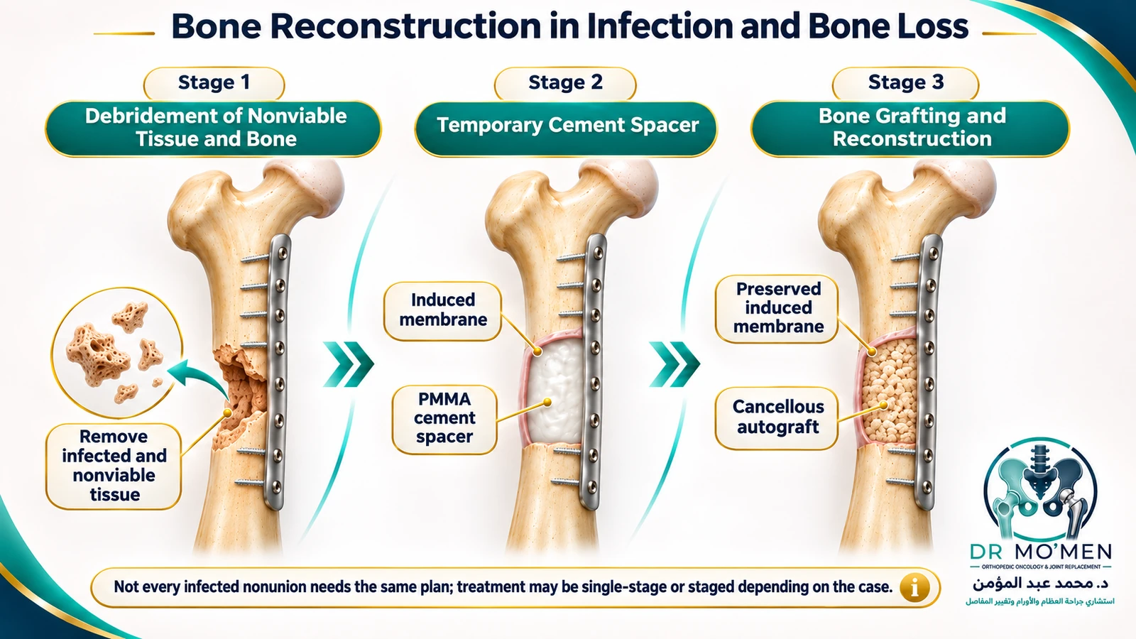
FRI requires both infection control and a strategy for fracture stability and eventual union. Treatment is not automatically 'remove all metal and wait.' Depending on fracture healing, implant stability, infection duration, soft-tissue condition, organism, bone loss and host factors, strategies may include debridement with implant retention in selected stable constructs, implant exchange, one-stage reconstruction, staged reconstruction, local antibiotic strategies and soft-tissue coverage.
When major infected bone loss is present, staged induced-membrane reconstruction, external fixation, bone transport or other limb-reconstruction methods may be appropriate. Antibiotic therapy should be culture-directed whenever possible and coordinated with the surgical plan.
When Is Conversion to Joint Replacement Considered?
If failure occurs close to a major joint, another fixation operation is not always the best salvage option. Conversion arthroplasty becomes more relevant when the native joint is irreversibly damaged, fixation has repeatedly failed, bone stock is severely compromised, or reliable reconstruction of the articular segment is unlikely.
Examples include selected failed proximal-femoral fixation with femoral-head damage or screw cut-out, and selected distal-femoral or proximal-humeral failures in older patients with severe bone loss. The decision between preserving the native joint and replacing it is individualized and should include infection assessment.
What Is Recovery Like After Revision Reconstruction?
Recovery varies widely because a revision plate, exchange nail, circular frame, Masquelet reconstruction and conversion arthroplasty are fundamentally different operations.
Early care usually focuses on pain control, wound and soft-tissue management, prevention of complications, and maintaining movement in adjacent joints when safe. If deep cultures were taken, the antimicrobial plan may change when final microbiology and histopathology results are available.
Weight-bearing is not determined by a fixed calendar. It depends on the fixation construct, fracture location, bone quality, defect reconstruction, infection status and radiographic progression. Some reconstructions permit early loading; others require prolonged protection. Serial radiographs and clinical examination guide progression.
When Is a Specialist Second Opinion Useful?
A second opinion is particularly useful when the plate or nail has broken, previous revision surgery has failed, infection has not been adequately assessed, there is major deformity or bone loss, a pathological fracture is possible, or different surgeons have proposed fundamentally different salvage strategies.
For stable patients outside Cairo or Egypt, current X-rays, CT when available, old fracture images, previous operative reports, implant details and relevant infection investigations can be reviewed before travel. The purpose is to clarify the likely pathway and what additional assessment is needed; it does not replace examination or guarantee that surgery will be recommended.
Dr. Mohammed Abdelmoemen Abuelhadid is a Lecturer of Orthopedic Surgery at Ain Shams University and a consultant orthopedic surgeon. Complex fracture failure is assessed in the context of alignment, union, infection, bone stock, soft tissues and whether biological reconstruction or joint salvage/replacement is more appropriate.
If MRI is required for another question in a patient with existing hardware, see MRI safety with orthopedic plates and screws; implant type and the imaging question should be checked with the radiology team.
Selected Scientific References
- Andrzejowski P, Giannoudis PV. The 'diamond concept' for long bone non-union management. J Orthop Traumatol. 2019;20:21.
- Govaert GAM, Kuehl R, Atkins BL, et al. Diagnosing Fracture-Related Infection: Current Concepts and Recommendations. J Orthop Trauma. 2020;34(1):8-17.
- Metsemakers WJ, Morgenstern M, McNally MA, et al. Fracture-related infection: consensus definition. Injury. 2018;49(3):505-510.
- Dudareva M, Barrett LK, Morgenstern M, et al. Evidence base for tissue sampling and culture interpretation in suspected fracture-related infection. JBJS Am. 2021;103(11):977-983.
- Metsemakers WJ, Morgenstern M, Senneville E, et al. General treatment principles for fracture-related infection. Arch Orthop Trauma Surg. 2020;140(8):1013-1027.
- American Academy of Orthopaedic Surgeons. Nonunions. OrthoInfo.
- Fu CX, Gao H, Ren J, et al. Augmentative plating after intramedullary nailing for lower-extremity long-bone nonunion: systematic review and meta-analysis. Chin J Traumatol. 2025;28(3):164-174.
- Walcher MG, Day RE, Gesslein M, et al. Augmentative plating versus exchange intramedullary nailing for aseptic femoral-shaft nonunion: biomechanical study. J Pers Med. 2023;13(4):650.
- Liu P, Jin D, Zhang C, et al. Revision surgery after failed internal fixation of intertrochanteric femoral fracture. BMC Musculoskelet Disord. 2020;21:573.
- Induced membrane technique for large bone defects: systematic review and individual participant data meta-analysis. Medicine (Baltimore). 2022.
- Baertl S, Rupp M, Alt V. The DAIR procedure in fracture-related infection—When and how. Injury. 2024;55 Suppl 6:111977.
- Gerrand C, Amary F, Anwar HA, et al. UK guidelines for the management of bone sarcomas. Br J Cancer. 2025;132(1):32-48.