
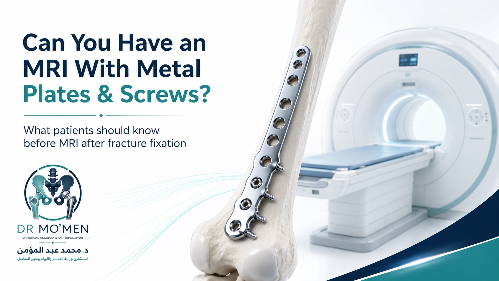
A common concern after fracture surgery is: “I have a metal plate and screws. Does that mean I can never have an MRI?” In many cases, the answer is no. The presence of orthopedic metal does not automatically prohibit MRI. The decision depends on the exact implant, its labeling, the scanner conditions, implant location, and the clinical question being investigated. [1,2]
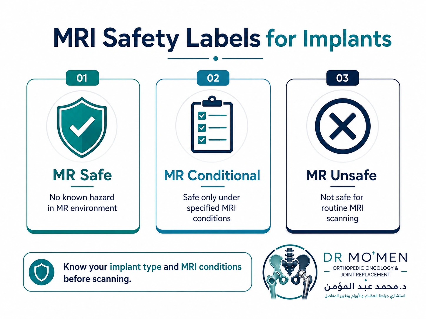
Patients should always tell the MRI team about any plate, screws, joint replacement, or implanted device before entering the MR environment. The standardized terms are MR Safe, MR Conditional and MR Unsafe; the older phrase “MRI compatible” is less precise. [1]
What Is the Actual Concern With Metal During MRI?
MRI uses a powerful static magnetic field, changing gradient fields and radiofrequency energy. For an implant, three separate issues matter:
- Potential magnetic force or torque on the device.
- Radiofrequency-related heating under specific scanning conditions.
- Image distortion or signal loss caused by metal artifact.
What Are Modern Orthopedic Plates Made From?
Titanium and Titanium Alloys
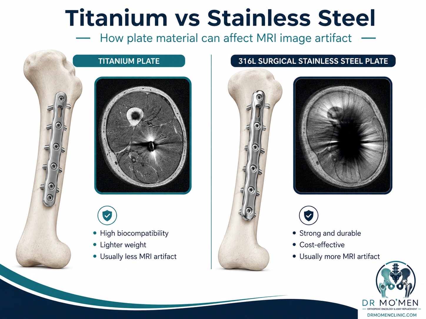
Titanium-based fixation systems are common in contemporary orthopedics. In experimental comparisons, titanium generally produces less susceptibility-related MRI artifact than stainless steel when similar implants are compared under similar conditions. Artifact is also affected by implant size, geometry, orientation, field strength and sequence choice. [3,4]
Surgical Stainless Steel
Implant-grade stainless steel remains an established fixation material, including alloys within the 316L family. Stainless steel can create larger signal void and distortion than titanium, particularly when the tissue of interest is immediately adjacent to the plate. Greater artifact does not, by itself, mean that MRI is unsafe. [3,4]
Cobalt-Chromium Alloys
Cobalt-chromium alloys are used more commonly in selected joint-replacement and high-strength orthopedic components. Their MRI assessment should be device-specific rather than based only on the alloy name.
MR Safe vs MR Conditional vs MR Unsafe
MR Safe
Under FDA terminology, an MR Safe item poses no known hazards in any MR environment and is made from nonmetallic, nonmagnetic and electrically nonconductive materials. A metallic orthopedic plate should therefore not simply be labeled “MR Safe.” [1]
MR Conditional
An MR Conditional device has demonstrated safety only when specified conditions are met. These conditions may relate to field strength, radiofrequency exposure, scanner operating mode, device configuration or other limits. [1]
MR Unsafe
An MR Unsafe device poses unacceptable risk in the MR environment. Routine scanning should not proceed with an MR Unsafe device unless a specialized safety pathway determines an alternative approach. [1,2]
Are Modern Orthopedic Plates Safe for MRI?
Testing of contemporary orthopedic plates and screws has been reassuring under defined conditions. In a study evaluating titanium-alloy and stainless-steel plates and screws at 1.5 T, magnetic interaction and heating were low, while artifact remained clinically relevant and could be reduced by appropriate sequence and parameter selection. That result should not be extrapolated to every implant, every scanner or every field strength. [3]
Metal Can Affect the Image — Not Necessarily Safety
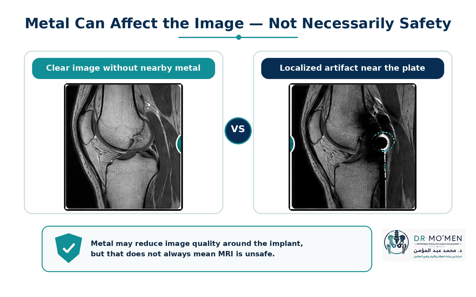
Metal can distort the local magnetic field and create signal void, image warping, bright or dark bands, and loss of detail around the implant. Titanium tends to create less artifact than stainless steel in many experimental comparisons, but the clinical image also depends on field strength, sequence, implant dimensions and orientation. [3,4]
Modern metal-artifact reduction approaches can improve postoperative MRI in selected situations. If MRI cannot answer the clinical question adequately, radiographs, CT or ultrasound may be more useful depending on what is being investigated.
What If I Do Not Know Which Plate Was Implanted?
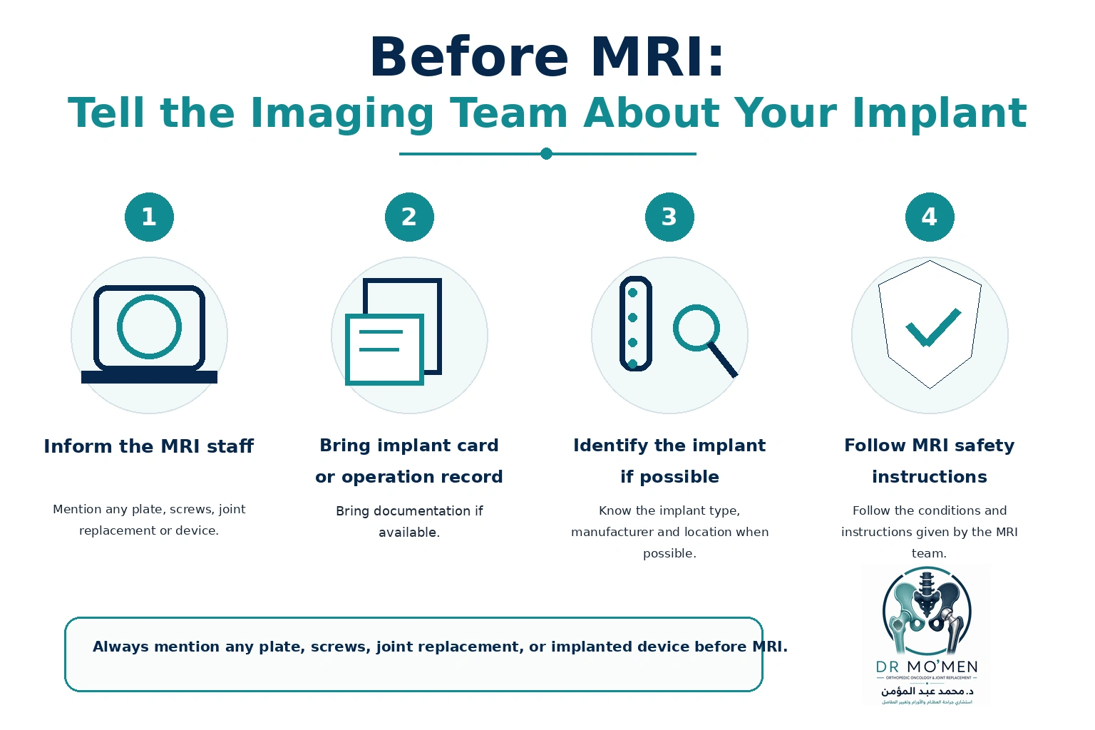
- Tell the MRI staff about any plate, screws, joint replacement or implanted device.
- Bring your implant card or operative report if available.
- Try to identify the manufacturer, model and date of implantation.
- Bring previous X-rays that may show the hardware configuration.
- Follow the MRI facility’s safety protocol rather than assuming the implant is safe or unsafe.
When an implant cannot be identified, the MRI facility should follow its local safety process. FDA and ACR guidance emphasizes documented device information and structured safety screening before scanning. [1,2]
Are Pacemakers and Electronic Implants the Same as Bone Plates?
No. Plates and screws are usually passive implants. Pacemakers, neurostimulators, implanted pumps and other active electronic devices require a different safety assessment. It is also no longer accurate to say that every pacemaker prohibits MRI; MR Conditional systems exist, but they may require programming, monitoring and a device-specific scanning protocol. [1,2]
When Is Specialist Advice Useful?
- The implant cannot be identified.
- An electronic, programmable or magnetically controlled device is present.
- There is an external fixator or unusual fixation system.
- A metallic foreign body is suspected.
- The tissue of interest is directly adjacent to extensive hardware and artifact may limit diagnosis.
- The patient has complex reconstruction, tumor surgery or a large joint-replacement construct.
Dr. Mo’men’s Role in Complex Orthopedic Cases
In complex fracture fixation, failed hardware, joint replacement, tumor reconstruction or limb-salvage surgery, the question is broader than “is there metal?” Assessment may require review of current and previous imaging, the operative history, implant location and configuration, the suspected complication, and whether MRI is actually the best investigation.
When a bone or soft-tissue tumor is suspected, imaging should be planned before biopsy or unplanned surgery. Accurate diagnosis, biopsy planning and multidisciplinary care are central to safe orthopedic-oncology decision making.
Frequently Asked Questions
Can I have a knee MRI if I have a plate in my femur or tibia?
Potentially yes. The implant’s MRI conditions should be verified first, and the radiology team should then consider whether local artifact will limit the study.
Is a titanium plate completely safe in MRI?
Titanium generally has favorable magnetic properties and usually produces less artifact than stainless steel, but safety should still be based on the specific implant and its labeling.
Does stainless steel mean I cannot have an MRI?
No. Stainless-steel orthopedic hardware is not automatically a contraindication, although it may produce more artifact than titanium. Device-specific conditions still matter.
Can MRI pull screws out of the bone?
This should not be answered from the word “metal” alone. Magnetic force, torque and heating are part of device testing, so the implant’s MRI classification and conditions should be followed.
Should I remove my plate before MRI?
A well-functioning plate is generally not removed simply to obtain MRI. Hardware removal is a separate surgical decision with its own indications and risks.
Key Takeaway
Having an orthopedic plate or screws does not automatically mean that MRI is prohibited. For many modern implants, the key issues are the device-specific scanning conditions and the amount of image artifact rather than simply the presence of metal. Identify the implant when possible, tell the MRI team and follow the applicable conditions.
This article is for patient education and does not replace clinical examination, imaging review or individualized medical advice.
For a broader discussion of when MRI findings should—and should not—drive treatment decisions, see specialist second-opinion review and fracture care.