
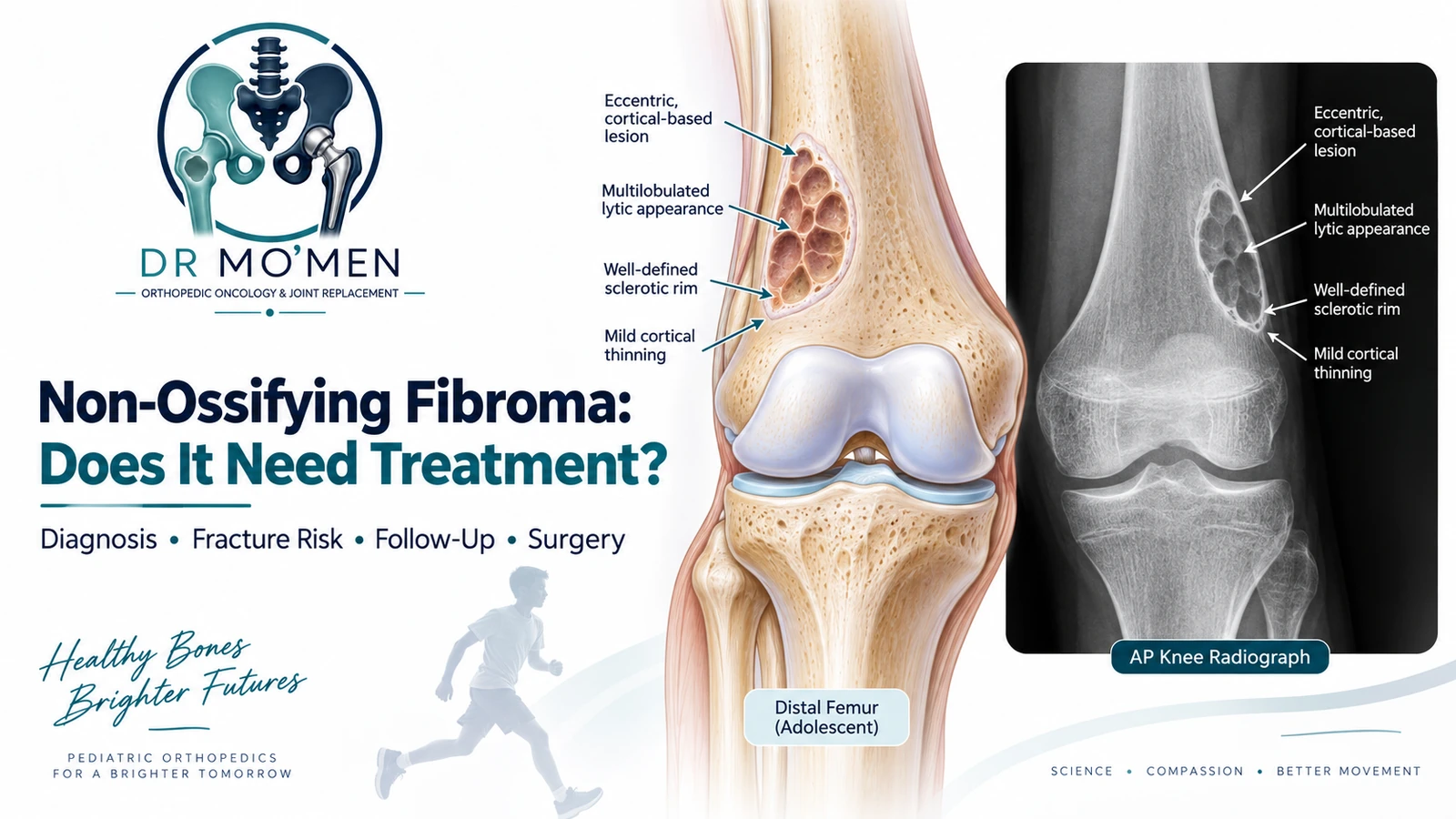
Finding a non-ossifying fibroma (NOF) on a child's X-ray can sound alarming, particularly because the radiology report may use terms such as:
- bone lesion
- lytic lesion
- cortical defect
- fibrous lesion
However, a typical non-ossifying fibroma is a benign developmental bone lesion and is one of the most common incidental skeletal findings in children and adolescents.
Most NOFs:
- cause no symptoms
- have a characteristic X-ray appearance
- do not require biopsy
- do not require surgery
- gradually become sclerotic and regress as the skeleton matures
The main clinical issue is usually not cancer risk.
It is determining whether a larger lesion has weakened the bone enough to increase the risk of a pathological fracture.
What Is a Non-Ossifying Fibroma?
A non-ossifying fibroma is a benign fibrous lesion that develops within the cortex of a growing bone.
It most commonly occurs in children and adolescents.
NOFs are usually located in the metaphyseal region of long bones, particularly around the knee.
Common sites include:
- distal femur
- proximal tibia
- distal tibia
- fibula
A typical lesion is often discovered accidentally when an X-ray is taken after an injury.
The lesion itself may have nothing to do with the reason the X-ray was obtained.
Is a Non-Ossifying Fibroma a Bone Cancer?
No.
A characteristic NOF is a benign lesion and does not behave like an aggressive malignant bone tumor.
It typically follows a natural progression toward increasing sclerosis and eventual healing.
The important caveat is that the diagnosis should genuinely fit the typical clinical and radiographic pattern.
If imaging is unusual, destructive, or inconsistent with a classic NOF, the lesion should be reassessed rather than assuming every eccentric lytic lesion is harmless.
What Is the Difference Between a Fibrous Cortical Defect and an NOF?
The terms fibrous cortical defect (FCD) and non-ossifying fibroma describe closely related lesions and are often considered part of the same biological spectrum.
A fibrous cortical defect is generally:
- smaller
- confined primarily to the cortex
- discovered incidentally
- clinically insignificant
An NOF is generally:
- larger
- more expansile
- capable of extending farther into the medullary cavity
- more relevant to mechanical strength when extensive
For the patient, the practical issue is usually not which label is used.
The important question is:
How large is the lesion, how much cortex remains, and is the bone mechanically safe?
Who Gets Non-Ossifying Fibromas?
NOFs are predominantly seen during skeletal growth.
They are frequently found in:
- school-aged children
- adolescents
Many patients never know they had one because the lesion may resolve without ever causing symptoms.
NOF is sufficiently common that it should not automatically be approached as a rare tumor requiring advanced testing.
What Symptoms Does an NOF Cause?
Most NOFs are asymptomatic.
They are commonly discovered after imaging for:
- sports injury
- knee pain
- ankle sprain
- minor trauma
- another unrelated orthopedic problem
The presence of an NOF on the X-ray does not necessarily mean that it is causing the patient's pain.
Can an NOF Cause Pain?
Sometimes.
Larger lesions may occasionally be associated with:
- activity-related discomfort
- local tenderness
- pain associated with stress injury
- pain related to a pathological fracture
However, pain should not automatically be attributed to an incidental NOF.
For example, a child with knee pain may have an NOF in the distal femur but actually have:
- patellofemoral pain
- overuse symptoms
- ligament injury
- meniscal pathology
- another cause
Symptoms and imaging should be correlated.
What Does a Non-Ossifying Fibroma Look Like on X-Ray?
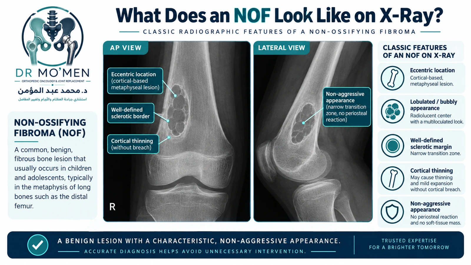
A classic NOF has a very characteristic appearance.
It is usually:
- eccentric
- cortically based
- located in the metaphysis or metadiaphyseal region
- radiolucent or lytic
- lobulated or “bubbly”
- surrounded by a well-defined sclerotic border
- non-aggressive in appearance
This characteristic appearance is one reason that many NOFs can be diagnosed from plain radiographs alone.
Why Is the Sclerotic Border Important?
A sharply defined sclerotic border usually indicates that the bone has had time to respond to a slowly behaving lesion.
This contrasts with aggressive bone lesions that may show:
- poorly defined destruction
- a wide zone of transition
- aggressive periosteal reaction
- cortical destruction
- soft-tissue mass
A well-defined border does not replace specialist interpretation, but it is one of the reassuring characteristics of a typical NOF.
Does Every NOF Need MRI?
No.
For a characteristic NOF on plain radiographs, MRI is often unnecessary.
MRI may become useful when:
- the X-ray is not typical
- symptoms do not fit the lesion
- another diagnosis is being considered
- the lesion has unusual characteristics
- an associated injury is being assessed
MRI should therefore answer a specific clinical question rather than simply being ordered because a benign lesion appears on an X-ray.
Does Every NOF Need CT?
No.
CT is not routinely necessary for a classic NOF, particularly because children should not be exposed to additional radiation without a clinical reason.
However, CT can occasionally help assess:
- remaining cortical thickness
- cortical breach
- exact lesion dimensions
- fracture risk in selected large lesions
This is useful in selected mechanically important lesions, but it should not be interpreted as a reason to perform CT on every child with an NOF.
Does a Non-Ossifying Fibroma Need Biopsy?
Usually no when the imaging appearance is classic.
A typical NOF is one of the bone lesions that may often be diagnosed radiographically.
Biopsy becomes more relevant if:
- imaging is atypical
- the lesion does not occur in the expected location
- the borders are unusually aggressive
- cortical destruction cannot be explained mechanically
- a soft-tissue component is present
- the lesion behaves unexpectedly
- another tumor diagnosis remains possible
If biopsy is genuinely required, it should be appropriately planned.
Related: Bone Tumor Biopsy: Core Needle vs Open Biopsy
Does an NOF Grow?
It can enlarge during skeletal growth.
This does not automatically indicate aggressive tumor behavior.
NOFs have a characteristic radiographic natural history.
As the child grows, lesions may initially become larger and then gradually develop increasing sclerosis before eventually becoming much less conspicuous.
What Are the Ritschl Stages of an NOF?
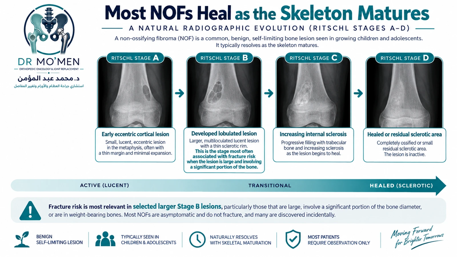
A commonly described radiographic staging system divides the natural evolution of NOF into four stages.
Stage A
An early eccentric lesion near the growth plate.
Stage B
The lesion becomes more clearly developed with a thin sclerotic border.
This is often the most mechanically relevant stage for larger lesions.
Stage C
The lesion begins to show increasing sclerosis.
Stage D
The lesion becomes extensively or completely sclerotic.
Pathological fractures have been reported predominantly among larger stage B lesions, although most stage B lesions still do not fracture.
That distinction is important.
Stage B does not mean a fracture is inevitable.
It simply indicates that lesion stage should be considered together with size, location and cortical integrity.
Can an NOF Cause a Pathological Fracture?
Yes, but most NOFs do not.
A pathological fracture means the bone fractures through an area weakened by an underlying lesion.
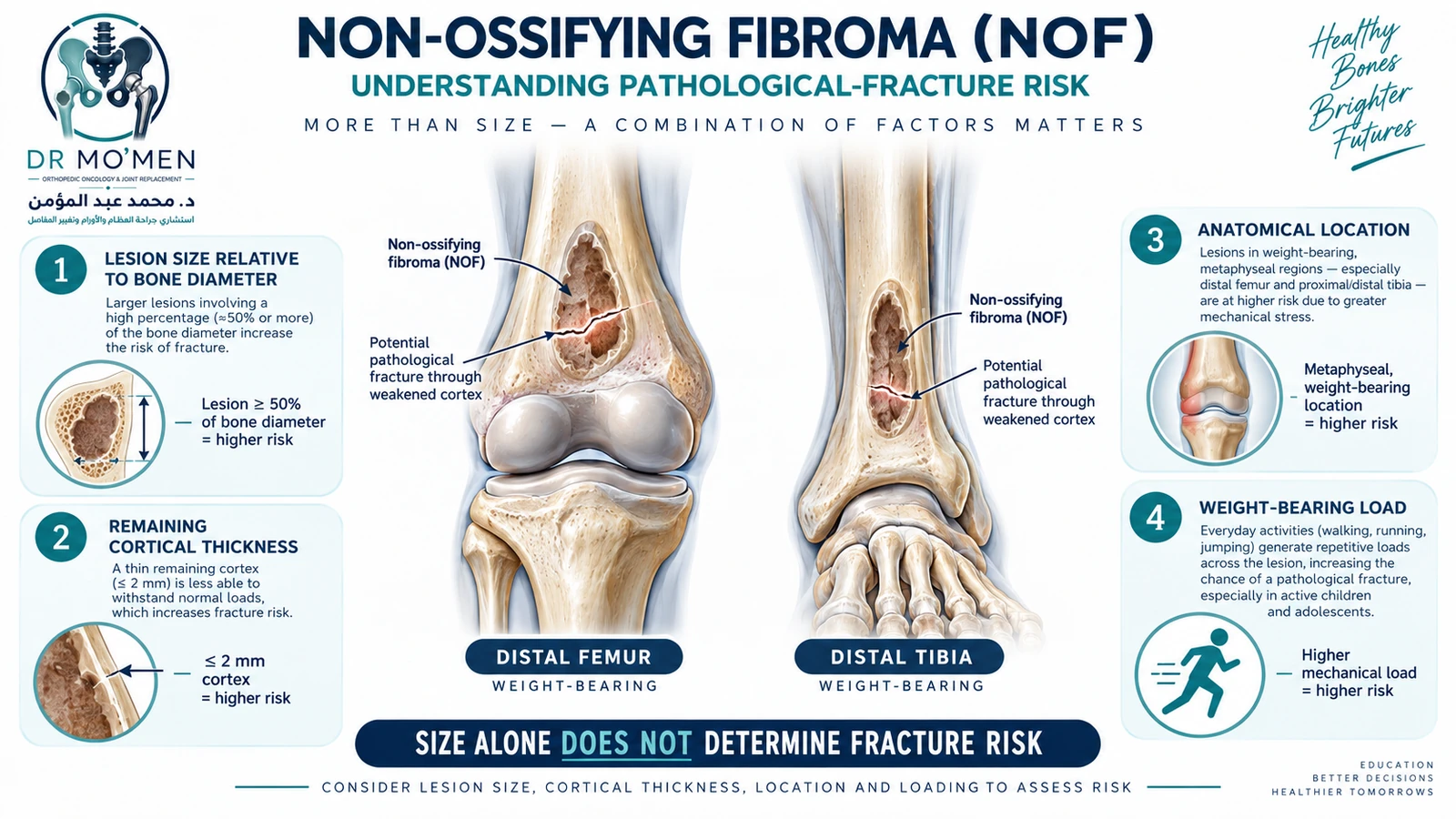
Fracture risk becomes more relevant when an NOF is:
- large
- located in a weight-bearing bone
- associated with marked cortical thinning
- occupying a substantial proportion of the bone diameter
- in a mechanically vulnerable location
Larger stage B lesions have been associated with fracture, particularly in the distal tibia.
Does Size Alone Predict Fracture?
No.
This is an important correction to an oversimplified approach.
Historically, clinicians used thresholds such as:
- lesion involving more than approximately half of the bone diameter
- large lesion length in a weight-bearing bone
as warning signs.
But these should not be treated as automatic indications for surgery.
Many lesions exceeding proposed size thresholds never fracture.
Modern assessment should consider:
size + location + cortical integrity + symptoms + skeletal maturity + lesion stage + activity
rather than one measurement alone.
Why Does Location Matter?
Two NOFs of the same size may not carry the same mechanical risk.
For example, a lesion in the distal tibia may behave differently from one in another metaphyseal location.
Fracture risk is influenced by size, stage, cortical thinning and site rather than size alone.
This reinforces the principle that mechanical context matters.
What Happens if an NOF Fractures?
A pathological fracture through an NOF does not mean that the lesion has become malignant.
Many such fractures can heal normally.
Treatment depends primarily on:
- fracture displacement
- fracture stability
- anatomical location
- patient age
- residual mechanical weakness
Pathological fractures through large NOFs can often heal successfully with appropriate fracture treatment, including non-operative management in selected cases.
The residual NOF may remain visible after the fracture has healed.
That does not automatically mean treatment failed.
Does the NOF Need to Be Removed When a Fracture Occurs?
Not necessarily.
The first priority is appropriate fracture treatment.
After union, the lesion can be reassessed.
If the remaining bone is mechanically satisfactory and the lesion is beginning to ossify, further surgery may not be necessary.
Surgery becomes more relevant when substantial structural weakness persists or the fracture itself requires operative stabilization.
When Can an NOF Be Observed?
Observation is appropriate for the majority of typical NOFs.
This is especially true when the lesion:
- has characteristic imaging
- causes no clear symptoms
- does not significantly compromise cortical strength
- has not fractured
- is showing progressive sclerosis
- has low mechanical risk
The natural history is generally spontaneous ossification as skeletal maturity approaches.
Does Every NOF Need Regular X-Ray Follow-Up?
No.
Small, characteristic lesions with negligible mechanical risk may not require repeated imaging indefinitely.
Follow-up becomes more relevant when the lesion is:
- large
- stage B
- located in a weight-bearing bone
- associated with substantial cortical thinning
- symptomatic
- mechanically concerning
A 6–12 month interval may be considered for selected larger stage B lesions until sclerosis progresses, but follow-up should be individualized rather than used as a rigid rule for every patient.
Can Children With an NOF Play Sports?
Often, yes.
A small typical NOF does not automatically mean that a child should stop sport.
Activity recommendations depend on:
- lesion location
- lesion size
- cortical strength
- previous fracture
- sport type
- impact/contact level
- symptoms
For a large lesion with substantial cortical thinning or high mechanical risk, temporary activity modification may be appropriate until the fracture risk is better defined.
The goal should be reasonable risk management rather than unnecessary restriction from activity.
When Is Surgery Considered?
Surgery is uncommon for routine NOFs.
It may be considered when there is:
- significant structural weakening
- recurrent or high-risk pathological fracture
- a fracture requiring surgical stabilization
- persistent lesion-related symptoms in a mechanically significant lesion
- diagnostic uncertainty
- selected large lesions where the consequences of fracture would be substantial
Importantly:
large lesion ≠ automatic surgery.
The mechanical risk must justify intervention.
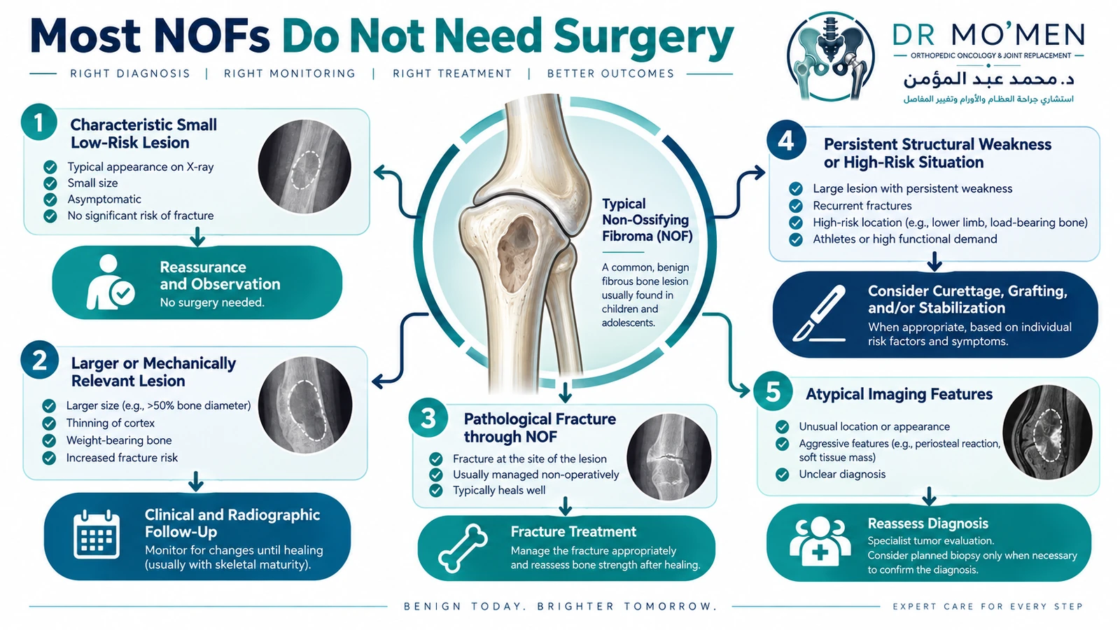
What Surgery Is Performed for an NOF?
When treatment of the lesion itself is required, the typical surgical strategy may involve:
Curettage
The fibrous tissue is removed from inside the lesion.
Filling the defect
Depending on the size and location, the cavity may be filled with:
- bone graft
- bone substitute
Internal fixation
Fixation may be added when:
- a significant pathological fracture is present
- bone strength is substantially compromised
- immediate mechanical protection is required
The exact procedure depends on the lesion and fracture—not simply the diagnosis “NOF.”
Does an NOF Come Back After Surgery?
Persistence or recurrence can occur, particularly if treatment is performed before skeletal maturity or if the lesion remains biologically active.
However, recurrence should not be interpreted as malignant transformation.
It should prompt review of:
- imaging
- diagnosis
- mechanical status
- previous surgery
Can an NOF Become Cancer?
A characteristic non-ossifying fibroma has no expected malignant behavior.
The clinical reason for follow-up is primarily fracture risk and diagnostic certainty—not concern that every NOF will transform into cancer.
If a lesion develops genuinely atypical features, the question should instead be:
Was the original diagnosis correct?
rather than assuming a routine NOF has suddenly transformed.
What Imaging Features Are Not Typical of an NOF?
Further review is appropriate when imaging demonstrates features such as:
- poorly defined margins
- aggressive bone destruction
- unexplained periosteal reaction
- substantial soft-tissue mass
- unusual location
- rapid unexpected change
- appearance inconsistent with an eccentric metaphyseal cortical lesion
These do not automatically indicate malignancy, but they should prompt reconsideration of the diagnosis.
NOF vs Fibrous Dysplasia
These are different lesions.
NOF
Usually:
- eccentric
- cortical-based
- metaphyseal
- lobulated
- surrounded by sclerosis
Fibrous dysplasia
Typically:
- intramedullary
- often more central
- fibro-osseous
- may show a ground-glass matrix
Related: Fibrous Dysplasia of Bone
NOF vs Simple Bone Cyst
Again, they are different.
NOF
Usually:
- eccentric
- cortical-based
- fibrous
Simple bone cyst
Usually:
- centrally located
- fluid-filled
- intramedullary
- particularly common in the proximal humerus and femur
NOF vs Enchondroma
An enchondroma is a cartilage-forming lesion.
It may demonstrate:
- central medullary location
- rings-and-arcs chondroid mineralization
A typical NOF does not have a cartilage matrix.
Related: Enchondroma: When Does It Need Treatment?
When Should You Seek Specialist Assessment?
Specialist orthopedic assessment is particularly appropriate when:
- the lesion is unusually large
- significant cortical thinning is present
- a weight-bearing bone is substantially affected
- the child develops persistent lesion-related pain
- a pathological fracture occurs
- recurrent fracture develops
- imaging is atypical
- the diagnosis remains uncertain
- surgery has been recommended
- previous treatment has failed
The Role of Dr. Mo’men in Non-Ossifying Fibroma Cases
Dr. Mohammed Abdelmoemen Abuelhadid evaluates benign and uncertain bone lesions with particular attention to:
- confirming whether imaging is genuinely typical of an NOF
- distinguishing NOF from other benign or aggressive bone lesions
- reviewing the actual X-rays rather than relying only on the report
- assessing lesion size and remaining cortex
- estimating pathological-fracture risk
- reviewing weight-bearing lesions carefully
- determining whether MRI or CT would add meaningful information
- avoiding unnecessary biopsy
- avoiding unnecessary surgery
- treating pathological fractures when they occur
- planning curettage or stabilization only when mechanically justified
The main goal is not to remove every NOF.
It is to identify the small subset of patients in whom the lesion creates a meaningful mechanical or diagnostic problem.
Patients outside Egypt may send available X-rays and medical reports for an initial review before deciding whether in-person assessment is appropriate.
Orthopedic Oncology: /en/services/orthopedic-oncology/
Bone Biopsy: /en/services/bone-biopsy/
Fractures: /en/services/fractures/
Second Opinion: /en/second-opinion/
International Patients: /en/international-patients/
For appointments or imaging review: 01021690693