
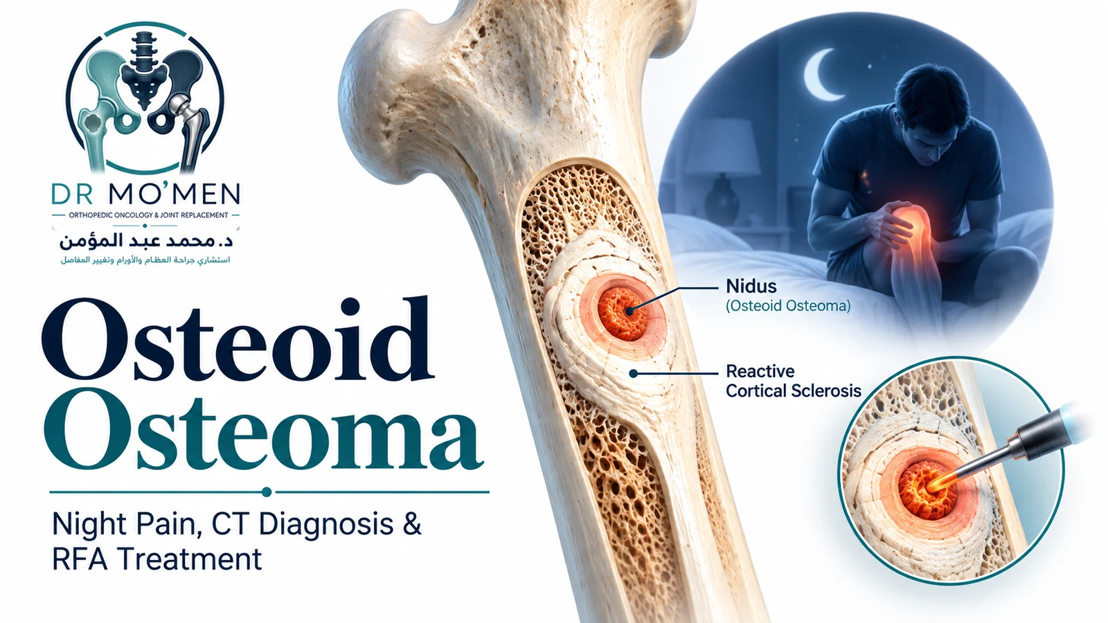
An osteoid osteoma is a small, benign bone-forming tumor that most often affects children, adolescents, and young adults. It commonly causes a deep, localized ache that becomes more noticeable at night and often improves markedly with nonsteroidal anti-inflammatory drugs (NSAIDs). The lesion contains a small central focus called the nidus, usually surrounded by reactive cortical sclerosis.
Key Takeaways
- Osteoid osteoma is benign and does not metastasize.
- The typical pain pattern is a localized ache that is worse at night and often improves substantially with NSAIDs.
- The femur and tibia are common sites, but the lesion can occur in other bones, including the posterior elements of the spine.
- Plain X-rays may show marked reactive sclerosis; thin-slice CT usually shows the small nidus more clearly.
- MRI is useful for marrow and soft-tissue reaction but can sometimes make the lesion look more aggressive because of extensive edema.
- Observation with medically supervised NSAID treatment is reasonable in selected patients; image-guided ablation is commonly used when definitive treatment is desired or symptoms remain significant.
- Atypical imaging, uncertain diagnosis, unusual size, aggressive features, or a soft-tissue component should trigger specialist tumor review before ablation or surgery.
What Is an Osteoid Osteoma?
Osteoid osteoma is a benign osteoblastic tumor characterized by a small nidus composed of osteoid and woven bone within a vascular stroma, surrounded by a variable degree of reactive sclerosis. AAOS OrthoInfo describes these lesions as typically smaller than about 1.5 cm, although published definitions commonly discuss lesions under approximately 2 cm.
The precise cause is unknown. It is not considered the result of ordinary trauma, dietary deficiency, or infection. It is most common in younger patients and shows a male predominance.
A larger osteoid-producing lesion raises the differential diagnosis of osteoblastoma, but size alone should not be used as an automatic reclassification. Imaging pattern, location, biological behavior, and pathology all matter.
What Are the Typical Symptoms?
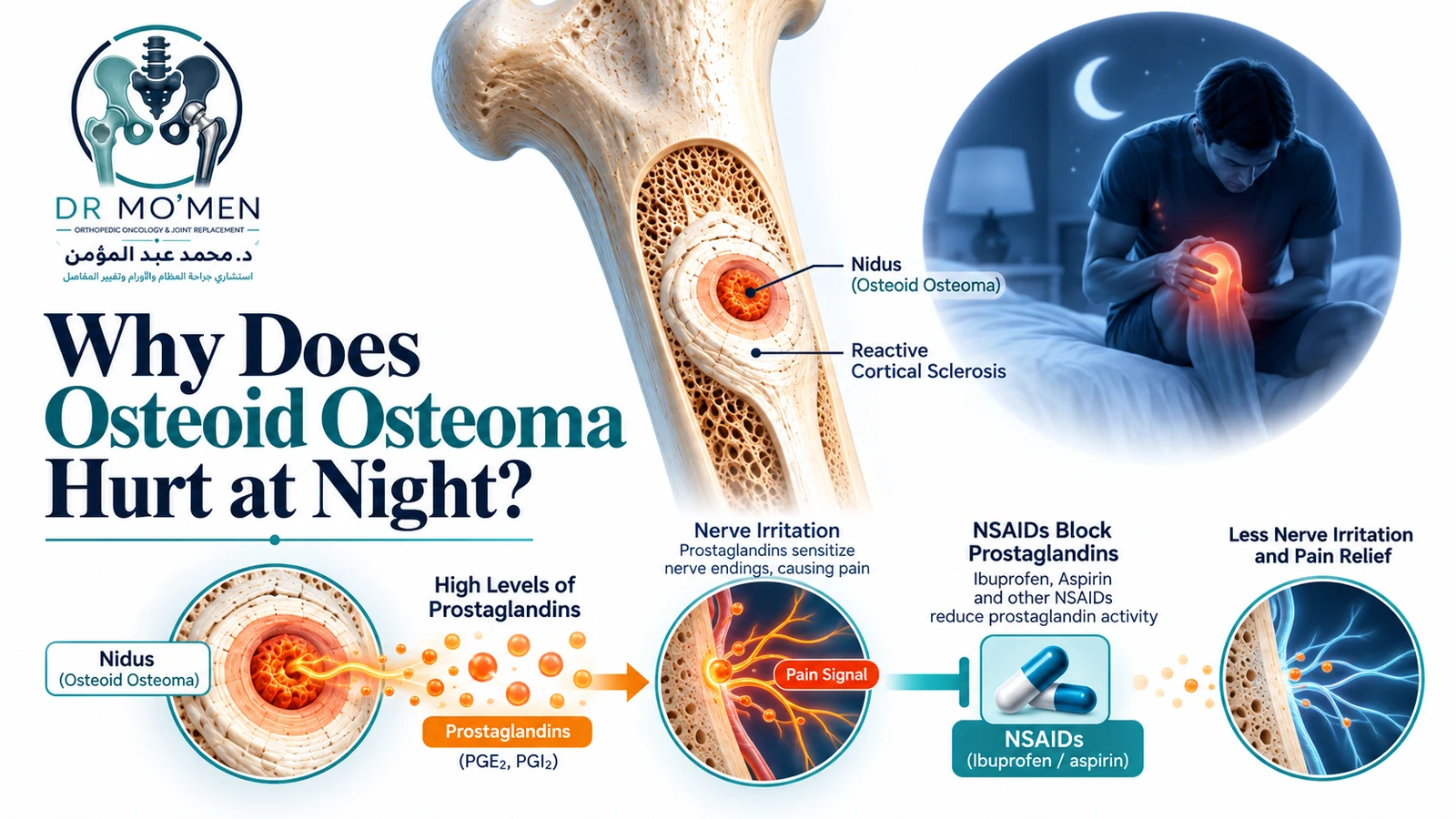
The clinical pattern can be highly suggestive. Patients often describe a deep, localized, aching pain that becomes worse at night and may interrupt sleep. The pain is usually not driven by activity alone. For a broader differential, see night bone pain causes and red flags.
- Night pain: often the dominant symptom.
- Marked NSAID response: pain may improve substantially after an anti-inflammatory such as ibuprofen or naproxen when medically appropriate.
- Localized tenderness or swelling: more noticeable in superficial lesions.
- Joint symptoms: intra-articular lesions, especially around the hip, may cause synovitis, stiffness, a limp, or reduced range of motion and can mimic inflammatory joint disease.
- Painful scoliosis: spinal osteoid osteoma may cause muscle spasm and a painful, relatively rigid curve.
How Is Osteoid Osteoma Diagnosed?
Diagnosis should combine the pain pattern, physical examination, and imaging. The main goal is to identify the nidus and make sure the imaging behavior is compatible with a benign osteoid osteoma rather than infection, stress injury, osteoblastoma, or an aggressive tumor.
1. Plain X-rays
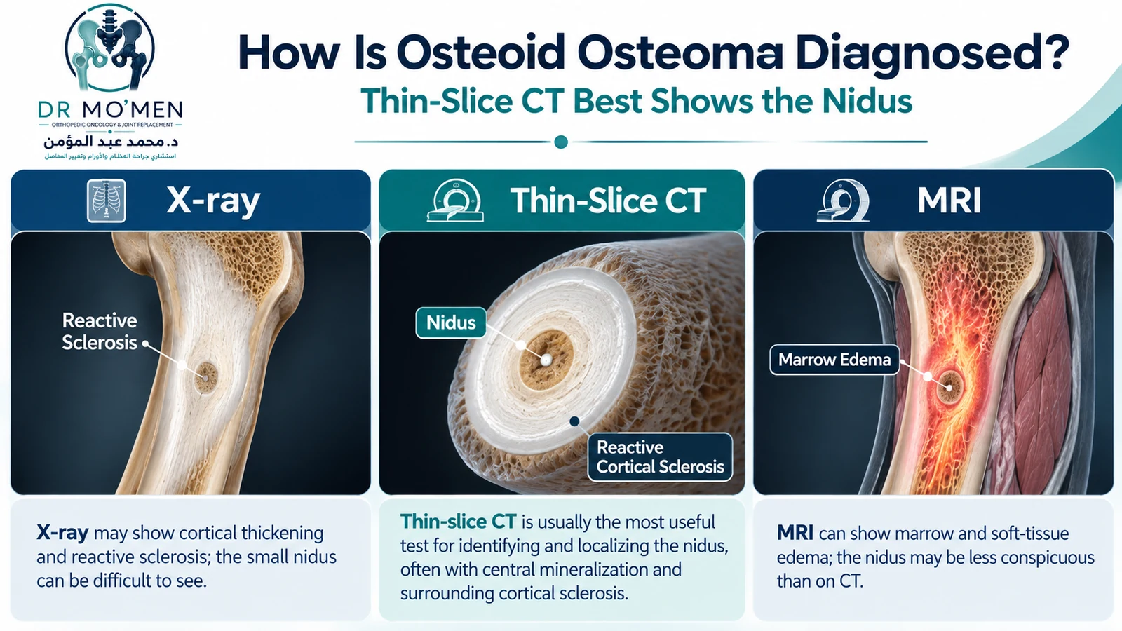
X-rays are an appropriate first test. A cortical lesion may show dense reactive sclerosis and cortical thickening around a small lucent focus. The reactive bone can sometimes be much more conspicuous than the nidus itself.
2. Thin-Slice CT
Thin-slice CT is usually the most useful imaging test for identifying and localizing the nidus, particularly in cortical lesions or anatomically complex regions. The nidus may be round or oval and can contain central mineralization.
3. MRI
MRI can demonstrate marrow edema, synovitis, and soft-tissue inflammatory reaction. These secondary changes may be striking and can occasionally obscure the nidus or make a benign lesion appear more aggressive. MRI findings should therefore be correlated with radiographs and CT when osteoid osteoma is suspected. For the broader role of imaging in bone tumors, see X-ray vs MRI for bone tumors.
4. Bone Scintigraphy / SPECT-CT
When localization is difficult, bone scintigraphy or SPECT/CT can show intense focal uptake. The classic “double-density sign” is a supportive imaging feature, but it should be interpreted with anatomical imaging rather than treated as a stand-alone diagnosis.
When Is Biopsy Needed?
A classic osteoid osteoma can often be diagnosed from the clinical picture and characteristic imaging, so biopsy is not mandatory in every patient. Biopsy becomes more important when the lesion is atypical, the diagnosis is uncertain, imaging and symptoms do not match, or another tumor or infection remains a realistic possibility.
What Conditions Can Mimic Osteoid Osteoma?
| Condition | Clues that may help distinguish it |
|---|---|
| Stress fracture | Linear fracture pattern, periosteal/callus response, and a history or biomechanics consistent with repetitive loading rather than a discrete round nidus. |
| Brodie abscess / subacute osteomyelitis | Clinical context and imaging may show an abscess cavity, serpiginous margin or other infection features; inflammatory markers can be normal, so normal ESR/CRP does not exclude it. |
| Osteoblastoma | Often larger, may be more expansile, and is more common in the spine; distinction uses imaging, behavior and pathology rather than size alone. |
| Inflammatory or intra-articular disease | Hip or joint-adjacent osteoid osteoma can cause synovitis and stiffness and may mimic juvenile inflammatory disease. |
| Aggressive bone tumor | Atypical size, ill-defined destruction, a substantial soft-tissue mass, unexpected progression or discordant symptoms should prompt specialist tumor assessment. |
Treatment Options
Treatment is individualized according to pain severity, lesion location, patient age, imaging certainty, access to image-guided treatment, medication tolerance, and patient preference.
1. Observation and NSAID-Based Symptom Control
Osteoid osteoma is self-limiting in many patients, and symptoms may eventually resolve over years. For a patient with a classic diagnosis whose pain is acceptably controlled and who can use NSAIDs safely, supervised conservative management is a reasonable option.
Long-term daily NSAID use is not appropriate for everyone. Gastrointestinal bleeding, kidney injury, asthma sensitivity, medication interactions, and other contraindications must be considered.
2. CT-Guided Radiofrequency Ablation (RFA)
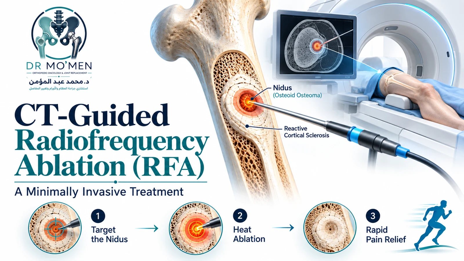
For a symptomatic lesion that is suitable for percutaneous access, CT-guided RFA is one of the most widely used minimally invasive definitive treatments. A probe is placed accurately into the nidus under image guidance and thermal energy is used to ablate the target. Exact temperature and duration depend on the ablation system and institutional protocol.
Large clinical series and systematic reviews report first-treatment clinical success above 90% overall. A 2020 systematic review of 3,023 patients reported a primary treatment-failure rate of 8.3%, falling to 3.1% after repeat treatment in appropriate cases. Review the PubMed abstract.
The main practical advantages are a very small access site, limited disruption of normal bone, and relatively rapid return to daily activities in many cases. Risks include skin or soft-tissue thermal injury, infection, injury to nearby nerves or vessels, and treatment failure. Location matters.
3. Cryoablation and Other Image-Guided Techniques
Cryoablation is an alternative image-guided treatment used in selected centers. The choice between RFA, cryoablation, and other percutaneous techniques depends on lesion location, operator experience, surrounding structures, equipment, and the clinical situation. Recent published series continue to support CT-guided cryoablation as a viable selected option, but RFA has the broader historical evidence base.
4. Surgical Excision or Curettage
Open or minimally invasive surgical removal remains appropriate in selected cases, including diagnostic uncertainty requiring tissue, anatomy that is unsuitable for percutaneous ablation, structural problems requiring simultaneous reconstruction, or persistent symptoms after image-guided treatment.
Spinal lesions deserve special attention. RFA can be performed successfully in selected spinal osteoid osteomas, but proximity to neural structures may require protective techniques and specialist expertise. A 2023 systematic review and meta-analysis found high success with both RFA and surgery in spinal lesions; the choice remains anatomy- and expertise-dependent. PubMed. There is no universal 1–1.5 mm cutoff that determines treatment for every patient.
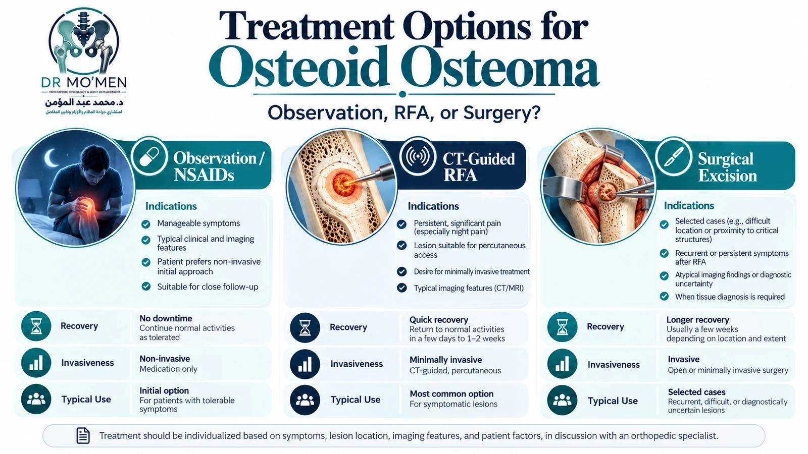
Treatment Comparison
| Feature | Observation / NSAIDs | CT-Guided RFA | Surgical Excision |
|---|---|---|---|
| Invasiveness | Non-procedural | Percutaneous, minimally invasive | Operative |
| Best suited to | Classic diagnosis with manageable symptoms and safe medication use | Symptomatic lesion suitable for percutaneous access | Selected difficult, recurrent, structurally complex or diagnostically uncertain lesions |
| Expected symptom course | Symptoms may remain controlled while the lesion resolves over time | Pain often improves rapidly after successful treatment | Pain improves after complete removal, with recovery depending on the operation |
| Recovery | No procedural recovery; medication monitoring may be needed | Often return to daily activity within days, but site-specific restrictions vary | Usually longer and dependent on the amount of bone/soft-tissue surgery |
| Main limitations | Years of symptoms or long-term medication exposure may be unacceptable | Thermal injury, recurrence/persistence, access limitations near critical structures | Larger incision, more bone disruption, wound and structural risks |
Recovery After RFA
After successful RFA, the characteristic night pain often improves within the first day or several days. Return to school, work, walking, gym activity, or sport should be individualized according to the treated bone, cortical access, symptoms, and the treating team’s instructions. A universal “full activity immediately” rule is not appropriate for every lesion.
When Should You Seek Specialist Assessment?
- Focal bone pain that repeatedly wakes you from sleep.
- Pain that consistently and markedly improves with an NSAID.
- Unexplained limp, persistent joint stiffness, or painful scoliosis in a child or young adult.
- Persistent localized tenderness with no clear injury.
- Imaging showing a small cortical lesion, unexplained sclerosis, or a suspected nidus.
- Atypical imaging, rapid change, a soft-tissue component, or uncertainty between tumor and infection.
Urgent Red Flags
Emergency or urgent assessment is appropriate when bone pain is accompanied by high fever or systemic illness, rapidly increasing swelling, inability to bear weight after an acute event, a new neurological deficit, or severe spinal pain with weakness, numbness, or bladder/bowel symptoms. These features are not typical of uncomplicated osteoid osteoma and require broader evaluation.
Dr. Mo’men’s Role in Complex or Unclear Cases
Dr. Mohammed Abdelmoemen Abuelhadid is a Lecturer of Orthopedic Surgery at Ain Shams University with a clinical focus on orthopedic oncology, limb salvage, bone and soft-tissue tumors, joint replacement, and complex orthopedic reconstruction. In a suspected osteoid osteoma, the priority is to confirm that the clinical pattern and imaging fit the diagnosis, determine whether conservative treatment is reasonable, and decide whether image-guided ablation, surgery, or further investigation is more appropriate.
For patients outside Cairo or Egypt, available X-rays, CT/MRI studies and reports can be reviewed as part of a preliminary second-opinion pathway before travel. Remote review does not replace examination when a physical assessment is needed.
Evidence & Sources
- AAOS OrthoInfo — Osteoid Osteoma
- Rosenthal DI et al. Radiology. 2003;229(1):171-175.
- Rimondi E et al. Eur Radiol. 2012;22(1):181-188.
- Tordjman M et al. CT-guided radiofrequency ablation for osteoid osteomas: a systematic review. Eur Radiol. 2020.
- Sagoo NS et al. Radiofrequency ablation for spinal osteoid osteoma: systematic review. Surg Oncol. 2022.
- Sangiorgio A et al. RFA vs surgical excision for spinal osteoid osteoma: systematic review and meta-analysis. Eur Spine J. 2023.
- Michailidis A et al. Cryoablation of osteoid osteomas. World J Radiol. 2024.
- Jaffe HL. Osteoid-osteoma. Arch Surg. 1935;31:709-728.
Related guide: Osteoblastoma: symptoms, diagnosis, biopsy and treatment explains how this benign osteogenic tumor differs from osteoid osteoma and how treatment is selected.