
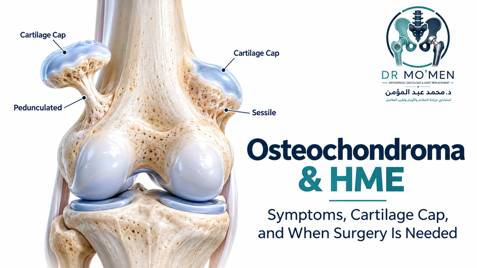
A hard, fixed bump near a child’s knee or shoulder can be worrying. One common explanation is an osteochondroma: a benign cartilage-capped outgrowth that develops near the growth plate. Most solitary osteochondromas never require surgery. The important questions are whether the imaging is typical, whether the lump is causing complications, and whether it has changed after skeletal maturity.
What Is an Osteochondroma?
An osteochondroma develops on the surface of bone, usually close to a growth plate, and consists of bone covered by cartilage. The defining imaging feature is continuity of both cortex and medullary cavity between the outgrowth and the underlying bone. Many lesions enlarge during childhood and stabilize after skeletal maturity; renewed growth in an adult warrants investigation.
Lesions may be pedunculated (on a stalk) or sessile (broad based). Common locations include the distal femur, proximal tibia and proximal humerus. Osteochondroma is widely described as the most common benign bone tumor; describing it as “not a true neoplasm” is unnecessarily categorical.
What Symptoms Can It Cause?
- A hard, immobile bump, often discovered by a parent or incidentally on an X-ray.
- Mechanical discomfort, tendon snapping, or an inflamed overlying bursa.
- Restricted movement or deformity when a lesion affects joint mechanics or growth.
- Tingling or weakness from nerve compression; proximal fibular lesions can occasionally affect foot dorsiflexion.
- Rare vascular complications, particularly around the knee.
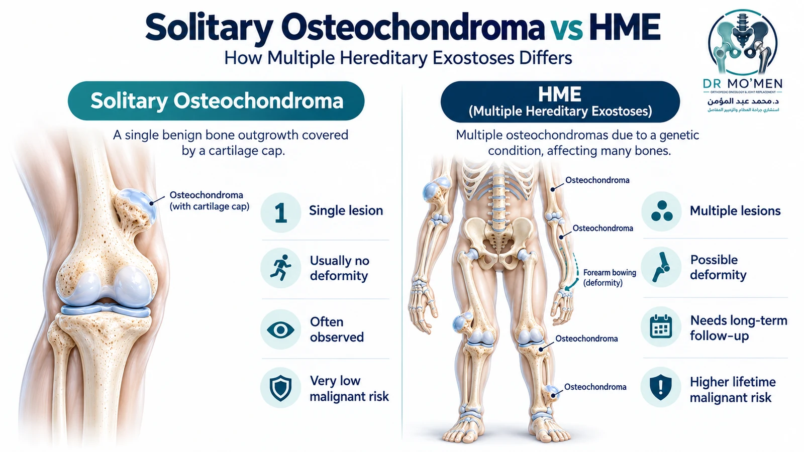
Solitary Osteochondroma vs Hereditary Multiple Osteochondromas (HMO/HME)
A solitary osteochondroma is usually an isolated finding. Hereditary multiple osteochondromas (HMO; also called HME/MHE) is generally an autosomal dominant disorder associated with pathogenic variants in EXT1 or EXT2. Multiple lesions can cause forearm or leg bowing, limb-length difference, restricted motion and reduced height. If a parent carries a causative variant, each child has a 50% chance of inheriting it.
Malignant transformation is very uncommon in a typical solitary lesion and is higher, but still uncommon, in HMO. The 2026 GeneReviews update cites a lifetime risk of approximately 2–10% in HMO; surveillance should therefore be individualized rather than assuming every patient needs the same annual MRI protocol. GeneReviews: Hereditary Multiple Osteochondromas.
Can Osteochondroma Turn Into Cancer?
Malignant transformation is rare in a solitary osteochondroma and more frequent in HMO. New growth after skeletal maturity, persistent deep pain or a new soft-tissue mass needs evaluation, usually including MRI. These symptoms can also have benign mechanical causes. If secondary peripheral chondrosarcoma is suspected, biopsy and definitive surgery should be planned by an orthopedic-oncology team—not by unplanned excision. See the chondrosarcoma guide.
How Is Osteochondroma Diagnosed?
X-ray: cortical and medullary continuity
Plain radiographs are often sufficient when the lesion looks characteristic and there are no concerning symptoms. The cortex and marrow of the osteochondroma should appear continuous with those of the parent bone. X-rays also help assess lesion direction, alignment and adjacent bone changes.
MRI: what does cartilage-cap thickness mean?
MRI shows the non-mineralized cartilage cap, an overlying bursa, nearby nerves and vessels, and suspicious soft-tissue change. It is particularly useful for unexplained pain, growth after skeletal maturity, deep or anatomically complex lesions and suspected malignant transformation. It is not automatically required for every classic, painless childhood osteochondroma.
Cartilage-cap thickness should be measured at the thickest point perpendicular to the osseous surface, without mistaking adjacent bursal fluid for cartilage. In a skeletally mature adult, a cartilage cap around or above 2 cm is an important suspicious finding in published imaging series, but it is not diagnostic by itself. Children may normally have thicker caps, so adult thresholds should not be applied uncritically. Bernard et al., Radiology 2010.
| Clinical situation | Interpretation | Usual next step |
|---|---|---|
| Typical painless lesion in a growing child | A relatively thick cartilage cap may reflect ongoing growth | Clinical assessment and observation; imaging according to circumstances |
| Stable, typical lesion in an adult | Small cap and no interval change are reassuring | No routine surgery solely because the bump exists |
| New pain or continued growth after maturity | Mechanical, vascular and neoplastic causes must be considered | Orthopedic-oncology review, often MRI |
| Adult cap around or above 2 cm or suspicious mass | Raises concern for secondary peripheral chondrosarcoma | MDT imaging review; planned biopsy when indicated |
No cartilage-cap threshold alone can confirm or exclude malignancy. Age, symptoms, anatomical site and the rest of the imaging pattern still matter.
Does Every Osteochondroma Need a Biopsy?
No. A classic benign osteochondroma can often be diagnosed without biopsy. If imaging is atypical or secondary peripheral chondrosarcoma is suspected, the case should be evaluated by an orthopedic-oncology/sarcoma team before any biopsy or operation. When biopsy is necessary, its trajectory must be planned in the context of any later definitive resection. Avoid unplanned local excision. Read more about planned bone-tumor biopsy.
When Is Surgery Appropriate?
Observation is the usual strategy for an asymptomatic solitary osteochondroma. Excision may be considered for persistent attributable mechanical symptoms, nerve or vessel impingement, loss of motion, symptomatic stalk fracture, progressive deformity, selected functional/cosmetic problems or suspicious change after maturity.
For a confidently benign lesion, surgery typically removes the bony outgrowth together with the cartilage cap and perichondrium while protecting nearby nerves, vessels and growth plates. A lesion suspicious for malignancy requires a different oncologic pathway; wide resection must not be assumed merely from a cartilage-cap measurement.
Documented Pediatric Proximal Humerus Case
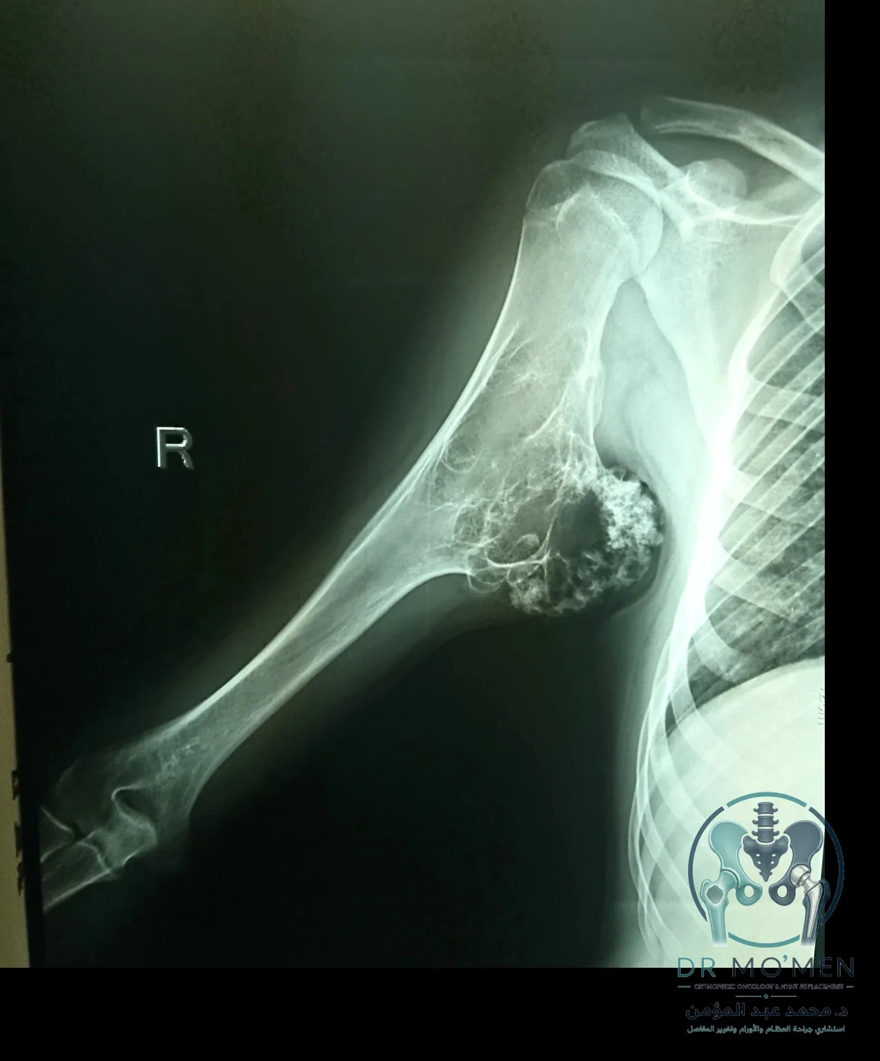
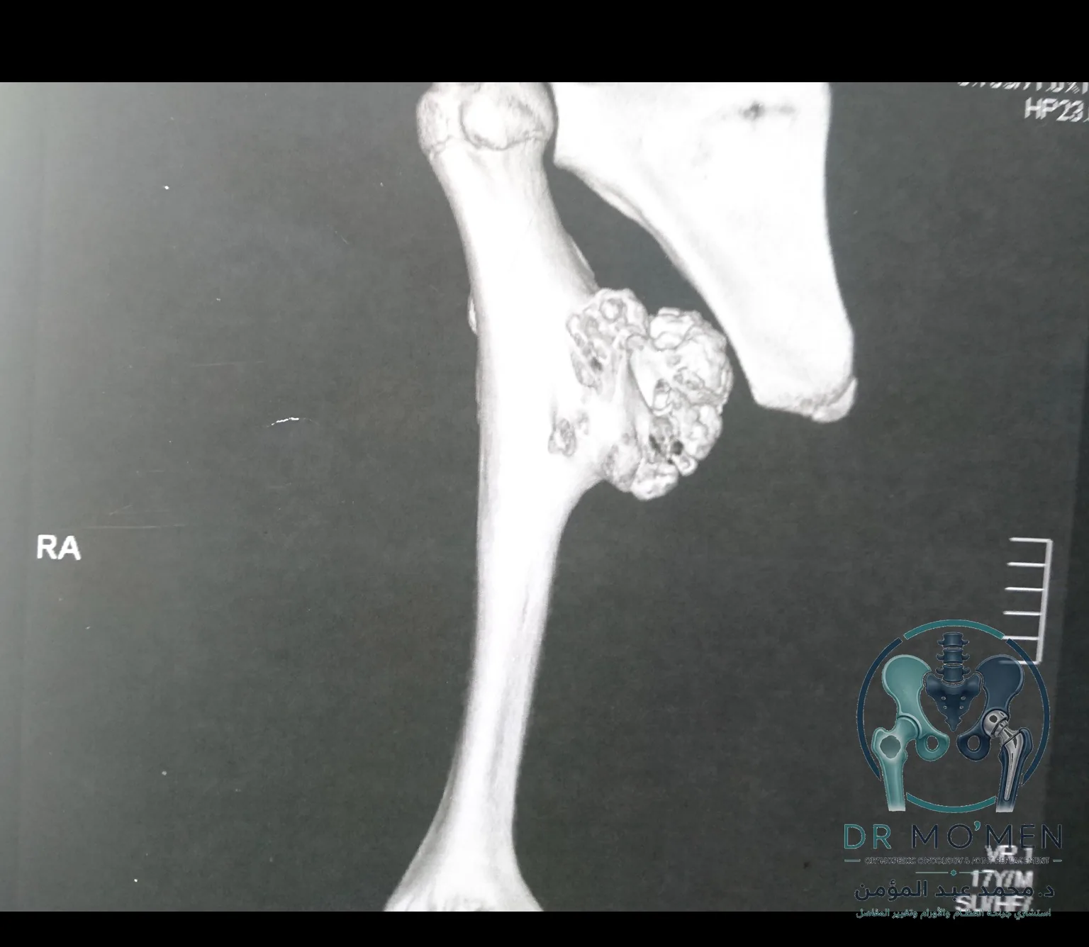
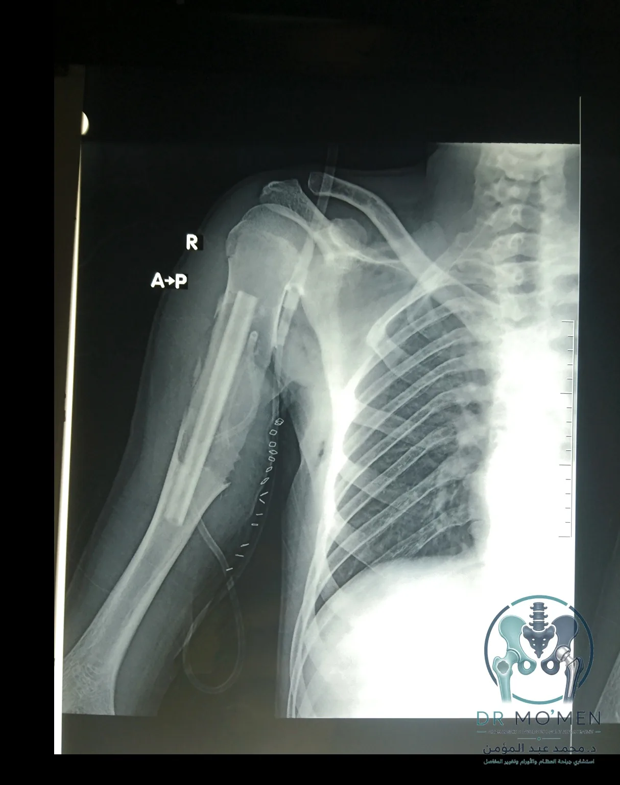
The MoemenOrtho Case Library includes a de-identified 12-year-old with a large medial proximal humerus osteochondroma. The anatomy required careful planning around nearby nerves and vessels. The published case includes real preoperative radiographs, 3D CT and postoperative radiographs with internal fixation. Histopathology details and long-term functional outcomes are not published there, so they should not be inferred. View the documented case.
What Happens After an Operation?
Rehabilitation and weight-bearing or activity restrictions depend on lesion location, defect size, whether fixation or reconstruction was required and the patient’s age. A small superficial excision and a large deep lesion near a neurovascular bundle have very different recovery needs. There is no reliable universal same-day discharge or fixed 2–4-week recovery promise for every case.
When Should You Seek Specialist Orthopedic-Oncology Assessment?
- A hard lump first noticed in a child when the diagnosis is uncertain.
- New or persistent pain, particularly after growth plates have closed.
- An enlarging lump in adulthood, unusual MRI report, or a pelvic/scapular lesion.
- More than one osteochondroma, limb deformity, or a family history of HME.
- Numbness, weakness, limited movement, or vascular symptoms.
A structured orthopedic-oncology review considers the original X-rays and MRI, symptoms, growth status, proximity to nerves and vessels, and whether observation, additional imaging, genetics input, planned biopsy or surgery is appropriate. Patients outside Egypt may use the second-opinion pathway for preliminary imaging and report review before travel.