What is chordoma, and where does it develop?
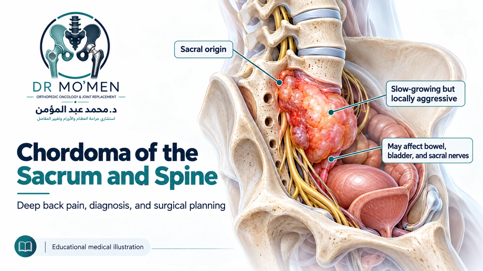
Chordoma arises from cells associated with the notochord, an embryonic structure involved in development of the spine. Conventional chordoma may grow relatively slowly, but it is malignant and can invade nearby bone, nerves and soft tissues or recur years after treatment. It occurs along the central axis, particularly the sacrum, skull base (clivus) and mobile spine.
Brachyury (TBXT) is an important pathological marker that can support the diagnosis. Imaging or a single tumor marker does not replace integrated pathology and radiology review.
What symptoms can a sacral or spinal chordoma cause?
Sacral chordoma: tailbone or pelvic symptoms
Symptoms may include persistent deep pain around the sacrum, buttocks or tailbone, pain while sitting, a gradually enlarging deep pelvic mass, altered sensation, constipation, changes in urinary emptying or sexual dysfunction. None of these symptoms alone diagnoses cancer.
Mobile-spine chordoma
Cervical, thoracic or lumbar chordoma can cause progressive localized pain, radiating symptoms, limb weakness, numbness or balance difficulty. Cervical tumors may occasionally affect swallowing depending on their location.
How is chordoma diagnosed?
1. MRI: define the tumor and neural relationships
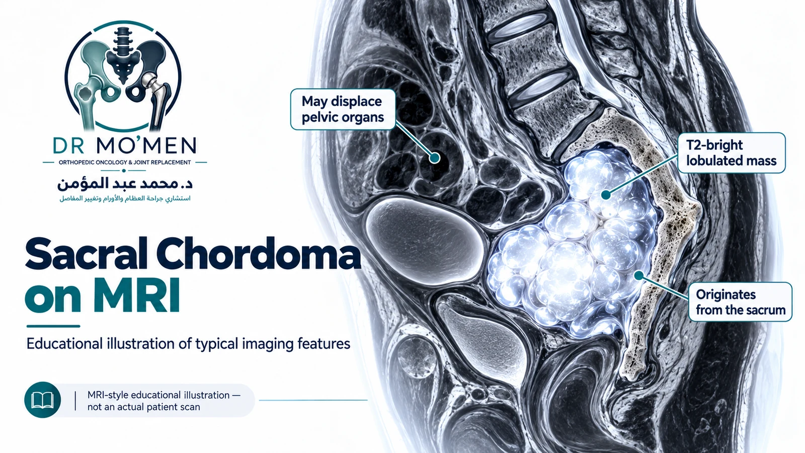
MRI is central for mapping tumor extent in bone, soft tissue and around sacral nerve roots or other neural structures. Chordoma often appears T2-hyperintense, but this is not specific enough to establish the diagnosis by itself.
2. CT and staging investigations
CT complements MRI by showing bone destruction, mineralization and anatomy relevant to biopsy or surgery. Staging may include chest and other cross-sectional imaging because chordoma can metastasize during its course. PET-CT is not mandatory for every patient.
3. Biopsy: why the route matters
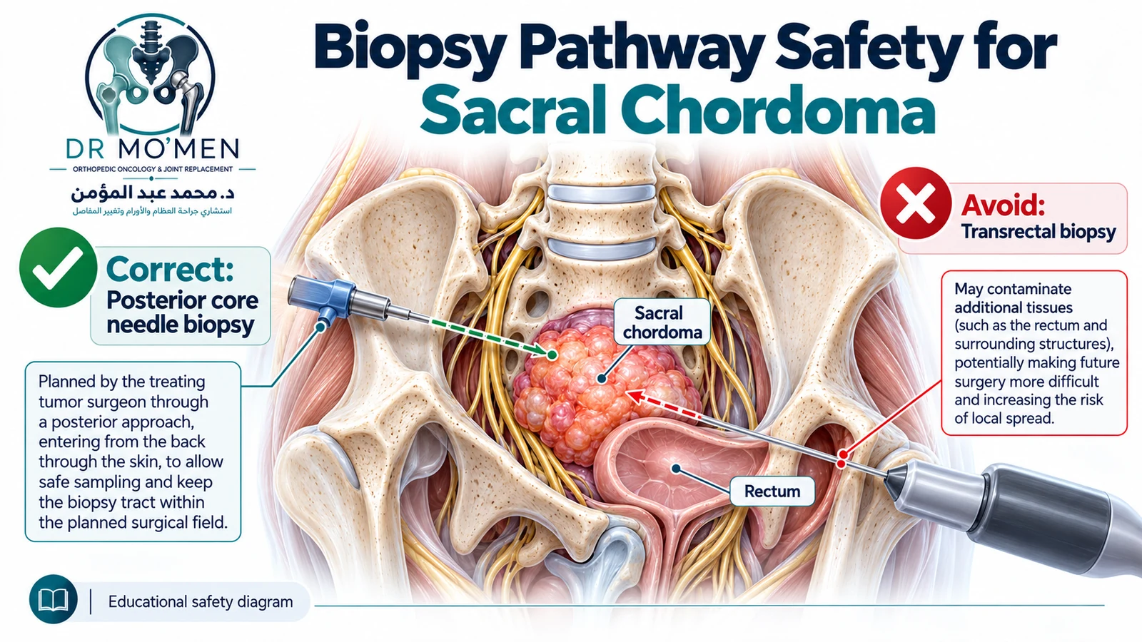
When tissue confirmation is required and the lesion can be sampled safely, a CT-guided core biopsy is commonly used for sacral and mobile-spine lesions. The orthopedic oncology/spine tumor team, radiologist and pathologist should agree on the route before the needle is placed. A posterior route that can be incorporated into later surgery is generally preferred. Transrectal or transvaginal approaches are generally avoided because they may contaminate otherwise uninvolved pelvic compartments.
Read more about planned bone-tumor biopsy and tract planning.
4. Expert pathology and imaging correlation
Diagnosis should be confirmed by a pathologist experienced in bone tumors using morphology and relevant immunohistochemistry, including brachyury where appropriate. Chondrosarcoma, giant cell tumor, plasma-cell tumors, metastasis and benign notochordal cell tumors may enter the differential. A biopsy result that does not fit the imaging should prompt specialist re-evaluation rather than unplanned excision.
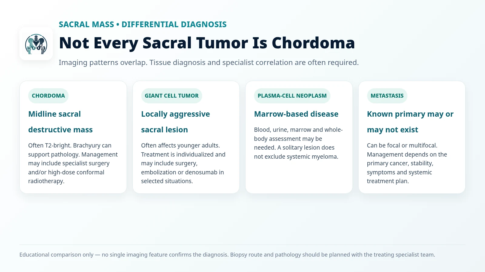
Chordoma versus other tumors of the sacrum
| Condition | Clues that may raise suspicion | Management principle |
|---|---|---|
| Chordoma | Midline sacral destructive mass; often T2-bright; brachyury can support pathology. | Specialist surgery and/or high-dose conformal radiotherapy. |
| Giant cell tumor of bone | Often younger adults; locally aggressive sacral lesion. | Individualized surgery, embolization and/or denosumab in selected situations. |
| Solitary plasmacytoma / myeloma | Marrow-based lesion; blood, urine and marrow testing may be needed. | Hematology assessment; radiation and/or systemic treatment according to diagnosis. |
| Metastatic disease | Known primary cancer may or may not be present; focal or multifocal lesions. | Identify the primary cancer and combine site-specific and systemic treatment as appropriate. |
Related guides: giant cell tumor of bone, solitary plasmacytoma, and pelvic bone tumor diagnosis and biopsy.
What are the treatment options for sacral and spinal chordoma?
En bloc resection: when is surgery appropriate?
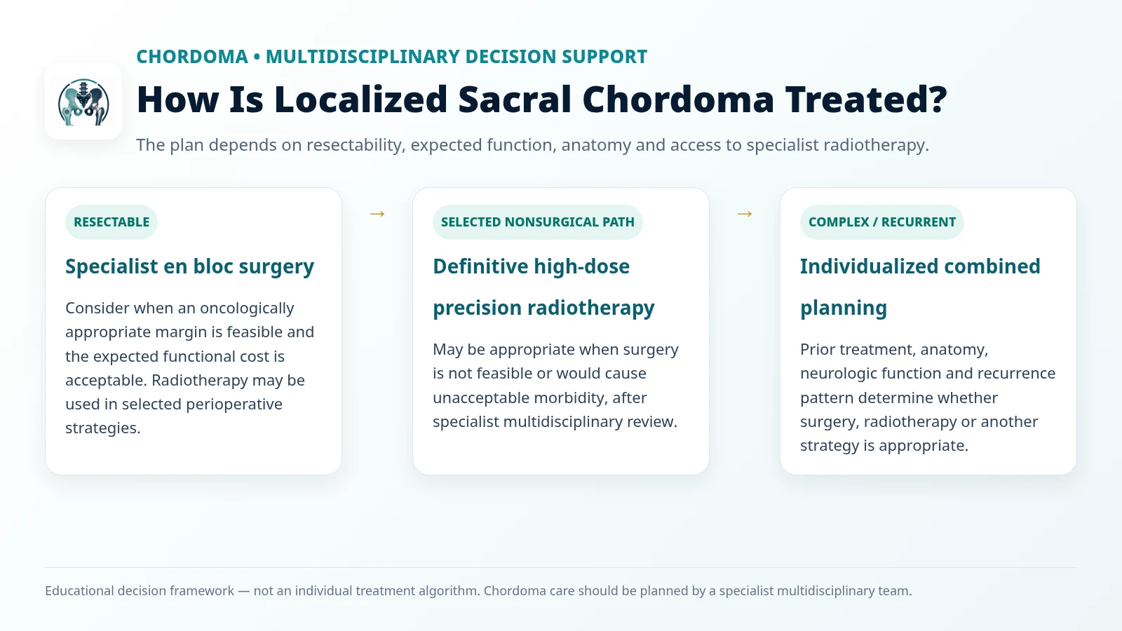
For many resectable sacral or mobile-spine chordomas, the surgical objective is removal in one piece with an oncologically appropriate margin when feasible. Piecemeal or intralesional surgery can increase local recurrence risk, but recurrence is not inevitable in every patient. The decision depends on tumor level, prior treatment, resectability, health status and the expected functional cost.
Partial or total sacrectomy and reconstruction
Higher sacral resections carry greater risk to sacral nerve function. Preserving bilateral S2 and especially S3 roots is associated with better bowel and bladder outcomes, but a named level cannot guarantee normal function. L5/S1 motor function is important for mobility. Some patients require lumbopelvic reconstruction, prolonged rehabilitation and bowel/bladder support; bowel diversion is not universally required.
Can radiotherapy replace surgery?
Yes, in selected patients. The 2026 global consensus recognizes definitive high-dose, highly conformal radiotherapy—including proton or carbon-ion approaches where appropriate—as a potential primary local treatment when surgery is unsuitable or would cause unacceptable functional loss. Highly conformal photon techniques may also be considered in selected settings. Radiotherapy may also be used around surgery according to the specialist plan.
Do chemotherapy or targeted drugs help?
Conventional chemotherapy has limited activity in most conventional chordomas and is not the usual first-line treatment for localized disease. Selected recurrent, unresectable or metastatic cases may be considered for targeted therapy, other systemic treatments or clinical trials through specialist oncology teams.
What are the major risks of sacral chordoma surgery?
Potential complications include nerve injury, bowel or bladder dysfunction, sexual dysfunction, blood loss, infection, wound-healing problems, vascular or visceral injury, cerebrospinal-fluid leak in some operations, pelvic instability and fixation failure. Functional and rehabilitation planning should begin before surgery.
Recovery, surveillance and long-term care
Recovery depends on the extent of surgery, neurological status, reconstruction and radiotherapy. Physiotherapy, occupational therapy, pain management, bowel/bladder care and psychological support may be required. Because chordoma can recur after a long interval, long-term risk-adapted clinical and imaging surveillance is important.
When should you request a specialist second opinion?
Specialist assessment is particularly useful before a planned biopsy, operation or radiotherapy course when MRI shows a suspicious sacral mass, the lesion lies near pelvic nerves/organs, or proposed treatment may permanently affect movement or continence. For patients outside Egypt, existing imaging and pathology can be reviewed before travel, but final recommendations require direct specialist and multidisciplinary assessment.
Evidence and further reading
- Radaelli S, Frezza AM, Fossati P, et al. Global Consensus on the Management of Primary Localized Chordoma. JAMA Oncology. 2026.
- Chordoma Foundation — Symptoms and diagnosis
- Chordoma Foundation — Surgery and treatment options
- Residual neurological function after sacral root resection during en-bloc sacrectomy: systematic review
- Bowel and bladder function after major sacral resection