
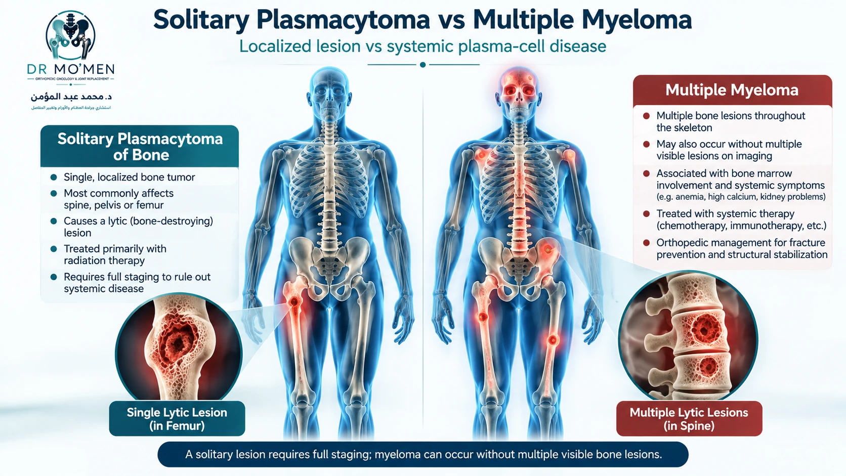
A painful hole in a bone on an X-ray can raise two immediate questions: Is the lesion confined to one site, and is the bone strong enough to avoid a fracture? A plasmacytoma and multiple myeloma both arise from abnormal plasma cells, but they require different staging and treatment pathways.
Quick answer: Solitary bone plasmacytoma is a biopsy-confirmed plasma-cell tumor at one skeletal site without myeloma-defining systemic disease. Multiple myeloma is a systemic plasma-cell disorder and can be diagnosed even before multiple bone lesions appear. Diagnosis combines lesion pathology, bone marrow testing, blood/urine studies, and appropriate whole-body imaging. Radiotherapy is central for solitary plasmacytoma; orthopedic surgery is considered when a bone is fractured, mechanically unstable, or threatening neural structures.
Key points: A solitary lesion does not establish solitary plasmacytoma until other disease is excluded. Not all lesions require orthopedic surgery. A painful weight-bearing bone or new neurological deficit warrants prompt assessment. Long-term hematology follow-up remains important after local radiotherapy.
What are solitary bone plasmacytoma and multiple myeloma?
Plasma cells are immune cells in bone marrow that normally make antibodies. In solitary bone plasmacytoma (SBP), clonal plasma cells form one tumor in bone. A related presentation, solitary plasmacytoma with minimal marrow involvement, has fewer than 10% clonal plasma cells outside the lesion on bone marrow assessment. Both require the exclusion of myeloma-defining events and additional sites of disease.
Multiple myeloma (MM) is a systemic plasma-cell malignancy. It may cause lytic bone lesions, fractures, anemia, kidney impairment and increased calcium, but a patient does not need to have all these findings or many visible lesions to meet diagnostic criteria. Hematologists also assess specific myeloma-defining biomarkers (the SLiM-CRAB framework).
The distinction cannot be made by lesion count on one X-ray alone. It requires pathology, whole-body staging and marrow investigations.
What symptoms should raise concern?
Persistent focal bone or back pain, pain during weight bearing, a fracture after minor trauma, loss of height related to vertebral collapse, and new nerve symptoms can prompt investigation. These features are not specific to plasma-cell disease; infection, osteoporosis, metastasis and other bone tumors may appear similarly. See bone tumor warning symptoms.
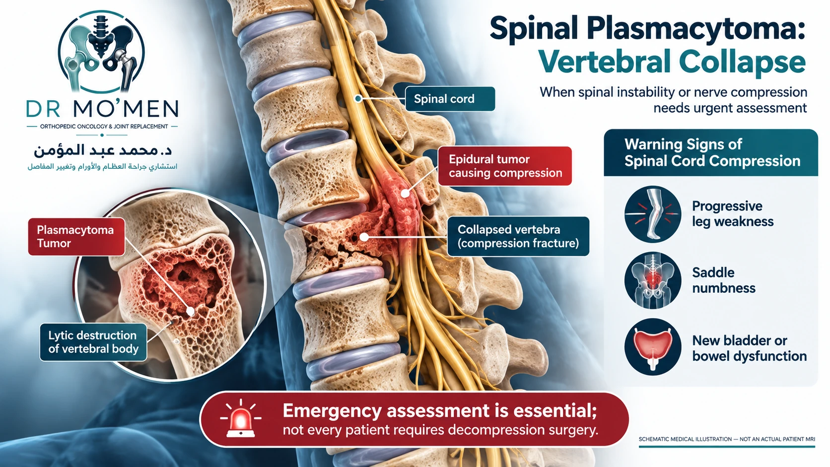
Urgent warning signs: New or progressive leg weakness, saddle numbness, urinary retention or new bladder/bowel dysfunction accompanying spinal pain needs emergency assessment for possible spinal cord or cauda equina compression. A sudden inability to bear weight with severe limb pain may represent a fracture and requires urgent care; avoid loading that limb until assessed.
How is the diagnosis established?
X-ray and CT: the shape and strength of the bone
Radiographs may show well-defined lytic (sometimes 'punched-out') lesions, but this classic appearance is more characteristic of myeloma than a universal appearance of solitary plasmacytoma. CT helps characterize cortical destruction, fracture configuration and fixation planning. Normal plain films do not exclude marrow disease.

MRI and FDG PET-CT: finding disease beyond the visible lesion
MRI is especially useful for marrow infiltration and assessing possible neural compression. Whole-body diffusion-weighted MRI and FDG PET-CT are recommended staging approaches; local MRI may also be needed for operative planning. These modalities may provide complementary information, but both tests are not automatically required in every patient. PET uptake alone should not be equated with an osteolytic lesion without appropriate anatomical correlation.
Lesion biopsy and separate bone marrow biopsy
An appropriately planned image-guided core needle biopsy of a suspicious bone lesion often confirms a clonal plasma-cell neoplasm using histology and immunophenotyping. The biopsy route should be planned with the orthopedic oncology and radiology team, particularly when the original differential includes a primary bone malignancy. Read when and how bone tumor biopsy is performed.
A separate iliac-crest marrow aspirate and trephine biopsy, ordinarily arranged by hematology, measures clonal plasma-cell involvement and helps distinguish isolated from systemic disease. One biopsy does not substitute for the other.
Blood and urine investigations
Initial work-up commonly includes CBC, calcium, creatinine and renal function; serum protein electrophoresis and immunofixation; quantitative immunoglobulins; serum free light chains; and appropriate urine studies including urine electrophoresis/immunofixation. Additional tests are individualized by hematology. A monoclonal protein result on its own does not establish active multiple myeloma.
Solitary plasmacytoma vs multiple myeloma: what is different?
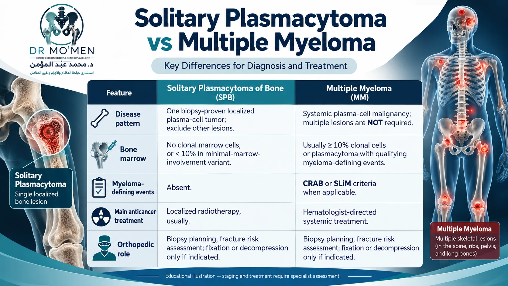
| Clinical feature | Solitary bone plasmacytoma | Multiple myeloma |
|---|---|---|
| Disease distribution | One biopsy-proven tumor; advanced imaging excludes other sites | Systemic plasma-cell disease; multiple visible bone lesions are not mandatory |
| Clonal plasma cells in uninvolved marrow | None, or minimal (<10%) in separately classified cases | Usually ≥10% marrow involvement, or biopsy-proven plasmacytoma with qualifying myeloma-defining criteria |
| Organ damage/biomarkers | No myeloma-defining events attributable to the disorder | At least one qualifying CRAB feature or myeloma-defining biomarker with appropriate clonal plasma-cell evidence |
| Main anticancer treatment | Local radiotherapy with curative intent for most patients | Hematology-led systemic treatment; regimen varies by fitness and disease features |
| Orthopedic treatment | Only when biopsy, instability, fracture or neural compression warrants it | Fixation/reconstruction/decompression when structurally or neurologically indicated |
CRAB describes hypercalcemia, renal impairment, anemia and bone disease attributable to the plasma-cell disorder. SLiM describes additional biomarkers: sufficiently high marrow plasma-cell percentage, markedly abnormal involved/uninvolved free-light-chain ratio with an absolute level requirement, and more than one focal MRI lesion of a qualifying size. Interpretation requires a hematologist, rather than self-diagnosis from a single abnormal value.
Which treatments are used, and when is orthopedic surgery needed?
Radiotherapy for solitary plasmacytoma
Definitive local radiotherapy is the standard main treatment for most confirmed solitary plasmacytomas. The 2026 IMWG recommendations describe radiotherapy as the treatment of choice, generally recommending 40–50 Gy in appropriately planned fractions. The radiation oncologist determines the prescription and field; selected circumstances warrant individualized discussion. Surgery is not routinely needed simply to remove a radiosensitive lesion.
Hematology-directed treatment for myeloma
Active multiple myeloma usually requires a systemic combination selected by hematology and oncology, potentially including targeted agents, immunomodulatory drugs, corticosteroids, antibodies and, in eligible patients, autologous stem-cell transplantation. Support for bone health often includes zoledronic acid or denosumab when indicated, with renal, calcium and dental precautions. The orthopedic procedure does not replace the systemic treatment.
Preventing or treating a pathological fracture
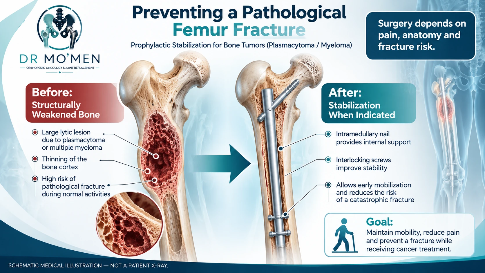
A lytic lesion may impair the structural integrity of the femur, humerus or another long bone. The decision for prophylactic fixation integrates functional pain, site, lesion size and location, cortical destruction, fracture pattern, expected treatment response and general health. Mirels score may contribute, but was developed for metastatic long-bone disease and is not a stand-alone validated myeloma prediction rule.
Depending on anatomy, options can include an intramedullary nail, plate fixation, cement augmentation, or reconstruction. Not every lesion with substantial cortical loss requires immediate fixation, and curettage is not a routine requirement before fixing a plasma-cell lesion. See the separate overview of impending pathological fracture and reconstructive options; the principles overlap, although myeloma is not a solid-tumor metastasis.
Vertebral collapse and cord compression
Spinal lesions may cause pain, instability and/or nerve compression. Suspected spinal cord compression is an emergency, but emergency evaluation does not necessarily mean emergency laminectomy for everyone: radiosensitive tumor without significant mechanical instability may be managed differently from a retropulsed bony fragment, unstable collapse or progressive neurological compromise. The spine surgery, hematology and radiation oncology teams coordinate urgent imaging and treatment.
Vertebral augmentation (such as kyphoplasty) can be considered for selected painful vertebral compression fractures; significant posterior wall defects, instability, neural compression and cement-leak risk alter suitability. Risks of surgery include infection, bleeding, thrombosis, fixation failure and the need for revision; risks of cement augmentation include cement leakage or adjacent fracture, depending on anatomy and context.
Will the bone heal, and what happens after treatment?
Effective treatment of the plasma-cell disease can control symptoms and may allow some bone repair, but cortical strength does not normalize immediately, and some defects remain mechanically important. Radiotherapy is not a substitute for fixation when the bone is unstable. Rehabilitation and weight-bearing instructions depend on the actual operation and imaging; there is no reliable universal 2–4-week recovery period.
The 2026 IMWG solitary plasmacytoma recommendations advise combined clinical, blood/urine and imaging follow-up. Advanced response imaging is generally assessed 6–9 months after radiotherapy, with subsequent monitoring individualized. The paper reports approximately half of solitary plasmacytoma patients progressing to symptomatic myeloma within five years across studied cohorts, but risk differs with marrow involvement and other factors. Regular hematology surveillance is therefore essential.
When should you seek specialist orthopedic oncology assessment?
Seek assessment when a bone lesion has uncertain pathology, when pain occurs with loading, when imaging suggests major cortical damage, or when a fracture is present or suspected. Before biopsy or fixation of an unexplained lesion, arranging coordinated review can avoid an unplanned procedure. A second opinion can help determine whether the bone needs structural surgery while hematology organizes disease-directed treatment.
For patients outside Egypt, reports and existing X-rays/MRI/PET-CT can be discussed before travel through the second-opinion pathway. Dr. Mohammed Abdelmoemen Abuelhadid evaluates the orthopedic oncology aspects, including biopsy planning, fracture prevention and reconstruction, in coordination with the hematology/radiation team when needed. See orthopedic oncology services.
For appointments or imaging review: +20 102 169 0693 (01021690693).
Evidence & Sources
- International Myeloma Working Group (IMWG), 2026 — Recommendations for the Diagnosis and Management of Solitary Plasmacytomas.
- International Myeloma Working Group — Diagnostic criteria for active multiple myeloma.
- NICE NG234 — Spinal metastases and metastatic spinal cord compression: emergency treatment principles.
- National Cancer Institute (NCI) PDQ — Plasma Cell Neoplasms (Including Multiple Myeloma) Treatment.
Frequently asked questions
Is solitary plasmacytoma of bone curable?
Radiotherapy may provide durable local control and is commonly delivered with curative intent. However, some patients later develop multiple myeloma, so long-term monitoring is important. The prognosis depends on staging and individual risk factors.
Does one bone lesion mean I do not have multiple myeloma?
No. Lesion count alone cannot establish the diagnosis. Hematology assesses bone marrow, CRAB features, myeloma-defining biomarkers and advanced imaging.
Are the tumor biopsy and the bone marrow biopsy the same test?
No. The lesion biopsy establishes the tumor type. A separate marrow sample assesses systemic plasma-cell involvement. Both may be required.
Can radiation alone prevent a weakened femur from breaking?
Radiotherapy treats radiosensitive tumor cells but does not immediately restore mechanical stability. Pain on weight bearing or significant cortical loss calls for an orthopedic fracture-risk assessment.
Does every myeloma-related vertebral fracture need surgery?
No. Treatment depends on pain, stability, neurological signs, imaging and the hematology/radiotherapy plan. New weakness or bladder/bowel dysfunction requires emergency assessment.
Medical disclaimer: This article is for patient education and does not replace an individual medical examination or review of imaging and investigations.