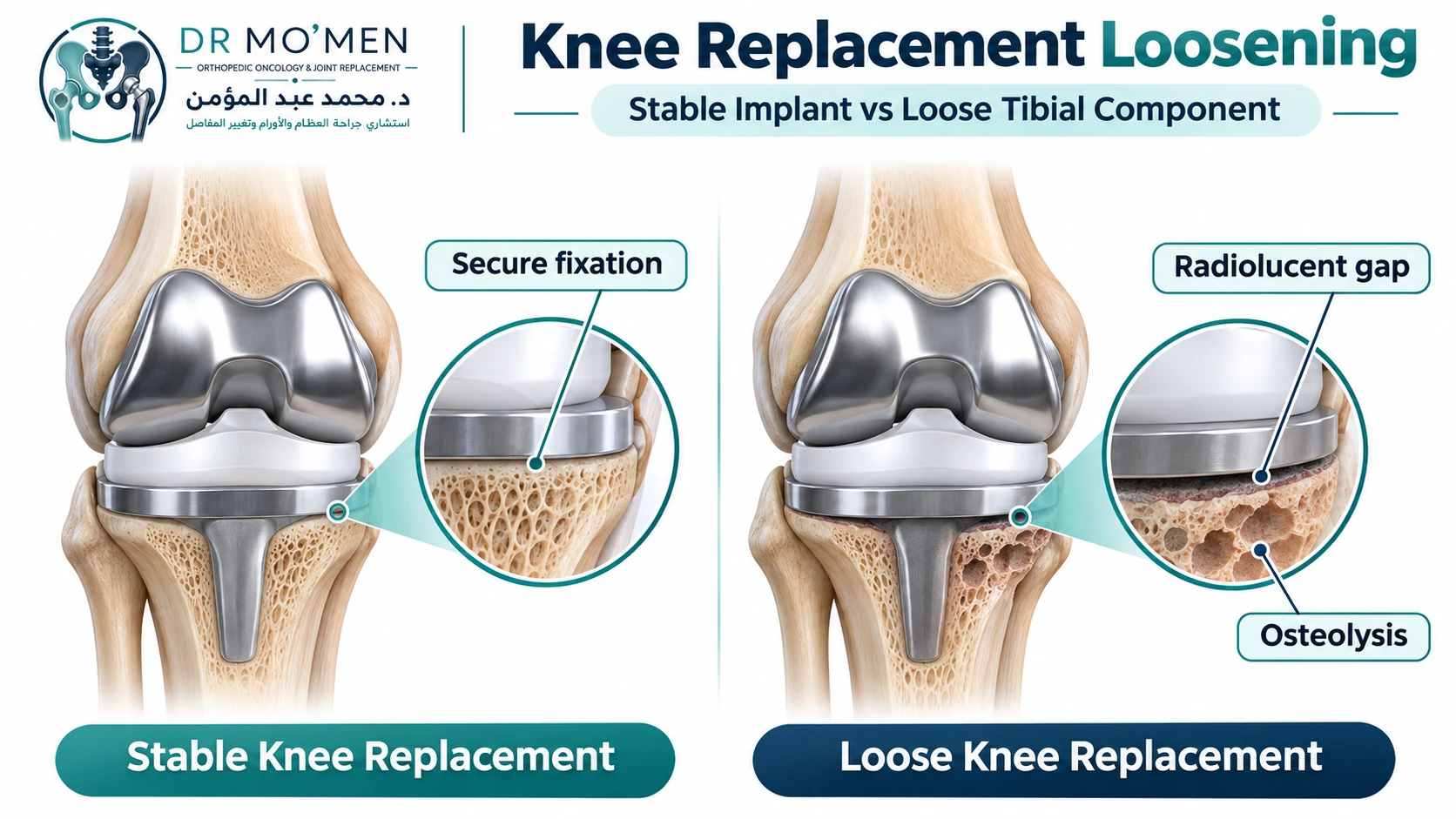
Pain after a knee replacement does not always mean that the implant is loose. However, new weight-bearing pain, recurrent swelling, progressive changes on X-rays, or a change in the position of the implant may indicate loosening and should be assessed by a surgeon experienced in revision knee replacement.
Knee replacement loosening occurs when one or more prosthetic components lose stable fixation to the supporting bone. The cause may be mechanical—known as aseptic loosening—or related to a periprosthetic joint infection. The distinction is critical because the diagnostic pathway and treatment are different.
No single symptom, blood test, or imaging finding should be interpreted in isolation.
Key Takeaways
- Weight-bearing pain is one of the most important clinical features associated with knee implant loosening.
- Progressive radiolucent lines, component migration, subsidence and radiolucency greater than 2 mm on serial X-rays or CT are important radiological findings supporting the diagnosis.
- Infection should be considered before labeling a failed knee replacement as purely aseptic.
- Comparing current X-rays with older postoperative films is often more informative than looking at one X-ray alone.
- Bone loss may progress while a loose implant continues to move, potentially making later revision surgery more complex.
- Revision surgery is individualized according to which components are loose, the amount of remaining bone, implant alignment and the condition of the collateral ligaments.
What Is Knee Replacement Loosening?
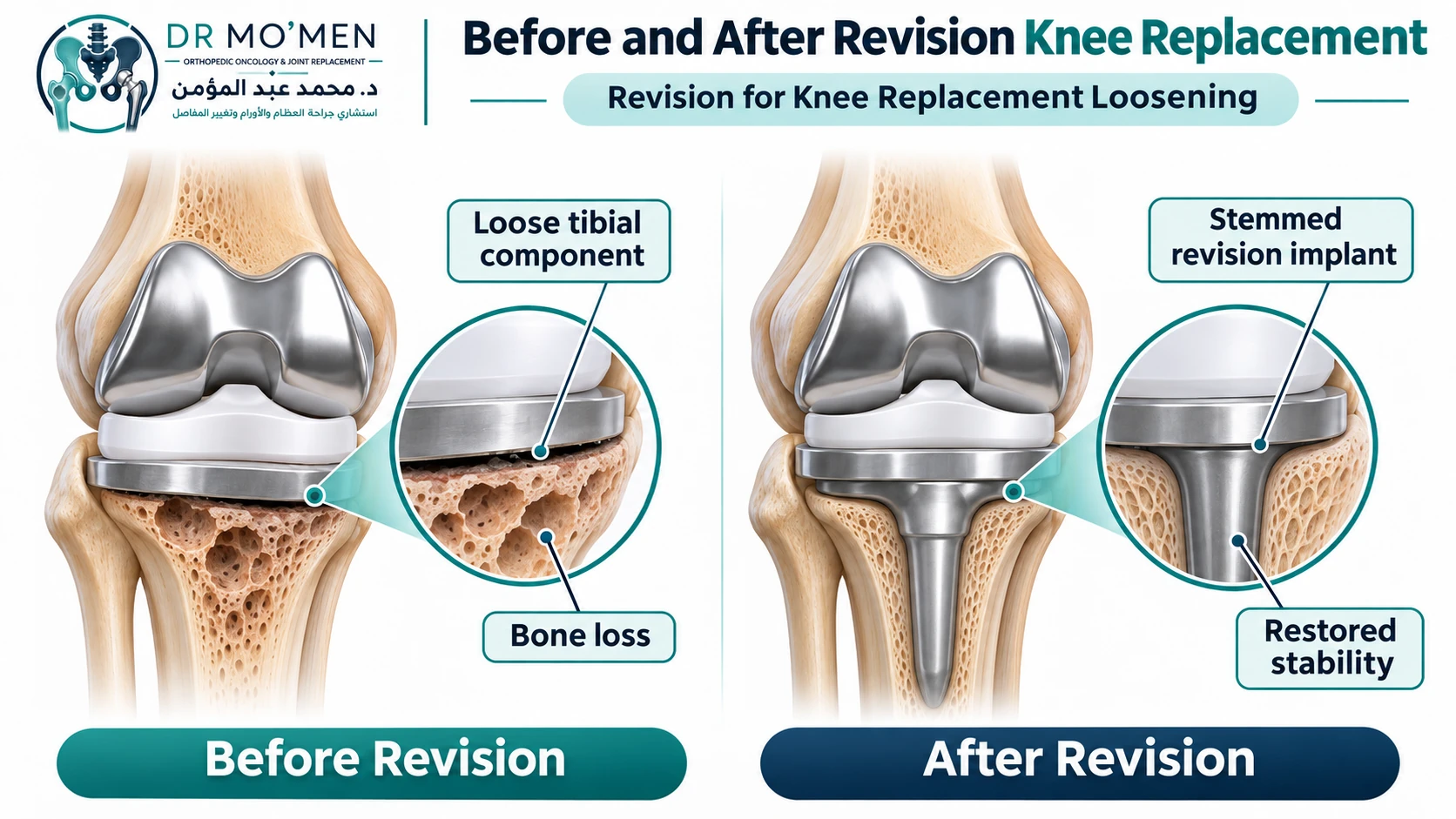
A knee replacement depends on stable fixation between the prosthetic components and the patient’s bone. Fixation may be achieved using bone cement or, in selected cementless implants, through biological bone ingrowth. When this fixation fails, the femoral or tibial component may develop microscopic or visible movement relative to the supporting bone. This process is called implant loosening.
Some patients develop loosening gradually over many years. Others develop earlier failure because of inadequate initial fixation, mechanical overload, malalignment, bone loss or infection. The tibial component is commonly involved, but either the tibial or femoral component—and occasionally both—may become loose.
What Causes a Knee Replacement to Become Loose?
Aseptic loosening
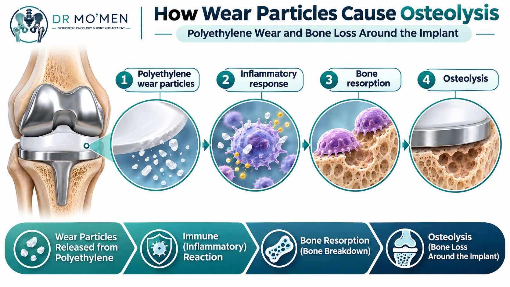
Aseptic loosening means loss of implant fixation without evidence of periprosthetic joint infection. It is usually multifactorial.
- Progressive mechanical failure at the bone–implant or bone–cement interface.
- Polyethylene wear particles that trigger an inflammatory response and periprosthetic osteolysis.
- Malalignment or abnormal load distribution across the prosthesis.
- Ligament imbalance that generates repeated abnormal forces.
- Poor bone stock or progressive bone loss.
- Failure of biological fixation in a cementless component.
Polyethylene wear is important, but not every case of aseptic loosening is caused primarily by polyethylene debris.
Septic loosening
A periprosthetic joint infection can also cause progressive destruction of bone around a knee replacement and loss of fixation. This is clinically important because a low-grade infection may present mainly as pain or loosening without obvious fever, redness or drainage. For this reason, infection should be actively considered during the evaluation of a painful or apparently loose knee replacement.
What Are the Symptoms of a Loose Knee Replacement?
Weight-bearing pain
Pain when standing or walking is particularly important. A 2025 international Delphi consensus among revision-knee specialists identified weight-bearing pain as the clinical criterion with the strongest consensus for implant loosening.
Start-up pain
Some patients describe pain during the first steps after standing from a chair or getting out of bed. Start-up pain may increase suspicion of mechanical loosening, particularly when accompanied by other clinical or radiological findings, but it should not be treated as a diagnostic sign on its own.
Recurrent swelling
Repeated joint effusions may develop because of mechanical irritation, polyethylene wear, synovitis or another underlying problem. Swelling is therefore not specific for loosening and infection must remain in the differential diagnosis.
Progressive loss of function
- Reduced walking distance.
- Pain on stairs.
- Increasing reliance on a cane or walker.
- Difficulty standing from a chair.
- Declining confidence in the operated knee.
Instability or giving way
A loose component can alter joint-line position and ligament tension. However, instability may also occur with completely well-fixed components. This is why knee replacement instability and implant loosening should be evaluated as related but separate diagnoses.
Change in limb alignment
Advanced component subsidence may lead to progressive varus or valgus deformity.
Which Symptoms Need Urgent Assessment?
Urgent orthopedic assessment is appropriate if a patient with a knee replacement develops:
- Sudden inability to bear weight.
- A new deformity after a fall or twisting injury.
- Fever with increasing knee pain or swelling.
- Marked warmth or spreading redness.
- Drainage from the surgical wound or an established sinus.
- Sudden severe mechanical locking or suspected implant failure.
These symptoms may indicate infection, periprosthetic fracture, major component failure, or another complication requiring prompt assessment.
How Is Knee Replacement Loosening Diagnosed?
The diagnosis is based on clinical findings, comparison of serial imaging, and exclusion of alternative causes of pain—particularly infection.
Step 1: Review the history and previous records
The timing and pattern of symptoms are important. The surgeon should determine:
- When the original knee replacement was performed.
- Whether the knee previously became pain-free.
- When the current pain started.
- Whether pain occurs predominantly during weight-bearing.
- Whether swelling or instability is present.
- Whether there has been a recent infection elsewhere in the body.
- Whether previous X-rays show progressive change.
The operative report and implant details can be useful, particularly if revision surgery is being considered.
Step 2: Rule out periprosthetic joint infection
A painful or loose knee replacement should not automatically be labeled aseptic. Screening commonly includes ESR (erythrocyte sedimentation rate) and CRP (C-reactive protein). Normal tests reduce the probability of infection but do not completely exclude every low-grade infection.
A 2025 systematic review and meta-analysis found that serum inflammatory markers do not have sufficient accuracy to reliably confirm or exclude PJI as stand-alone tests; synovial evaluation remains important when clinical suspicion persists. 2025 systematic review
If inflammatory markers are abnormal, or clinical suspicion remains significant, aspiration of the knee may be appropriate. Synovial-fluid assessment may include cell count, neutrophil percentage and microbiological cultures, with additional tests selected according to the clinical situation.
For the detailed infection pathway, see Infected Knee Replacement: DAIR, One-Stage or Two-Stage Revision?.
Step 3: Obtain standing radiographs and compare them with older films
Plain radiographs remain the first-line imaging study. Useful views may include standing AP, lateral and patellar/skyline views when relevant, plus long-leg alignment imaging in selected cases. The most important principle is comparison over time: a single radiograph may be inconclusive.
Which X-ray Findings Suggest Loosening?
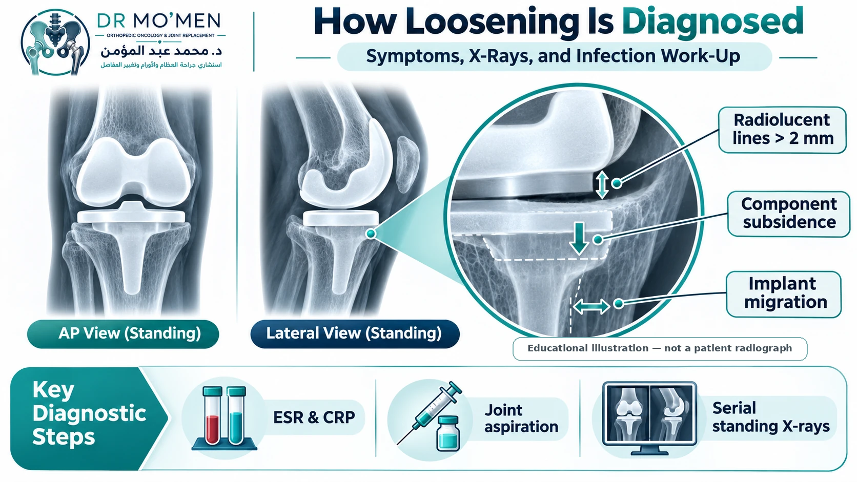
A 2025 international Delphi consensus among revision-knee specialists identified four major radiological findings supporting loosening:
1. Implant migration
A measurable change in component position compared with previous imaging is a strong sign of loss of fixation.
2. Progressive radiolucencies
Radiolucent lines that enlarge or progress on serial imaging are more concerning than a thin line that remains unchanged.
3. Component subsidence
A tibial or femoral component that gradually sinks into the supporting bone suggests failure of fixation.
4. Radiolucency greater than 2 mm
Radiolucency exceeding approximately 2 mm around the implant or bone–cement interface is an important finding, particularly when progressive or associated with migration or symptoms. These findings support the diagnosis but still need to be interpreted in clinical context.
Can a Knee Replacement Be Loose Even If the X-Ray Is Not Conclusive?
Yes. Early loosening or osteolysis can occasionally be difficult to define on standard radiographs because metal components obscure parts of the bone–implant interface. If symptoms remain concerning after appropriate infection assessment, additional imaging can be selected according to the clinical question.
CT with metal-artifact reduction
CT can help define osteolytic defects, bone stock, component position and rotation, subtle migration or subsidence, and fracture patterns. Modern CT protocols can reduce, although not completely eliminate, metallic artifact.
MRI with metal-artifact-reduction techniques
MRI may be useful in selected cases for assessing osteolysis, synovitis, soft tissues, the extensor mechanism and other causes of pain around the prosthetic knee.
Nuclear medicine imaging
Bone scintigraphy or SPECT/CT may occasionally provide additional information when conventional investigations remain inconclusive. Increased uptake is not specific for aseptic loosening and should not be interpreted without the clinical, laboratory and radiographic findings.
Aseptic Loosening vs. Infection vs. Instability
| Feature | Aseptic loosening | Periprosthetic joint infection | Instability |
|---|---|---|---|
| Typical pain | Frequently mechanical and weight-bearing | May be constant, mechanical or rest pain | Often associated with specific positions or activities |
| Swelling | May recur | May recur or persist | May occur but is less specific |
| Fever / redness | Usually absent | May be present but can be absent in chronic infection | Absent |
| ESR / CRP | Often normal | May be elevated; low-grade infection can have normal serum markers | Usually normal |
| Component fixation | May show migration, subsidence or progressive radiolucencies | May also cause loosening and bone loss | Components may remain well fixed |
| Key next question | How much fixation and bone stock remain? | Is infection present and what organism is involved? | Which ligament or gap is unstable? |
The categories may overlap; for example, infection can produce loosening, while a loose component may subsequently create instability.
Can a Loose Knee Replacement Heal Without Surgery?
A component that has genuinely lost mechanical fixation does not normally become securely fixed again simply through physiotherapy, medication or rest. However, not every radiolucent line requires surgery.
Observation may be reasonable when the patient has minimal or no symptoms, radiographic findings are stable rather than progressive, no meaningful component migration is present, or surgery carries disproportionate medical risk.
Conservative management may include activity modification, weight management, walking aids and physician-guided analgesia. These measures treat symptoms; they do not restore fixation of a genuinely loose component.
Intra-articular injections are not a treatment for mechanical loosening. Any proposed injection into a prosthetic knee should be discussed carefully with an arthroplasty specialist because of infection-related considerations.
When Is Revision Knee Replacement Needed?
Revision is generally considered when confirmed loosening is causing clinically important pain, progressive bone loss, component migration, instability, deformity or functional deterioration. The decision is not made from the X-ray report alone.
The surgeon assesses the patient’s symptoms and functional needs, infection status, which components are loose, implant alignment and rotation, remaining bone stock, ligament competence, the extensor mechanism and medical fitness for surgery.
For the broader decision pathway, see When Is Revision Knee Replacement Necessary?.
How Does Revision Surgery Treat a Loose Knee Replacement?
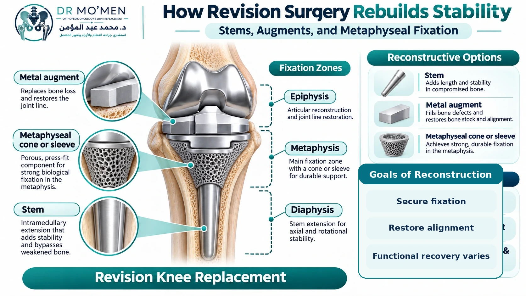
Revision knee replacement is more than simply removing one implant and inserting another. It is reconstructive surgery.
Can only the loose component be revised?
Sometimes. An isolated tibial or femoral revision may be considered when the opposite component is securely fixed, correctly aligned and rotated, undamaged, and compatible with the planned reconstruction. In other cases, revising both components gives the surgeon greater ability to restore alignment, joint line, flexion and extension gaps, and implant compatibility.
How is bone loss reconstructed?
The reconstruction depends on the location and size of the defect.
Stems: Stemmed components transfer part of the load into more reliable diaphyseal bone.
Metal augments: Blocks or wedges can restore localized missing bone and help re-establish joint position.
Metaphyseal cones or sleeves: Porous metaphyseal fixation may provide structural support when substantial cancellous bone has been lost.
Bone graft: Bone graft may be considered in selected defects.
The guiding principle is to obtain secure fixation in adequate remaining bone rather than relying on a severely damaged joint surface alone.
How is implant constraint selected?
Constraint depends primarily on ligament competence and the reconstruction. Options range from less-constrained revision designs to condylar-constrained knee implants when collateral-ligament support is inadequate but not completely absent, and rotating-hinge prostheses when severe ligament deficiency or major reconstruction requires linked mechanical stability.
A higher level of constraint is not automatically better; it should match the mechanical problem. See CCK vs Hinged Knee Replacement for a dedicated comparison.
What Is Recovery Like After Revision for Aseptic Loosening?
Recovery varies considerably more than after a routine primary knee replacement. Early mobilization is usually encouraged when medically and mechanically appropriate, but weight-bearing restrictions depend on the fixation obtained, severity of bone loss, use of bone graft, associated fracture, extensor-mechanism procedures and additional reconstructive techniques.
Physiotherapy typically focuses on regaining knee extension and flexion, quadriceps control, gait and functional independence. Meaningful recovery occurs over weeks to months rather than according to a single fixed timeline. Patients undergoing complex reconstruction may require a slower rehabilitation protocol.
What Should I Send for a Second Opinion About a Loose Knee Replacement?
A remote imaging review is more useful when the available information is complete. When possible, provide:
- Current standing knee X-rays.
- The earliest postoperative X-rays available for comparison.
- Recent CT or MRI images if already performed.
- The original operative report.
- Implant stickers, implant card or prosthesis details if available.
- ESR and CRP results.
- Aspiration and culture results if the knee has been aspirated.
- A short timeline describing when the pain began and whether it occurs during weight-bearing, at rest or at night.
For patients outside Egypt, reviewing these records before travel may help determine whether further investigations are required and whether revision surgery is likely to need specialist reconstruction. See the International Patients pathway or request a specialist second opinion.
When Should You Seek a Revision-Knee Specialist?
- Pain has returned after a previously well-functioning knee replacement.
- Serial X-rays suggest component migration or progressive radiolucencies.
- Significant osteolysis or bone loss is present.
- Infection has not been clearly excluded.
- There is uncertainty between loosening and instability.
- A first revision has already failed.
- Major bone reconstruction or a constrained/hinged implant may be required.
Dr. Mohammed Abdelmoemen Abuelhadid evaluates complex joint-replacement failures, revision arthroplasty and limb reconstruction at Ain Shams University and in specialist orthopedic practice in Cairo.