
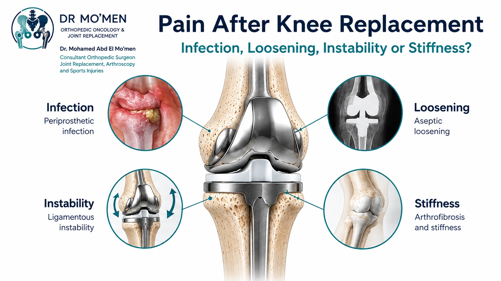
Pain and swelling are expected during the early recovery period after total knee replacement. A different problem exists when pain fails to improve progressively, returns after months or years of good function, becomes increasingly painful during weight bearing, or is associated with recurrent swelling, instability or progressive stiffness.
The key question is not simply how to suppress the pain. It is: Why is the knee replacement painful?
Recent systematic-review evidence identifies periprosthetic joint infection and aseptic loosening among the leading documented causes of revision TKA, with instability, stiffness and periprosthetic fracture also contributing to failure.[1]
For infection-specific treatment options, read: Infected Knee Replacement: DAIR vs One- or Two-Stage Revision
Is Pain After Knee Replacement Normal?
Some pain, swelling and difficulty with motion are expected early after surgery. The pattern becomes more concerning when pain does not improve, worsens after initial recovery, or returns after a previously well-functioning period.
Earlier assessment is appropriate when pain is associated with drainage, increasing redness, fever or chills, sudden severe swelling, marked new pain after trauma, or inability to bear weight. Severe calf swelling, chest pain or shortness of breath requires urgent medical assessment.
Important Causes of a Painful Knee Replacement
1. Periprosthetic Joint Infection
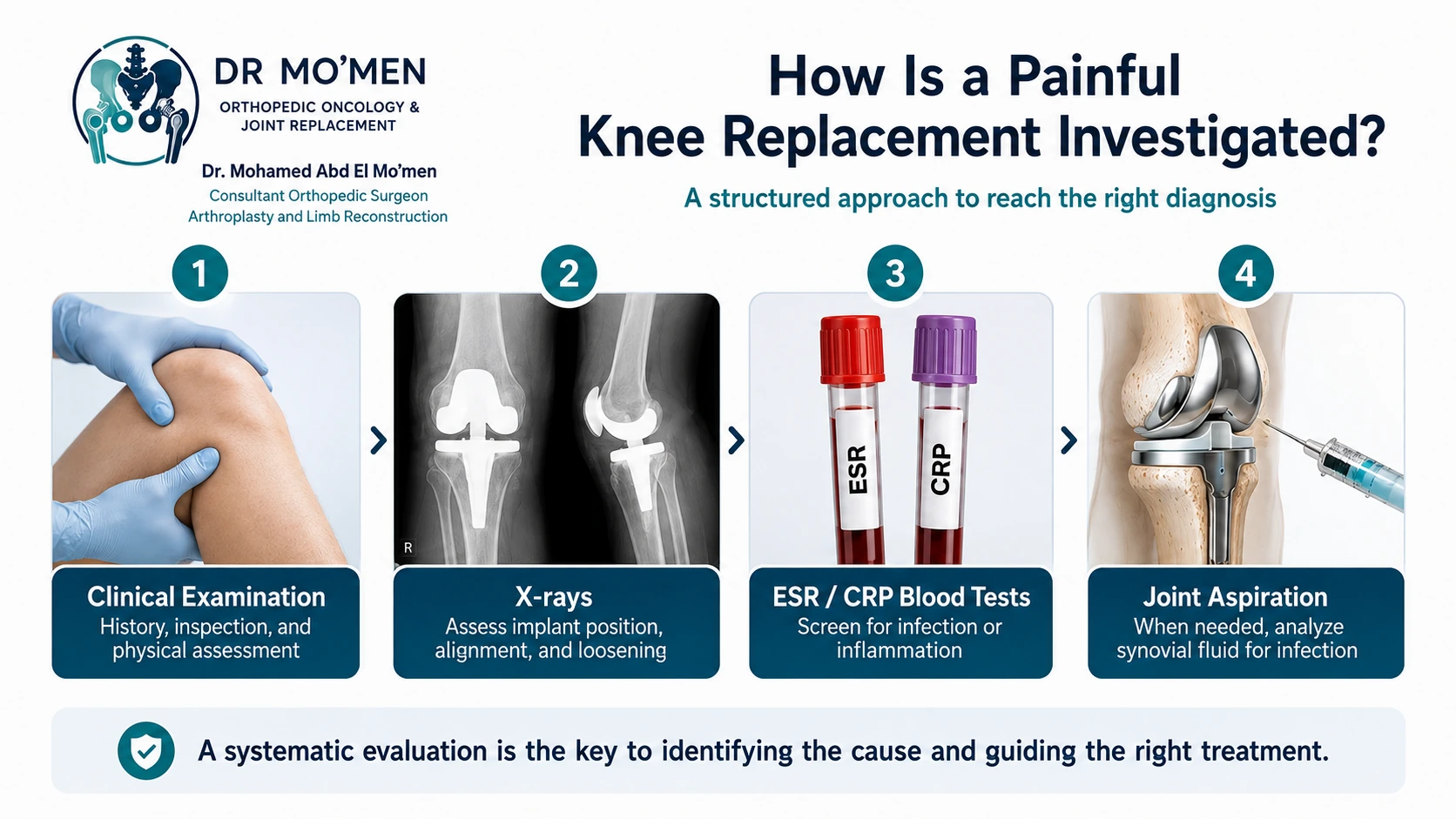
PJI can present with drainage, erythema, swelling, warmth and increasing pain, but chronic infection may be much less obvious and can sometimes present mainly as persistent pain.
When infection is suspected, evaluation may include ESR and CRP, followed when appropriate by joint aspiration for synovial white-cell analysis, differential, culture and other selected tests. AAOS guidance supports serum inflammatory markers and synovial-fluid assessment as important components of the diagnostic pathway.[2]
In a clinically stable patient, starting antibiotics before appropriate aspiration and cultures may reduce diagnostic yield. Patients with systemic illness or severe infection require urgent assessment and treatment.
2. Aseptic Loosening
A femoral or tibial component can gradually lose fixation to bone. Patients may notice progressive weight-bearing pain, pain when starting to walk, or worsening discomfort with prolonged activity.
Symptoms alone cannot establish loosening. Serial radiographs may show component migration, progressive radiolucent lines, subsidence or osteolysis. An international Delphi consensus among revision-knee specialists identified weight-bearing pain together with migration, progressive radiolucencies, radiolucencies over 2 mm and subsidence on radiographs or CT as important diagnostic criteria.[3]
Comparison with older postoperative radiographs can therefore be very valuable.
3. Instability
Patients may describe giving way, recurrent swelling, difficulty descending stairs, insecurity when rising from a chair or abnormal movement within the knee.
Possible causes include collateral ligament insufficiency, flexion-extension gap imbalance, component malposition or malrotation, polyethylene problems, and bone loss. Simply increasing implant constraint does not correct an unrecognized mechanical problem.
4. Stiffness and Arthrofibrosis
A painful stiff knee may have limited flexion, loss of full extension or both. Causes can include fibrosis, pain-limited rehabilitation, component malposition, infection or instability.
Arthrofibrosis should be considered only after important secondary causes have been excluded. A 2026 review emphasizes a diagnosis-of-exclusion approach and staged management, including rehabilitation, manipulation under anesthesia, adhesiolysis or revision in selected cases.[4]
5. Component Malalignment or Malrotation
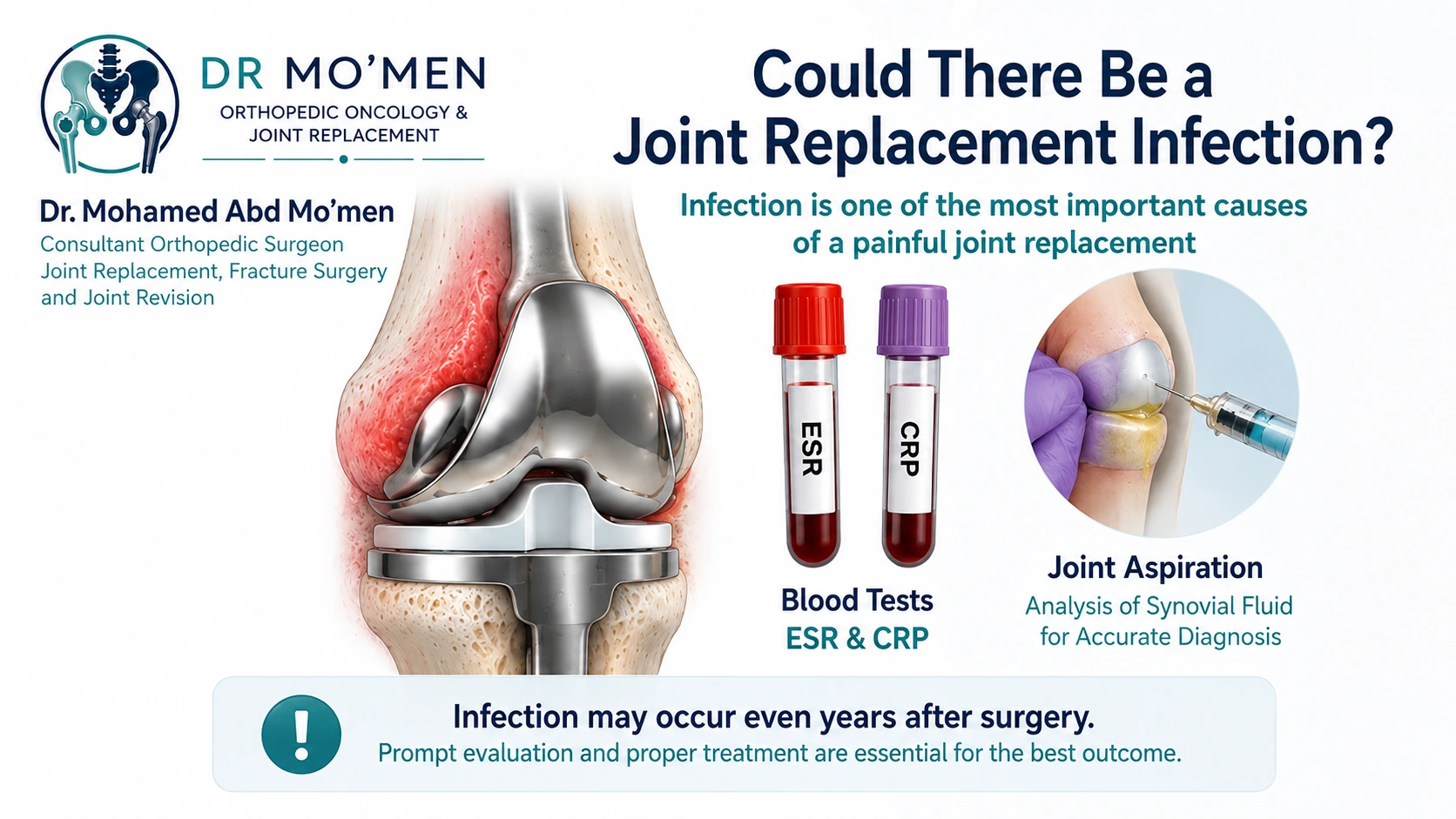
A prosthesis can remain fixed to bone yet still cause symptoms because of component alignment, rotation, joint-line problems, patellar tracking or soft-tissue imbalance.
Patients may experience anterior knee pain, stiffness, recurrent effusion or instability. CT can be useful in selected cases when component rotation or complex bone loss needs further assessment.
6. Patellofemoral Problems
Anterior knee pain can be related to patellar tracking, component position, soft-tissue balance, loosening, infection or other causes. It should not automatically be attributed to the kneecap alone.
7. Periprosthetic Fracture
A sudden increase in pain after a fall or injury, particularly with inability to bear weight, should raise concern for a fracture around the prosthesis.
Treatment may involve fracture fixation, revision of a loose component, or more extensive reconstruction depending on fracture pattern, implant fixation and remaining bone stock.
8. The Pain May Come From Somewhere Else
Not every painful knee replacement is caused by implant failure. Pain may originate from the hip, lumbar spine, peripheral nerves, tendons, soft tissues or other non-knee sources. Revision should not be undertaken simply because a prosthesis is present and the patient has pain.
Painful Knee Replacement: What Should Be Ruled Out First?
| Possible cause | Clues that may raise suspicion | What the work-up may focus on |
|---|---|---|
| Periprosthetic joint infection | Persistent or increasing pain, swelling, wound problems, systemic or laboratory concern | PJI assessment with inflammatory markers and aspiration when clinically indicated |
| Aseptic loosening | Weight-bearing pain, progressive radiolucency, migration or subsidence | Serial radiographs and fixation assessment; additional imaging selectively |
| Instability | Giving way, recurrent swelling, abnormal laxity | Ligament balance, component position and pattern of instability |
| Stiffness / arthrofibrosis | Restricted motion and functional limitation | Timing, scar restriction, component position and exclusion of infection or mechanical failure |
| Malalignment / malrotation / patellofemoral problem | Activity-specific pain, tracking symptoms or mechanical symptoms | Clinical alignment plus radiographs; CT may be useful for selected rotational questions |
| Extra-articular source | Pain pattern not explained by the knee reconstruction | Hip, spine, nerve, vascular or other causes when the clinical picture suggests them |
Does the Pain Pattern Help?
Pain pattern can provide clues but is not diagnostic on its own:
- Weight-bearing pain: may increase suspicion of mechanical failure or loosening.
- Giving way: raises concern for instability.
- Pain with severe restricted motion: requires a structured stiffness work-up.
- Pain with warmth or recurrent swelling: makes infection part of the differential.
- Anterior knee pain: may relate to patellofemoral mechanics, alignment or other causes.
How Is a Painful Knee Replacement Assessed?
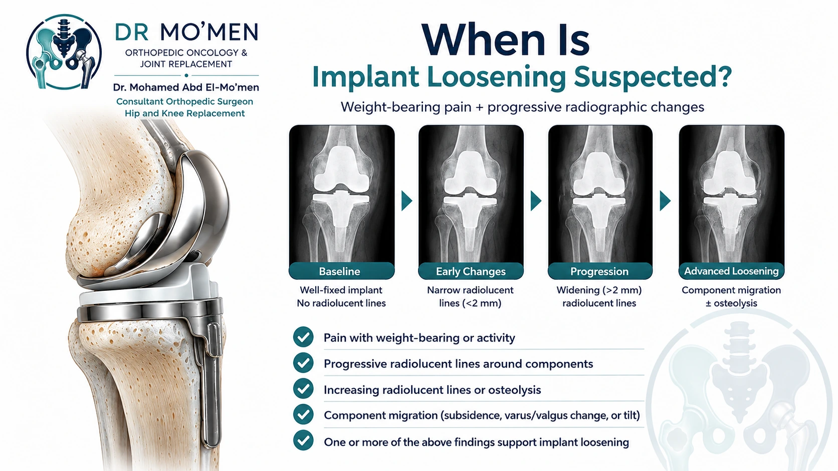
The evaluation begins with history and examination. Important questions include:
- When was the replacement performed?
- Was the knee ever significantly better after surgery?
- Is the pain related to weight bearing?
- Does the knee give way or swell repeatedly?
- Was there previous wound drainage or infection?
- Has there been trauma or a fall?
- Have there been multiple previous knee operations?
Examination should assess gait, alignment, range of motion, ligament stability, patellar tracking, the wound and soft tissues, with hip and spine assessment when appropriate.
Imaging and Laboratory Tests
X-rays
Standard radiographs remain central to evaluation and may include weight-bearing AP, lateral and patellofemoral views, plus full-length alignment imaging when indicated. Serial comparison can be more informative than a single study.
CT
CT may help when component malrotation, osteolysis, complex bone loss or fracture anatomy needs clarification. It is not required for every patient.
MRI
MRI is not usually the first imaging test for a painful knee prosthesis because metal can produce artifact, although modern metal-artifact-reduction techniques may help in selected situations.
Bone Scan or SPECT-CT
These tests can be useful in selected difficult cases but are not routine for every painful TKA. Nuclear-imaging findings must be interpreted in the clinical context and relative to the timing of arthroplasty.[5]
Does Every Painful Knee Need Revision Surgery?
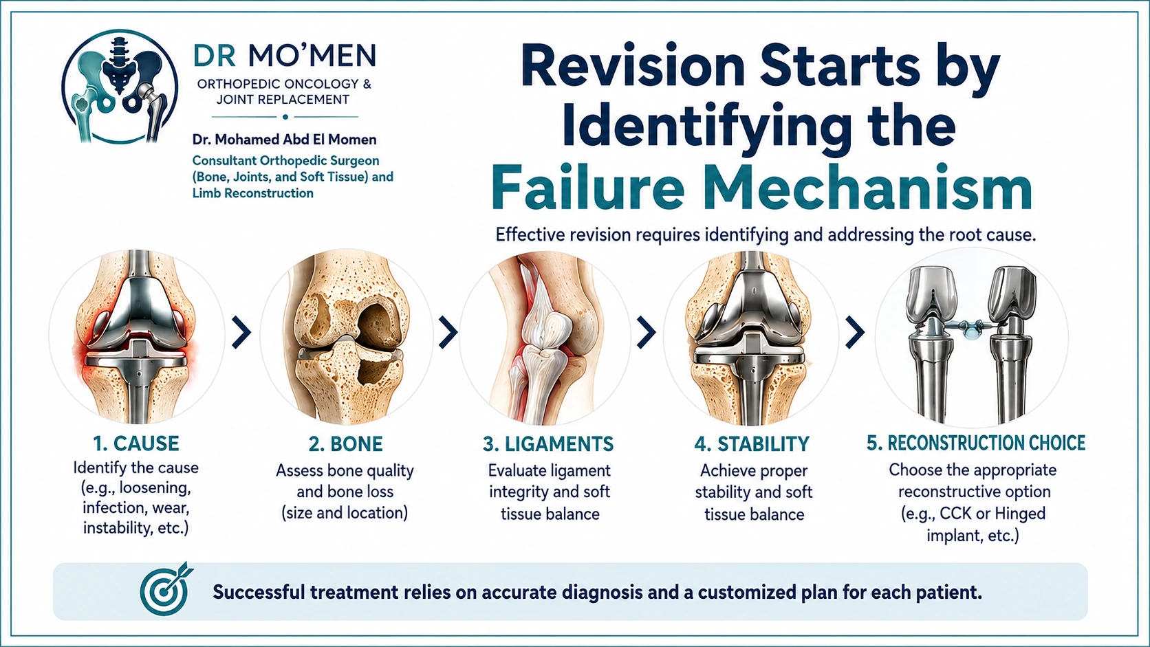
No. Revision should ideally target an identifiable and correctable failure mechanism.
- Infection: may require DAIR, single-stage or two-stage revision depending on timing, organism, implant fixation and soft-tissue condition.
- Loosening: may require revision of the loose component and reconstruction of bone loss.
- Instability: requires correction of the mechanical cause; selected patients may require CCK or rotating-hinge constraint.
- Stiffness: management depends on timing and cause and may include rehabilitation, MUA, adhesiolysis or revision.
- Periprosthetic fracture: treatment depends on fracture pattern, implant fixation and remaining bone stock.
The principle is: Diagnose the failure mechanism first, then treat that mechanism.
Where Do CCK and Rotating-Hinge Implants Fit?
They are tools for specific instability and reconstructive problems, not general treatments for unexplained pain. The degree of constraint should be selected according to ligament function, bone loss, instability pattern, fixation and the reason the previous replacement failed.
Related specialist article: CCK vs Hinged Knee Replacement: When Is More Constraint Needed?
When Should You Seek Specialist Review?
Revision-knee assessment is appropriate for persistent pain beyond expected recovery, new pain after a previously well-functioning knee, recurrent swelling, instability, progressive stiffness, suspected loosening, multiple previous operations or a proposed revision where a second opinion is desired.
Urgent assessment is warranted for wound drainage, rapidly progressive redness, systemic illness, sudden severe pain after trauma, inability to bear weight, severe calf swelling, chest pain or shortness of breath.
Role of Dr. Mo’men in Painful and Failed Knee Replacements
The evaluation starts with the failure mechanism, not the next implant. The central questions are: Is there infection? Is the implant fixed? Is the knee stable? Are alignment, bone stock and soft tissues adequate for function?
Assessment may include current and previous radiographs, operative reports, implant information, clinical stability testing, bone-loss assessment, infection investigations, joint aspiration when indicated, and CT for selected mechanical questions.
For appointments or imaging review: +20 102 1690693
Patients who already have a knee replacement should bring or send previous X-rays, the operative report, implant card if available, recent radiographs and relevant laboratory results.
International patients can use the second-opinion pathway for preliminary review before arranging travel to Egypt.
For the surgical decision and reconstruction options, read: When Is Revision Knee Replacement Necessary?
If pain is accompanied by persistent loss of motion, also see: Stiff Knee After Replacement: MUA, Arthrolysis or Revision
Medical References
- Datta S, et al. Why Are Primary Total Knee Arthroplasties Failing? A Systematic Review and Meta-Analysis. J Arthroplasty. 2025. PMID 40349863. https://pubmed.ncbi.nlm.nih.gov/40349863/
- American Academy of Orthopaedic Surgeons. Diagnosis and Prevention of Periprosthetic Joint Infections Clinical Practice Guideline. https://www.aaos.org/pji-cpg
- Buijs GS, et al. Weight-bearing pain and implant migration, progressive radiolucencies, radiolucency more than 2 mm and subsidence...: An international Delphi consensus study. Knee Surg Sports Traumatol Arthrosc. 2025. PMID 39126268. https://pubmed.ncbi.nlm.nih.gov/39126268/
- Hosanee S, et al. Reluctant knee: understanding and managing stiffness after total knee arthroplasty. Int Orthop. 2026. PMID 42342864. https://pubmed.ncbi.nlm.nih.gov/42342864/
- Diagnosis of loosening after knee arthroplasty. PMID 34694449. https://pubmed.ncbi.nlm.nih.gov/34694449/
This article is for patient education and does not replace clinical examination or review of appropriate imaging and investigations.