Key Takeaways
- This reconstruction replaces a large missing segment of the proximal femur; it is not simply a standard total hip replacement.
- Oncological clearance comes first. Reconstruction is selected only after the safe resection plan, neurovascular anatomy and remaining bone are defined.
- For metastatic disease, a megaprosthesis is not automatically required. Internal fixation may be preferable when adequate bone remains for durable stabilization.
- Hip stability depends on the bearing, remaining capsule, abductor mechanism and quality of soft-tissue reconstruction.
- Early mobilization is often possible because the reconstruction does not depend on fracture union, but weight-bearing and hip precautions must be individualized.
Background: megaprosthesis reconstruction after bone tumor resection.
What Conditions May Require a Proximal Femoral Megaprosthesis?
Related reading: fracture stabilization versus reconstruction in metastatic bone disease.
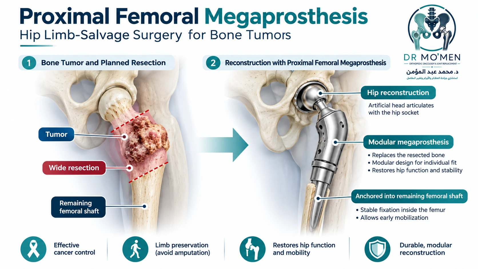
The proximal femur includes the femoral head and neck, greater and lesser trochanters, and the subtrochanteric region. A proximal femoral replacement is considered when disease or previous surgery has removed so much bone that a conventional implant, nail or plate cannot provide a dependable reconstruction.
1. Primary malignant bone tumors
Chondrosarcoma is an important indication when wide resection of the proximal femur is required. Osteosarcoma and Ewing sarcoma can also involve this region, although their anatomical distributions differ and treatment must follow histology-specific sarcoma protocols. Chemotherapy is integrated for chemosensitive tumors such as osteosarcoma and Ewing sarcoma, whereas conventional chondrosarcoma is managed primarily with adequate surgical resection when operable.
2. Selected metastatic bone disease and pathological fractures
The proximal femur is a common long-bone site for metastatic disease because of its high mechanical load. The key question is not simply whether a metastasis is present, but whether the remaining femoral head, neck, calcar and trochanteric bone can support a durable fixation. Extensive destruction, pathological collapse, selected solitary or oligometastatic lesions, or failed fixation may favor endoprosthetic replacement.
3. Severe non-oncological bone loss
Proximal femoral replacement can also be a salvage option in complex revision arthroplasty, including severe proximal femoral bone loss, selected Vancouver B3 periprosthetic fractures, recurrent reconstruction failure, or major bone loss after previous infection treatment. These cases often carry a higher complication burden than primary oncological reconstruction.
When Is a Megaprosthesis Chosen Over an Intramedullary Nail?
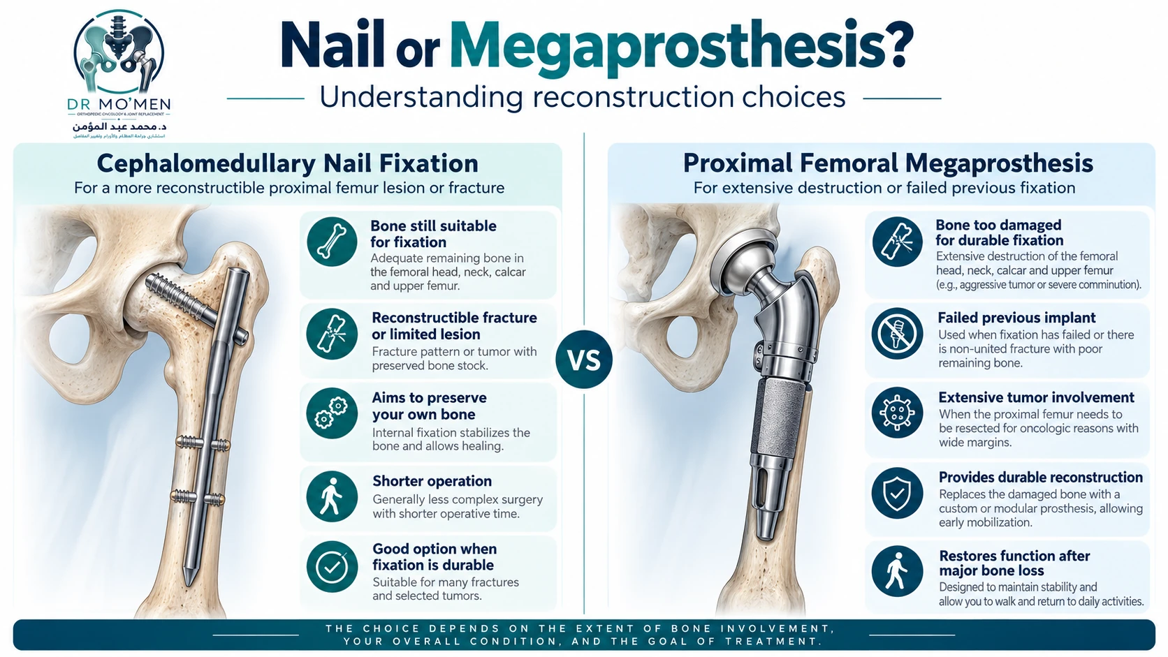
For metastatic or pathological fractures, the choice is individualized. A nail or plate may be appropriate when enough bone remains for a stable construct expected to last for the patient's clinical needs. A proximal femoral replacement may be appropriate when the head, neck, calcar or peritrochanteric bone is too damaged for reliable fixation, or when previous fixation has failed with major bone loss.
| Clinical factor | Fixation may be appropriate | Proximal femoral replacement may be appropriate |
|---|---|---|
| Femoral head / neck | Preserved | Extensively destroyed or not reconstructible |
| Calcar / trochanteric bone | Adequate for fixation | Severely deficient or unsalvageable |
| Remaining shaft | Stable nail or plate purchase possible | Large segmental defect must be bypassed |
| Existing fracture | Reconstructible | Pathological collapse or non-reconstructible fracture |
| Previous implant | Revision fixation remains feasible | Failed fixation with nonunion and major bone loss |
| Treatment goal | Preserve native bone with durable stabilization | Replace an unsalvageable segment with a reconstruction not dependent on union |
How Does Proximal Femoral Limb-Salvage Surgery Work?
Related reading: biopsy planning and surgery for pelvic bone tumors.
Biopsy planning and oncological resection
If the diagnosis is not already established, biopsy should be planned with the orthopedic oncology team so the tract can be removed with the definitive specimen. During tumor surgery, the prior biopsy tract is included in the resection. The safe bone-cut level is determined from preoperative imaging, and the tumor is removed without compromising the planned oncological margin. If joint contamination or acetabular involvement is suspected, the surgical strategy may need to change.
Reconstruction of length, offset and version
The remaining femoral canal is prepared for a cemented or uncemented modular stem according to bone quality and the reconstructive plan. Leg length, offset and femoral version are restored using pre-resection landmarks and intraoperative assessment; there is no single fixed version target that applies to every proximal femoral tumor reconstruction.
Reconstruction of the hip articulation
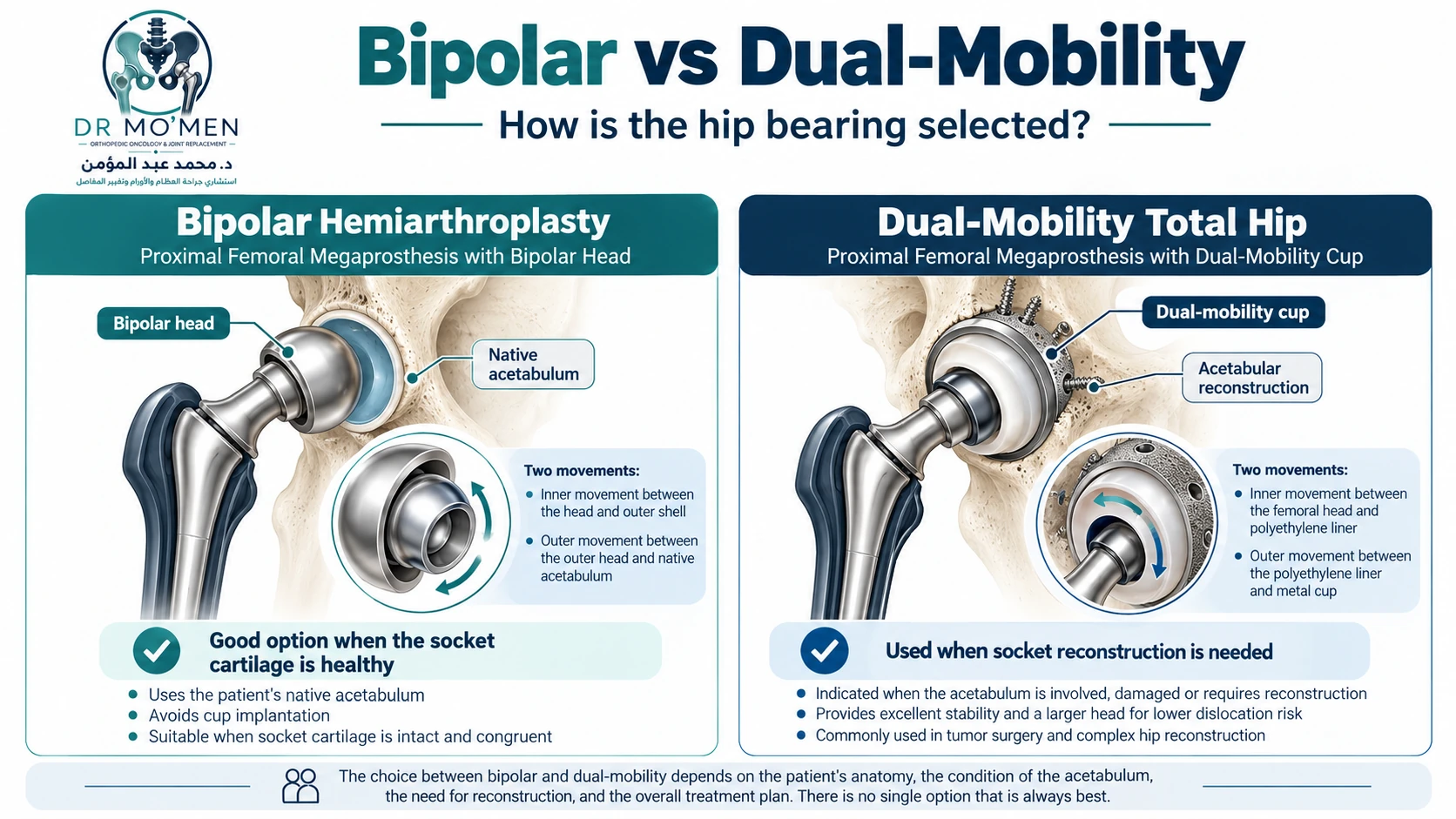
The megaprosthesis may articulate with a bipolar head in the native acetabulum or with an acetabular component when socket resurfacing is required. Bearing selection should consider acetabular cartilage, previous arthroplasty, pelvic irradiation, tumor involvement, instability risk and the patient's expected long-term needs.
Abductor and capsular reconstruction
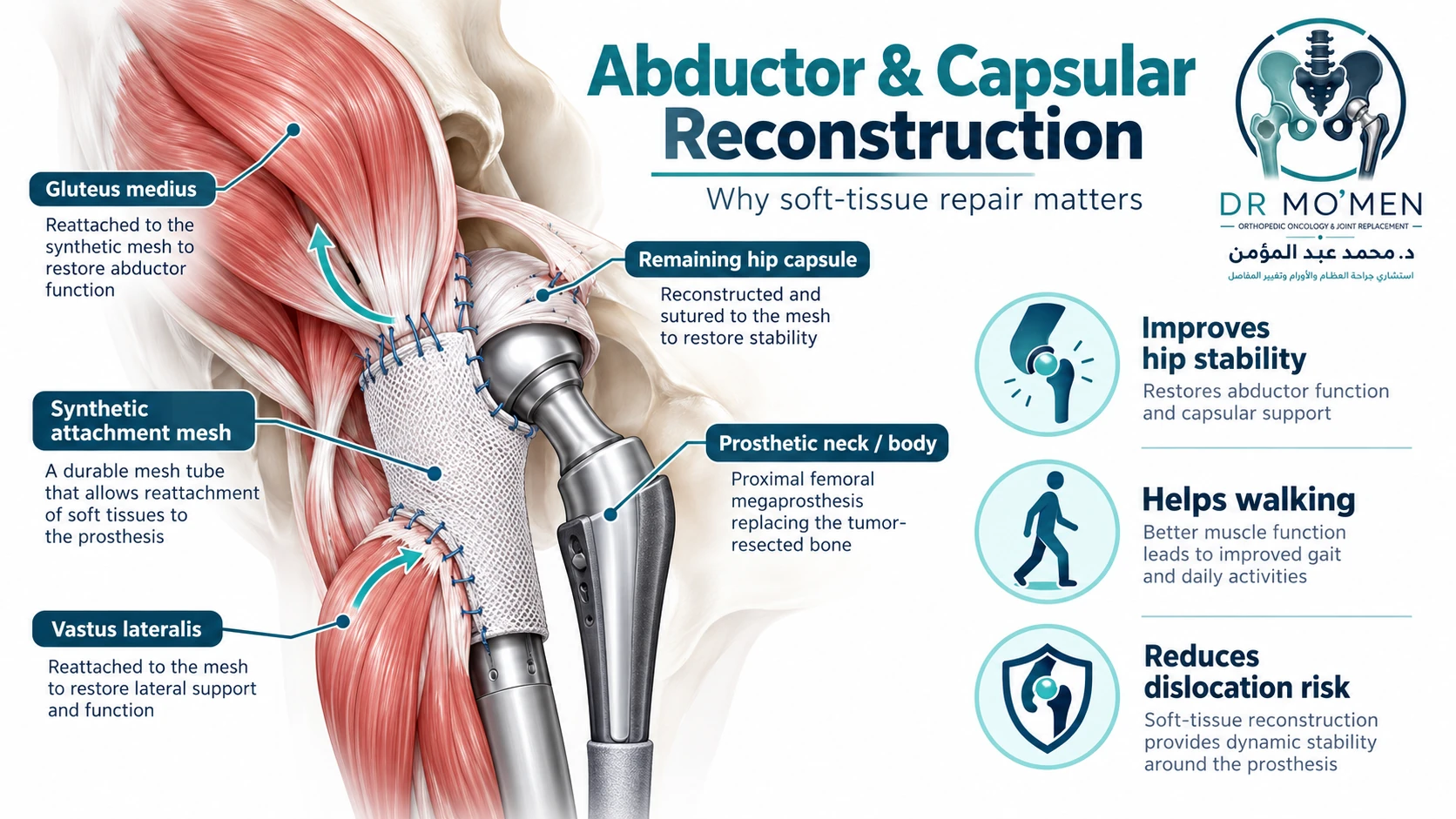
The greater trochanter and normal tendon insertions may be removed with the specimen. Therefore the remaining capsule, gluteus medius/minimus, vastus lateralis and other available soft tissues are reconstructed around the implant. A synthetic attachment tube or mesh may provide a surface for soft-tissue fixation. This step is central to hip stability and gait.
Bipolar Hemiarthroplasty vs Dual-Mobility Total Hip: How Is the Bearing Selected?
Related reading: hip replacement dislocation and instability.
There is no single bearing that is best for every patient. In oncological proximal femoral replacement with a healthy native acetabulum, bipolar hemiarthroplasty can avoid acetabular implantation and has shown low early dislocation rates in published series. Its trade-off is possible late acetabular cartilage wear in long-term survivors. When the socket itself requires reconstruction, a total hip articulation may be necessary; dual-mobility designs are frequently considered when instability risk is high, but they are not universally superior in every proximal femoral replacement.
| Bearing | Where it may fit | Main advantage | Main consideration |
|---|---|---|---|
| Bipolar hemiarthroplasty | Healthy native acetabulum | Avoids acetabular component; often stable in primary oncological PFR | Possible late acetabular wear / groin pain in long-term survivors |
| Dual-mobility THA | Acetabular reconstruction needed or selected high-instability-risk cases | Larger effective head and increased jump distance | Requires secure acetabular fixation; not proven universally superior in all PFRs |
| Constrained liner | Selected recurrent instability or severe soft-tissue deficiency | Mechanical constraint | Higher stresses at the cup / locking mechanism; not routine first choice |
What Is Recovery and Walking Like After Surgery?
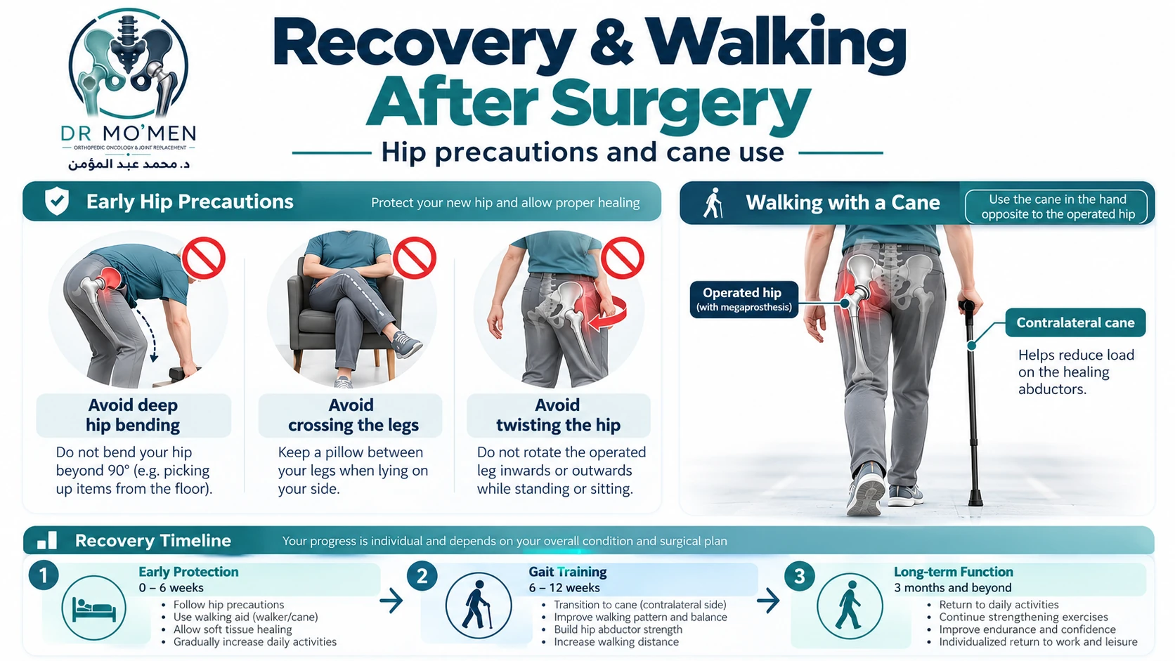
A major advantage of proximal femoral replacement is that mechanical stability does not depend on fracture union. Many patients can begin early mobilization, but the amount of weight-bearing and the hip-motion restrictions are individualized according to stem fixation, abductor and capsular repair, trochanteric reconstruction, wound healing and oncology treatment.
| Phase | Approximate period | Typical goals |
|---|---|---|
| Early protection | First 4–6 weeks | Protect soft-tissue repair; begin safe transfers and gait with walker/crutches; thrombosis prevention; maintain knee and ankle movement |
| Progressive gait | About weeks 6–12 | Advance walking aid as permitted; improve balance and trunk control; begin graded abductor rehabilitation |
| Long-term function | From about 3 months onward | Build endurance, walking confidence and daily function; continue strengthening; avoid high-impact activity unless specifically cleared |
Why the cane is usually held in the opposite hand
When a single cane is appropriate, using it in the hand opposite the operated hip reduces the hip abduction moment and lowers the demand on the healing abductor mechanism. This can improve balance and reduce a Trendelenburg-type limp. The timing of cane use should follow the rehabilitation plan.
What Complications Are Monitored During Follow-Up?
Related reading: megaprosthesis loosening and revision options.
Complication rates vary substantially according to the indication, previous operations, infection history, systemic treatment, implant design and follow-up. The Henderson failure framework is often used for tumor megaprostheses.
- Type 1 — soft-tissue failure: instability, dislocation, abductor insufficiency or other soft-tissue problems.
- Type 2 — aseptic loosening: loss of fixation without infection.
- Type 3 — structural failure: periprosthetic fracture, breakage or another structural implant problem.
- Type 4 — infection: deep periprosthetic infection remains one of the most important complications of large endoprosthetic reconstructions.
- Type 5 — tumor progression or recurrence in oncology patients.
- With bipolar hemiarthroplasty, late acetabular cartilage wear is an additional long-term consideration.
Urgent Red Flags After Surgery
Emergency: Sudden severe hip deformity or inability to bear weight, new limb circulation or nerve problems, chest pain or sudden breathlessness require immediate emergency-department assessment. Do not wait for a routine appointment or attempt to reposition the joint yourself.
Seek prompt medical assessment for:
- sudden severe hip pain, deformity, a new leg-length change or inability to bear weight, which may indicate dislocation or fracture;
- persistent fever, wound drainage, increasing redness, warmth or swelling;
- painful calf swelling, chest pain or sudden shortness of breath, which require urgent assessment for venous thromboembolism;
- a new mass, progressive swelling or unexplained worsening night pain near the surgical site.
Frequently Asked Questions
Can I walk normally after a proximal femoral megaprosthesis?
Many patients regain useful independent or assisted walking. Some continue to have abductor weakness or a Trendelenburg-type limp because the hip abductors must be reconstructed after the proximal femur is removed. Recovery depends on the tissues preserved, implant stability, oncology treatment and rehabilitation.
Why is dislocation a particular concern after proximal femoral replacement?
Tumor resection may remove the native capsule, greater trochanter and normal abductor insertions. This reduces the natural soft-tissue stability of the hip, so bearing choice and meticulous capsular and abductor reconstruction are important.
What is the difference between a bipolar head and a dual-mobility cup?
A bipolar head uses the patient's native acetabulum and avoids placing an acetabular cup. A dual-mobility total hip reconstruction includes an acetabular component and may be selected when the socket needs resurfacing or when instability risk is a major concern. The best choice depends on the individual anatomy and indication.
How long does a proximal femoral megaprosthesis last?
There is no single fixed lifespan. Many oncological reconstructions remain functional for years, but longevity varies with the reason for reconstruction, infection history, soft-tissue stability, fixation, mechanical complications, previous revision surgery and activity level. Long-term follow-up is essential.
Can it be used after a failed nail or plate?
Yes, proximal femoral replacement may be an important salvage option when previous fixation has failed with nonunion, severe bone loss or an unsalvageable proximal femur. It is not the only possible solution; revision fixation or another arthroplasty strategy may still be appropriate in selected cases.
Specialist Assessment and Second Opinion
Choosing between fixation, proximal femoral replacement and other reconstructive options requires review of full-length femur and pelvis radiographs, cross-sectional imaging when indicated, staging studies in oncological cases, and pathology when a tumor diagnosis is involved. Patients in Egypt and international patients may request specialist review with Dr. Mohammed Abdelmoemen Abuelhadid, Lecturer of Orthopedic Surgery at Ain Shams University.
For booking or imaging review: +20 102 1690693
Heliopolis clinic: 50 El Hegaz Street, Cairo. Saturday 3–5 PM, Monday 7–9 PM, Wednesday 5–7 PM.
Evidence & Sources
- Zucchini R, Sambri A, Fiore M, Giannini C, Donati DM, De Paolis M. Megaprosthesis Reconstruction of the Proximal Femur following Bone Tumour Resection: When Do We Need the Cup? Hip & Pelvis. 2021;33(3):147–153. doi:10.5371/hp.2021.33.3.147. — View publication
- Lex JR, Evans S, Parry MC, Jeys L, Stevenson JD. Acetabular complications are the most common cause for revision surgery following proximal femoral endoprosthetic replacement: what is the best bearing option in the primary and revision setting? Bone Joint J. 2021;103-B(10):1633–1640. doi:10.1302/0301-620X.103B10.BJJ-2020-2480.R1. — View publication
- Toepfer A, Straßer V, Ladurner A, et al. Different outcomes after proximal femoral replacement in oncologic and failed revision arthroplasty patients — a retrospective cohort study. BMC Musculoskelet Disord. 2021;22:813. doi:10.1186/s12891-021-04673-z. — View publication
- Roustemis AG, Gavriil P, Goumenos S, et al. Implant survival and risk factors for failure after proximal femoral megaprosthetic reconstruction. SICOT-J. 2025;11:50. doi:10.1051/sicotj/2025031. — View publication
- Strauss SJ, Frezza AM, Abecassis N, et al. Bone sarcomas: ESMO–EURACAN–GENTURIS–ERN PaedCan Clinical Practice Guideline for diagnosis, treatment and follow-up. Ann Oncol. 2021;32(12):1520–1536. doi:10.1016/j.annonc.2021.08.1995. — View publication
Medical disclaimer
This educational article is not a substitute for a clinical examination, specialist review of imaging and pathology, or an individualized treatment plan.