
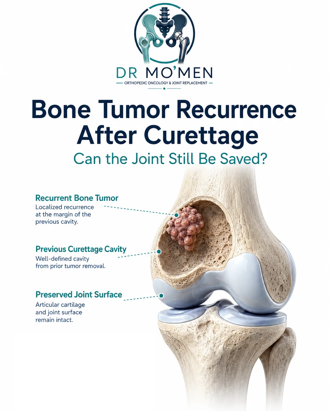
Hearing that a bone tumor has returned after curettage can be alarming. A common concern is: “Does recurrence mean that the joint now has to be removed?”
Not necessarily.
For several benign-aggressive or intermediate bone tumors, recurrence does not automatically mean that limb-salvage treatment has failed or that a megaprosthesis is required. In selected patients, another carefully planned operation may still allow repeat extended curettage, reconstruction of the bone defect, and preservation of the native joint.
This is not appropriate for every recurrence. The decision depends on the original diagnosis, location, remaining bone, condition of the articular surface, soft-tissue extension, previous operations and the biological behavior of the recurrent lesion. For a broader overview, see what to do when a bone tumor recurs.
What does local recurrence mean?
Local recurrence means that tumor tissue has appeared again at or close to the previously treated site.
It may be discovered because the patient develops:
- new or increasing pain;
- swelling;
- a palpable mass;
- reduced joint movement;
- changes on surveillance X-rays or MRI.
Sometimes recurrence is detected on imaging before major symptoms develop.
Which tumors can recur after curettage?
Curettage is used for several benign or locally aggressive bone lesions in appropriately selected cases, including:
- giant cell tumor of bone;
- chondroblastoma;
- aneurysmal bone cyst;
- selected other benign-aggressive lesions.
The recurrence risk is different for each diagnosis. Primary malignant bone tumors such as conventional osteosarcoma are generally not treated by simple curettage. Therefore, when a lesion previously treated by curettage returns, the original pathology should be reviewed before simply repeating the same operation.
Giant cell tumor of bone: the classic example
Giant cell tumor of bone often occurs near the end of a long bone and commonly affects young or middle-aged adults. Around the knee, preserving the native joint can have substantial long-term functional value.
Local recurrence can occur after extended curettage, but recurrence does not automatically require wide resection. Repeat joint-preserving treatment remains an important option for appropriately selected recurrent GCTB. For the broader disease pathway, read Giant Cell Tumor of Bone: Treatment and Recurrence.
Can chondroblastoma recur after curettage?
Yes. Chondroblastoma is usually a benign tumor occurring in younger patients, often close to a joint. Local recurrence is recognized after intralesional treatment, and repeat curettage can control selected recurrent lesions. More extensive recurrence can occasionally require wider reconstruction.
Some recent pediatric data have reported an association between larger tumor size and recurrence risk, but this should not be treated as a universal numerical threshold.
Why can a bone tumor recur after curettage?
Residual microscopic tumor
Even after apparently complete curettage, microscopic tumor cells may remain within irregular areas of cancellous bone. Extended curettage therefore goes beyond simply removing the visible tumor.
Difficult anatomical location
Some tumors lie very close to articular cartilage, a growth plate, major nerves or vessels, or in areas with limited surgical access. This can make complete mechanical removal more difficult.
Extensive or aggressive disease
A lesion with very thin remaining cortex, pathological fracture, soft-tissue extension or a large subchondral component can be more challenging to treat intralesionally.
Previous unplanned or inadequate surgery
An operation performed before the diagnosis or true extent of the lesion is understood can make subsequent treatment more complicated. This is why diagnosis and biopsy planning remain important even when a lesion initially appears benign. See Bone Tumor Biopsy: Core Needle vs Open Biopsy.
Does recurrence mean the first surgeon did something wrong?
Not necessarily. Some tumors have a recognized biological tendency to recur despite appropriate surgery. Recurrence should trigger a careful review of pathology, original imaging, surgical technique, reconstruction and current imaging rather than assuming that recurrence alone proves inadequate treatment.
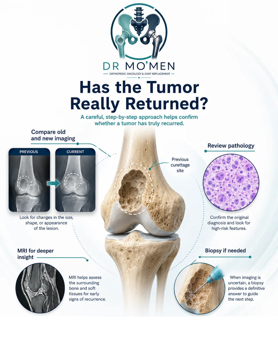
The first step: confirm what has actually recurred
Before deciding on another operation, the diagnosis should be reassessed, especially if the new lesion looks more aggressive, recurrence has occurred after a long interval, pathology was unusual or uncertain, imaging no longer resembles the original lesion, there is a large soft-tissue mass, or there have already been multiple recurrences.
The original pathology slides may need expert review. In selected cases, repeat biopsy may be necessary before definitive surgery, and the biopsy should again be planned with the tumor surgery team.
What imaging is usually needed?
X-rays: Plain radiographs help show recurrent bone destruction, remaining cortex, relationship to the joint, previous cement or graft, and pathological fracture. Comparison with previous X-rays is particularly useful.
MRI: MRI helps define the extent within bone, soft-tissue extension, and the relationship to cartilage, muscles and neurovascular structures. Interpretation may be more difficult after previous curettage, cement or surgery, so correlation with earlier imaging matters.
CT: CT can sometimes help assess cortical destruction, subchondral bone, cement margins, fracture and complex anatomy.
Can the joint still be preserved?
Often, yes — but not always.
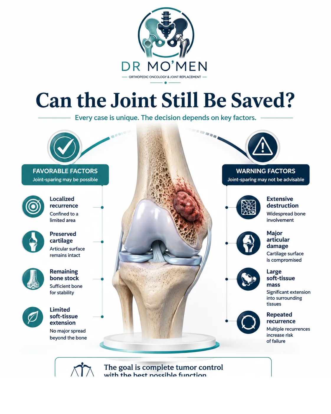
The key question is not simply, “Has the tumor recurred?” A more useful question is: Can adequate tumor clearance still be achieved while leaving enough bone and soft tissue to support a useful native joint?
When might repeat curettage be possible?
Repeat extended curettage may be considered when:
- the diagnosis remains suitable for intralesional treatment;
- recurrence remains localized;
- sufficient subchondral bone can be preserved or reconstructed;
- the joint surface remains salvageable;
- there is no uncontrollable soft-tissue disease;
- adequate surgical access can be obtained.
For recurrent GCTB in particular, repeat joint-preserving surgery remains an important option for appropriately selected patients.
What happens during repeat extended curettage?
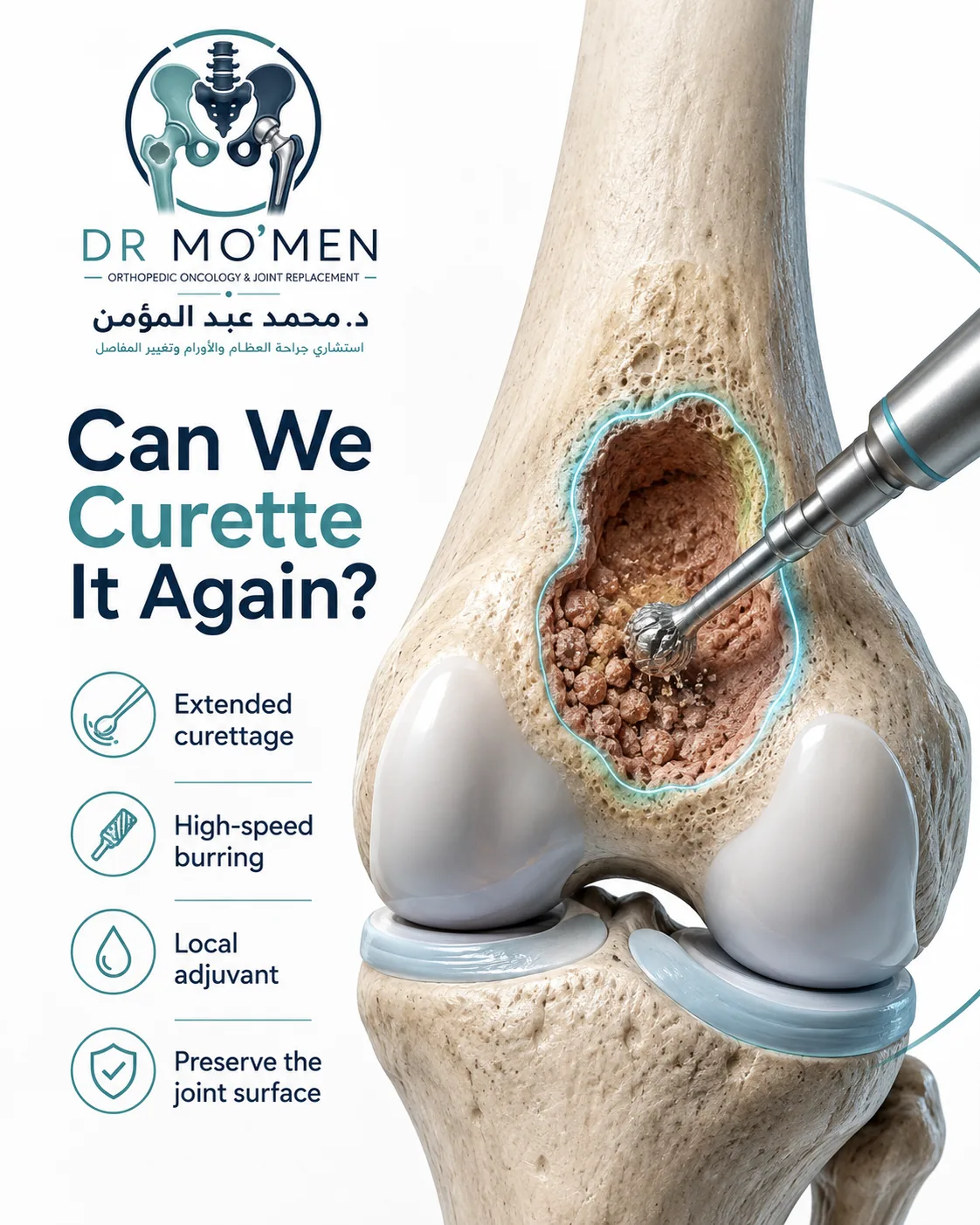
The exact procedure varies, but a typical strategy may include:
- reopening the previous surgical area with oncological planning;
- identifying the previous cavity and recurrent tumor;
- removing gross tumor;
- enlarging the cavity to expose hidden recesses;
- using a high-speed burr where appropriate;
- using a local adjuvant according to tumor type and the surgeon's strategy;
- reconstructing the resulting bone defect.
The aim is to improve local tumor clearance while preserving useful surrounding bone.
What are local adjuvants?
Various methods have been used after mechanical curettage to treat residual microscopic tumor at the cavity margin. Depending on tumor type and local practice, these may include high-speed burring, chemical agents, thermal techniques, cryotherapy or other local strategies.
No single adjuvant should be presented as universally superior for every tumor. Thorough mechanical tumor removal remains central to intralesional surgery.
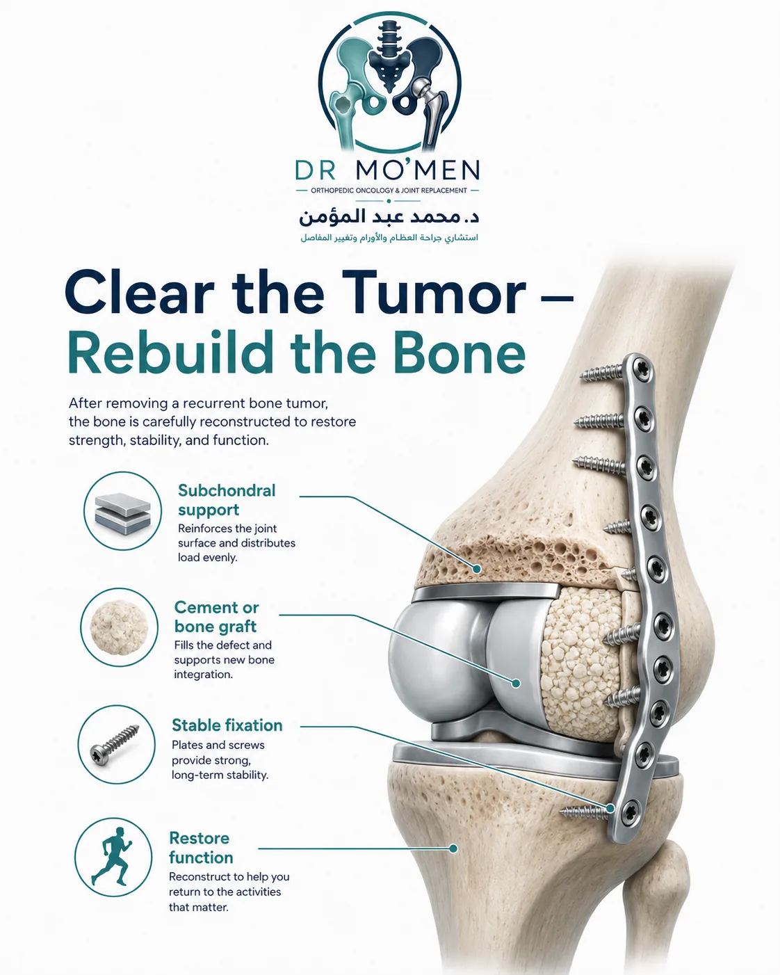
Cement or bone graft after repeat curettage?
Bone cement
Polymethylmethacrylate cement can provide immediate structural filling and is commonly used in selected GCTB reconstructions. It also creates a radiographically distinct cavity that may help comparison during follow-up.
Bone graft
Bone graft may be appropriate when biological bone restoration is desirable, particularly in younger patients or selected anatomical situations.
Combined reconstruction
Some defects require combinations of subchondral graft, cement, internal fixation or structural support. Reconstruction should be adapted to the remaining bone and joint surface.
What if the recurrence is directly beneath the cartilage?
This is one of the most difficult situations. The surgeon must balance tumor clearance, preservation of subchondral bone and protection of the articular surface.
If residual subchondral bone is very thin, additional reconstruction may be required to support the cartilage. In some cases preservation remains possible; in others the joint surface is too damaged or involved for durable joint salvage.
Does a pathological fracture automatically mean the joint cannot be saved?
No. A pathological fracture makes surgery more complicated but does not automatically eliminate the possibility of joint preservation. The decision depends on fracture pattern, displacement, articular damage, soft-tissue involvement, tumor type and remaining bone.
When is repeat curettage no longer the best option?
Wide resection becomes more likely when recurrence involves:
- extensive destruction of the joint surface;
- very limited remaining bone;
- major soft-tissue extension;
- repeated recurrence despite appropriate intralesional surgery;
- a lesion that cannot be adequately accessed by curettage;
- pathological findings suggesting malignant transformation;
- a diagnosis that requires oncological wide resection.
The objective is not to preserve the joint at any cost. The objective is safe oncological treatment while preserving function when that can be done reliably.
For location-specific context around the knee, see Bone Tumor Around the Knee: Diagnosis and Reconstruction.
What happens if the joint cannot be preserved?
The reconstruction depends on the anatomical location. Around the knee, options after wide resection may include distal femoral megaprosthesis, proximal tibial megaprosthesis, or other individualized reconstructions.
Does recurrence automatically mean a megaprosthesis?
No. The treatment spectrum may range from repeat curettage, to curettage with reconstruction or fixation, to wider resection, and finally megaprosthetic reconstruction when necessary. The choice depends on the biological and mechanical situation rather than on the word “recurrence” alone.
What about denosumab in recurrent giant cell tumor?
Denosumab can have an important role in selected patients with GCTB, particularly when surgery would otherwise be highly morbid or difficult. It should not be presented simply as a medication that prevents recurrence. Its use should be individualized within an orthopedic-oncology treatment plan.
What happens after repeat curettage?
Follow-up remains essential because a patient who has already developed one recurrence requires continued surveillance. Follow-up may include clinical examination, serial X-rays, MRI when indicated, and chest surveillance where appropriate for the specific diagnosis. The schedule depends on tumor type, previous recurrence, surgery and overall risk profile.
Can a bone tumor recur more than once?
Yes. Some locally aggressive tumors can recur more than once. A second recurrence does not automatically mean limb salvage is impossible, but repeated recurrence can progressively reduce bone stock, articular support, soft-tissue quality and reconstructive options.
When should the patient seek assessment quickly?
- new swelling at the previous surgical site;
- increasing pain after a stable period;
- a new palpable mass;
- progressive loss of joint movement;
- sudden pain or inability to bear weight;
- suspected pathological fracture;
- rapid change on surveillance imaging.
These symptoms do not prove recurrence, but they require evaluation.
When is a second opinion particularly useful?
- a recurrent tumor has been diagnosed;
- joint replacement has been recommended;
- wide resection has been proposed;
- the patient has already undergone more than one curettage;
- there is a pathological fracture;
- pathology is uncertain;
- imaging shows major subchondral or soft-tissue involvement;
- the patient wants to know whether joint-preserving surgery remains possible.
Role of Dr. Mohammed Abdelmoemen Abuelhadid in complex recurrent bone tumors
A recurrent bone tumor should not simply be treated by repeating the previous operation without reassessment. Evaluation may include review of the original diagnosis and pathology, comparison of initial and current imaging, assessment of the previous surgical cavity, evaluation of the articular surface and subchondral bone, and determining whether repeat curettage or wider resection is oncologically appropriate.
Dr. Mohammed Abdelmoemen Abuelhadid evaluates recurrent bone tumors, complex joint-preservation decisions, limb-salvage surgery and megaprosthetic reconstruction.
For patients outside Egypt, previous pathology reports, operative records and current and previous imaging can be reviewed initially before travel when appropriate.
For appointments or imaging review: +20 102 1690693
Frequently Asked Questions
Does recurrence after curettage mean the tumor has become malignant?
No. Local recurrence does not by itself mean malignant transformation. Unexpectedly aggressive or atypical recurrence may, however, require pathology review or repeat biopsy.
Can the same bone tumor be curetted again?
Yes, in selected recurrent benign-aggressive tumors. The decision depends on diagnosis, recurrence extent, available bone and the condition of the joint.
Does a recurrent giant cell tumor always need a megaprosthesis?
No. Many recurrent giant cell tumors of bone can still be treated with joint-preserving surgery when adequate tumor control and reconstruction remain feasible.
Is joint replacement better than repeat curettage?
Neither is automatically better. Repeat curettage preserves the native joint but may not be appropriate for extensive recurrent disease. Wider resection has different oncological and mechanical trade-offs.
Can recurrence happen again after the second operation?
Yes. Repeat surgery cannot guarantee that recurrence will never happen again, which is why continued surveillance is important.
This article is for patient education and does not replace clinical examination, pathology review, specialist assessment, or review of current and previous imaging.