
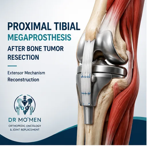
A bone tumor affecting the upper part of the tibia, immediately below the knee, can require removal of a substantial segment of bone. When adequate tumor clearance can be achieved while preserving the limb, the missing bone and knee joint may be reconstructed using a proximal tibial megaprosthesis.
Proximal tibial reconstruction is particularly demanding. The surgeon is not only replacing bone and the knee joint. The patellar tendon and extensor mechanism, which allow the patient to actively straighten the knee, frequently need to be reconstructed as well. The area also has relatively limited soft-tissue coverage, making wound healing and infection prevention important parts of surgical planning.
What is a proximal tibial megaprosthesis?
A proximal tibial megaprosthesis is a modular metallic reconstruction designed to replace a substantial part of the upper tibia together with the knee articulation after major bone resection. It generally includes:
- a stem fixed into the remaining tibia;
- a large metallic replacement for the resected proximal tibia;
- a constrained or rotating-hinge knee mechanism;
- a femoral component;
- fixation points or reconstruction material for surrounding tendons and soft tissues.
Unlike ordinary knee replacement, tumor surgery may remove several centimetres of bone together with muscle, capsule, tendon attachment and other tissues. The reconstruction therefore has to restore not only the joint but also the mechanical and soft-tissue continuity of the limb.
Why is proximal tibial reconstruction more challenging?
1. The patellar tendon attaches to the proximal tibia
The patellar tendon forms the lower part of the knee extensor mechanism. During proximal tibial tumor resection, its normal attachment to the tibial tuberosity may need to be removed. Without effective reconstruction, the quadriceps cannot efficiently transmit force through the patella to straighten the knee.
2. Soft-tissue coverage is limited
The front of the proximal tibia has relatively little muscle covering it. After a large oncological resection, the megaprosthesis can therefore be difficult to cover adequately.
3. Important nerves and blood vessels are nearby
The popliteal vessels, anterior tibial vessels and common peroneal nerve may lie close to the surgical field depending on tumor extent. Accurate MRI assessment and surgical planning are therefore essential.
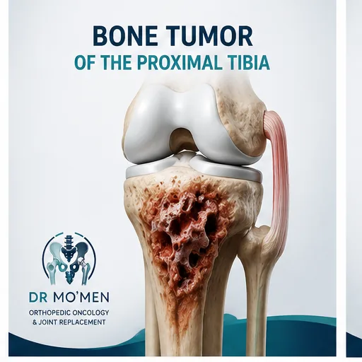
Which tumors may require proximal tibial replacement?
A proximal tibial megaprosthesis may be considered after wide resection of selected tumors such as:
- osteosarcoma;
- chondrosarcoma;
- Ewing sarcoma;
- other primary malignant bone tumors;
- recurrent aggressive tumors;
- selected metastatic lesions;
- selected destructive benign-aggressive tumors when joint preservation is not appropriate.
A tumor in the proximal tibia does not automatically require a megaprosthesis. Some tumors can be treated with curettage, cement, grafting, internal fixation or joint-preserving resection. The appropriate procedure depends on histological diagnosis, imaging, tumor extent, response to systemic treatment when relevant, remaining bone and soft tissues, and whether an adequate oncological margin can be achieved.
For the broader decision pathway, see bone tumors around the knee and the general guide to megaprosthesis after bone tumor resection.
For the specialist service pathway, see limb-salvage reconstruction after bone tumor resection and orthopedic oncology.
Diagnosis must come before reconstruction
A suspected malignant bone tumor should be treated as an oncological problem before it is treated as a reconstruction problem.
Imaging
Assessment commonly starts with plain X-rays followed by MRI of the entire relevant anatomical compartment. Depending on the suspected diagnosis, additional staging may include CT of the chest and other systemic investigations.
Biopsy planning
Biopsy should ideally be planned by, or in coordination with, the team that would perform definitive tumor surgery. The biopsy tract should be positioned so that it can subsequently be removed with the tumor. An unplanned incision or fixation through an undiagnosed primary bone malignancy may contaminate additional compartments and make limb-salvage surgery substantially more difficult.
What happens during proximal tibial megaprosthesis surgery?
Wide tumor resection
The tumor-bearing part of the proximal tibia is removed according to the required oncological margin. When necessary, parts of surrounding muscle or other structures may also have to be removed to obtain adequate tumor clearance. The oncological resection takes priority over reconstruction.
Implant reconstruction
The remaining tibia is prepared to receive the prosthetic stem. The modular prosthesis then replaces the missing proximal tibia and reconnects the lower leg with the knee. A constrained or rotating-hinge mechanism is commonly required because major ligamentous attachments may have been removed during tumor resection.
The key issue: rebuilding the extensor mechanism
This is the major difference between proximal tibial and many distal femoral reconstructions. The extensor mechanism can be summarized as:
Quadriceps → quadriceps tendon → patella → patellar tendon → tibia
If the patellar tendon attachment is removed with the tumor, the tendon must be securely reconstructed around or onto the new prosthesis. Without effective healing and appropriate tension, the patient may develop an extensor lag: the knee can be straightened passively, but the patient cannot completely straighten it using the quadriceps.
Reconstruction is individualized according to the remaining tendon, resection length, soft tissues, implant design and the surgeon’s reconstructive strategy.
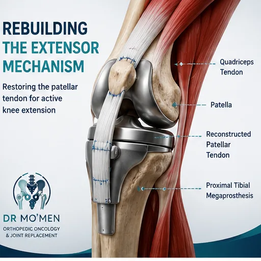
How can the patellar tendon be reconstructed?
Several techniques have been described and may use combinations of:
- direct tendon fixation;
- synthetic mesh;
- synthetic reconstruction tubes;
- autologous tissue;
- allograft tissue;
- iliotibial-band augmentation;
- muscle-flap reinforcement.
The exact technique should be selected according to the individual surgical defect rather than applying one reconstruction method to every patient.
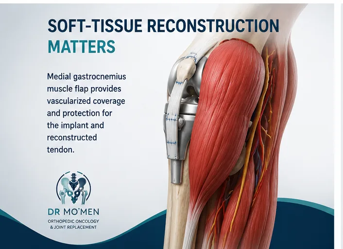
Why is the gastrocnemius muscle often important?
The medial gastrocnemius muscle is located behind the upper tibia. In many proximal tibial tumor reconstructions, it can be rotated forward to cover the prosthesis and reconstructed extensor mechanism. This may provide vascularized soft-tissue coverage, protect the prosthesis, support the reconstructed tendon and separate the implant from the skin.
Can the patient walk immediately after surgery?
A megaprosthesis can provide immediate skeletal stability, but that does not mean the reconstructed knee should be used without restriction immediately after surgery. The repaired patellar tendon and surrounding soft tissues need time to heal.
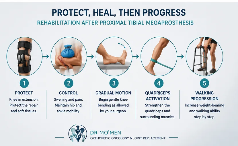
The knee may initially be protected in extension using a brace, while knee flexion, active extension and weight-bearing are introduced progressively according to the reconstruction used. There is no single rehabilitation timetable that is appropriate for every patient.
Rehabilitation after proximal tibial megaprosthesis
Early phase
The initial priorities usually include wound and flap protection, swelling control, prevention of thromboembolic complications when indicated, safe transfers, maintenance of hip and ankle movement, activation of appropriate muscle groups, and protection of the reconstructed extensor mechanism.
Progressive knee movement
Knee flexion is introduced gradually. Aggressive early flexion may place excessive tension on the reconstructed patellar tendon, while prolonged immobilization can contribute to stiffness. Rehabilitation therefore balances protection and mobility.
Quadriceps recovery
Quadriceps rehabilitation is particularly important because the reconstructed tendon must transmit force effectively. Active straight-leg raising may be delayed until the treating surgeon considers the repair sufficiently protected.
Walking
Patients may initially require a walker or crutches. Progression depends on muscle control, wound healing, balance, oncological treatment and the integrity of the reconstruction.
What is extensor lag?
Extensor lag means that a patient cannot actively achieve the full knee extension that is possible when the knee is moved passively. Contributors may include stretching of the reconstructed patellar tendon, quadriceps weakness, altered patellar height, scar formation, extensive muscle resection or tendon reconstruction failure.
What function can be expected?
Many patients can achieve useful walking and functional knee movement following rehabilitation. However, the final result varies substantially between individuals and depends on the extent of tumor resection, preservation of muscle, extensor mechanism reconstruction, nerve function, postoperative infection, knee stiffness, systemic oncological treatment, age, general health and rehabilitation.
No specific degree of knee flexion, walking level or functional score can be guaranteed for an individual patient.
What are the major complications?
Infection
A dedicated guide covers megaprosthesis infection diagnosis and treatment options.
Infection remains one of the most significant complications following proximal tibial megaprosthetic reconstruction. Warning signs include increasing pain, persistent wound discharge, redness, swelling, fever or development of a sinus. Treatment depends on timing, organism, implant stability and patient factors.
Wound or flap problems
Because soft-tissue coverage is limited, wound healing requires careful surveillance. Persistent drainage, wound separation or abnormal skin changes should be reviewed promptly.
Extensor mechanism failure
The reconstructed tendon or supporting tissue may stretch or fail, causing increasing extensor lag or difficulty actively straightening the knee.
Aseptic loosening
Implant-bone fixation can fail without infection. Progressive pain with weight-bearing may require serial radiographs and additional investigations.
Mechanical failure
Possible problems include component wear, hinge-related problems, implant breakage or periprosthetic fracture.
Knee stiffness
Protection of the extensor reconstruction can contribute to stiffness, which is why rehabilitation must balance biological healing with mobility.
Nerve dysfunction
Because the common peroneal nerve is close to the proximal tibia and fibula, temporary or persistent nerve dysfunction can occur in selected complex resections.
Local tumor recurrence
The megaprosthesis reconstructs the skeletal defect but does not replace oncological surveillance. New swelling, increasing pain or a new mass requires assessment.
How long does a proximal tibial megaprosthesis last?
There is no guaranteed lifespan. Some prostheses function for many years, while others require revision because of infection, loosening, mechanical failure, extensor mechanism problems or tumor-related factors. Younger patients are particularly likely to require decades of implant follow-up.
Are there alternatives?
Depending on the tumor and patient, alternatives may include biological reconstruction, osteoarticular allograft, allograft-prosthetic composite, joint-preserving resection, arthrodesis or other individualized reconstructions. No single reconstructive method is universally superior for every patient.
When should specialist assessment be urgent?
- persistent drainage or wound breakdown;
- increasing redness or swelling;
- fever associated with wound symptoms;
- sudden inability to actively straighten the knee;
- sudden loss of weight-bearing ability;
- new instability or painful mechanical symptoms;
- rapidly increasing pain;
- a new mass around the operated area.
Patients seeking review before major surgery can use the second-opinion pathway. Patients travelling from outside Egypt can also review the International Patients pathway.
Role of Dr. Mohammed Abdelmoemen Abuelhadid in complex cases
Proximal tibial tumor surgery requires the integration of oncological diagnosis, biopsy planning, resection margins, megaprosthetic reconstruction, extensor mechanism repair and rehabilitation.
Dr. Mohammed Abdelmoemen Abuelhadid evaluates patients with complex bone tumors, limb-salvage reconstruction and megaprostheses, including patients seeking a second medical opinion before major tumor surgery or revision. For patients outside Egypt, imaging and medical reports may be reviewed initially before travel when appropriate.
For appointments or imaging review: +20 102 1690693
Send Your Records Before You Travel
If you are outside Egypt, you can send existing X-rays, MRI or CT images, pathology reports, operative notes and implant information for an initial specialist review before arranging travel. This preliminary review can help clarify what additional evaluation may be needed and whether an in-person orthopedic oncology or complex-reconstruction consultation is appropriate.
Remote review does not replace physical examination, final diagnosis, infection work-up, pathology review or a definitive surgical plan when those are required.
How to send MRI, CT and X-ray files for review
International Patients Request a Specialist Second Opinion Send the file link by WhatsApp
Frequently Asked Questions
Is proximal tibial megaprosthesis the same as a normal knee replacement?
No. It replaces a large segment of the upper tibia as well as reconstructing the knee and often requires rebuilding the patellar tendon and surrounding soft tissues.
Why may I need a brace for longer after proximal tibial replacement?
The brace can protect the reconstructed extensor mechanism while the tendon and soft tissues heal. The exact duration depends on the operation and the treating surgeon’s instructions.
Will I be able to straighten my knee normally?
Many patients regain useful active extension, but some degree of extensor lag may remain. The result depends on tendon reconstruction, muscle preservation, complications and rehabilitation.
Why is a muscle flap sometimes required?
The proximal tibia has limited natural soft-tissue coverage. A gastrocnemius flap may provide vascularized coverage over the implant and help support the reconstruction.
Can the megaprosthesis be revised later?
Yes. Many complications can potentially be managed using modular revision surgery, although revision tumor prosthesis surgery can be complex.
Medical Disclaimer
This article is for patient education and does not replace clinical examination, specialist medical advice, or review of imaging and investigations.