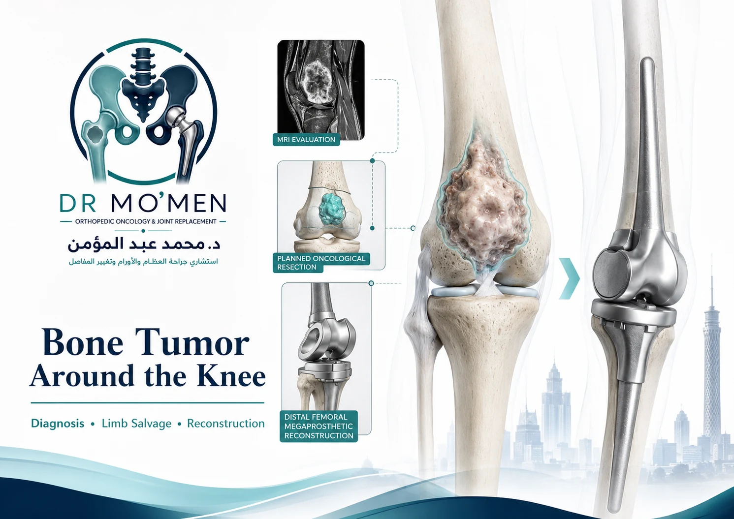
Discovering a bone lesion near the knee can immediately raise difficult questions:
Is it cancer? Does it need a biopsy? Can the knee joint be preserved? Will part of the bone need to be replaced?
The region around the knee is one of the most important areas in orthopedic oncology because tumors may develop in either the distal femur, above the knee, or the proximal tibia, below the knee.
Different tumors can occur in this region, ranging from benign or locally aggressive lesions to primary malignant bone tumors.
- giant cell tumor of bone
- osteosarcoma
- chondrosarcoma
- Ewing sarcoma
- metastatic bone disease
- other less common bone lesions
The treatment is therefore not determined by location alone. The correct strategy depends on the exact diagnosis, tumor grade and biological behavior, MRI findings, relationship to the knee joint, amount of remaining bone, soft-tissue extension, neurovascular involvement, pathological fracture, previous surgery or biopsy, and expected function after reconstruction.
Key principle
The first objective is accurate diagnosis and proper oncological planning before definitive surgery.
Why Are Bone Tumors Around the Knee Clinically Important?
The knee is surrounded by structures that are essential for normal walking and limb function, including articular cartilage, subchondral bone, the quadriceps mechanism, patellar tendon, major muscles, popliteal vessels and major nerves.
A tumor may be very close to these structures without necessarily involving all of them. Good treatment planning therefore aims to answer two separate questions:
- How much tissue must be removed to treat the tumor appropriately?
- How much useful anatomy can safely be preserved?
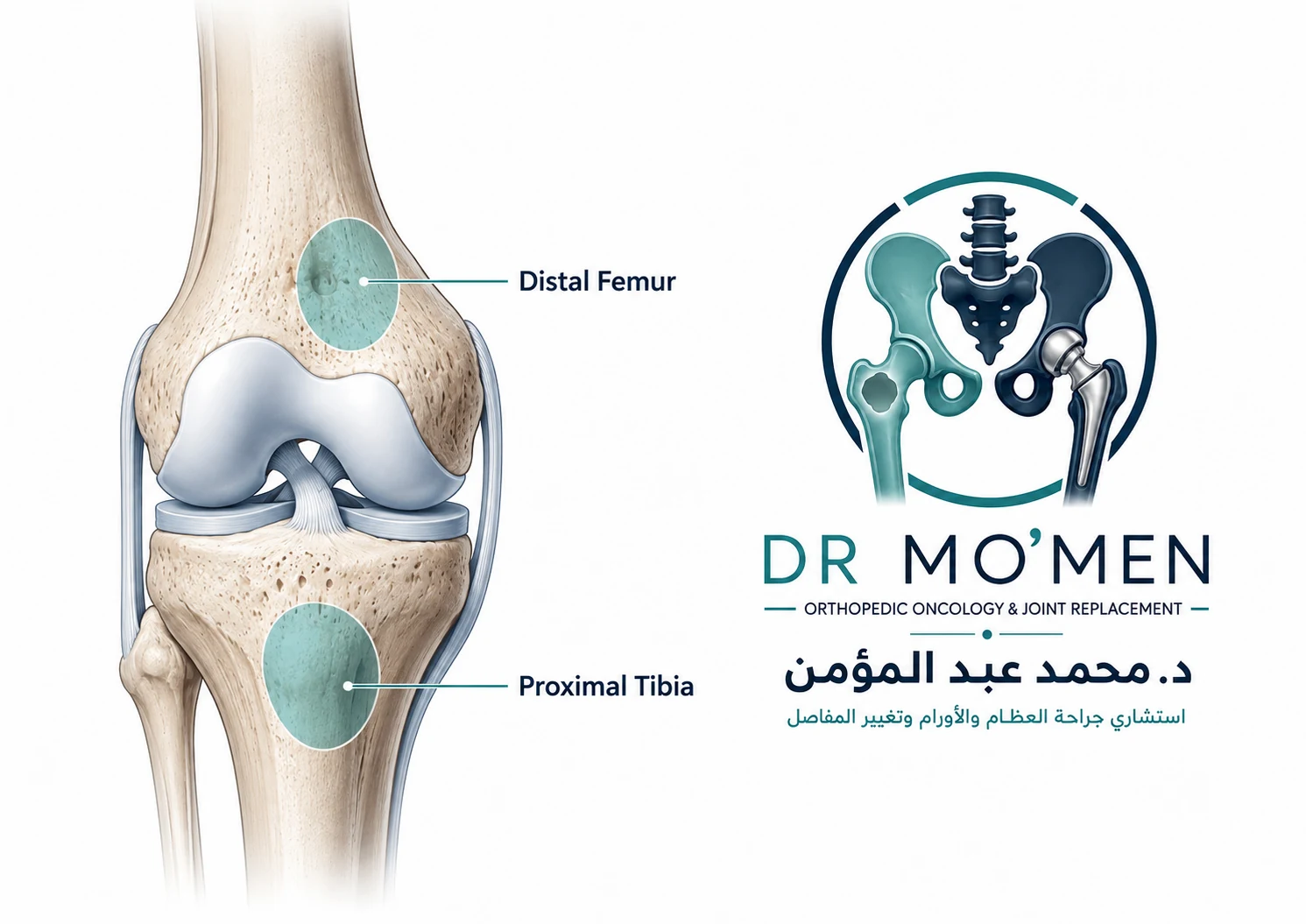
What Symptoms Can a Bone Tumor Around the Knee Cause?
- persistent pain
- increasing pain over weeks or months
- swelling
- visible or palpable mass
- reduced knee movement
- limping
- pain during weight-bearing
- night pain
- sudden pain after pathological fracture
These symptoms are not specific to tumors. Arthritis, sports injuries, infection and other orthopedic conditions can cause similar symptoms. Persistent or progressive symptoms therefore require appropriate imaging rather than diagnosis from symptoms alone.
Can a Bone Tumor Be Mistaken for Knee Arthritis?
Yes. Pain around the knee is extremely common, particularly in adults. A tumor may initially be mistaken for osteoarthritis, meniscal pain, sports injury, tendon pain or muscle strain.
Certain situations should prompt further investigation, such as unexplained progressive pain, swelling, pain that is inconsistent with routine arthritis, destructive changes on X-ray, rapidly worsening symptoms or a palpable mass.
Why Is the Plain X-Ray Important?
X-rays remain one of the most useful initial investigations for a suspected bone tumor. They can provide information about location, pattern of bone destruction, cortical involvement, mineralized tumor matrix, periosteal reaction, pathological fracture and relationship to the joint.
The radiographic pattern helps estimate whether a lesion appears non-aggressive, indeterminate or aggressive. However, X-ray alone does not usually establish the final diagnosis.
Why MRI Is Essential
MRI is particularly important for tumors around the knee because it defines the local anatomical extent. MRI can assess the length of tumor inside the bone, proximity to the knee joint, soft-tissue extension, quadriceps involvement, patellar tendon region, relationship to popliteal vessels and major nerves, surrounding muscles, pathological fracture and potential surgical resection level.
When a primary bone malignancy is suspected, MRI should ideally be completed before biopsy.
Does Every Bone Tumor Need a Biopsy?
No. Some lesions have sufficiently characteristic clinical and imaging features that the diagnostic pathway may differ. However, when malignancy is suspected, the diagnosis is uncertain, major surgery is being considered, or imaging could represent several different tumors, tissue diagnosis is often necessary. The decision should be individualized.
Why Biopsy Planning Is Critical
A biopsy is not simply a diagnostic test. In orthopedic oncology, the biopsy route can affect the definitive operation. The biopsy tract may potentially contain tumor cells and may need to be removed with the final specimen.
Poor biopsy placement can contaminate unnecessary tissue, cross muscle compartments, approach important nerves or vessels, complicate limb-salvage surgery and increase the required resection.
Safest biopsy principle
The biopsy should ideally be planned by or with the orthopedic oncology team responsible for definitive surgery.
Read more: bone tumor biopsy planning.
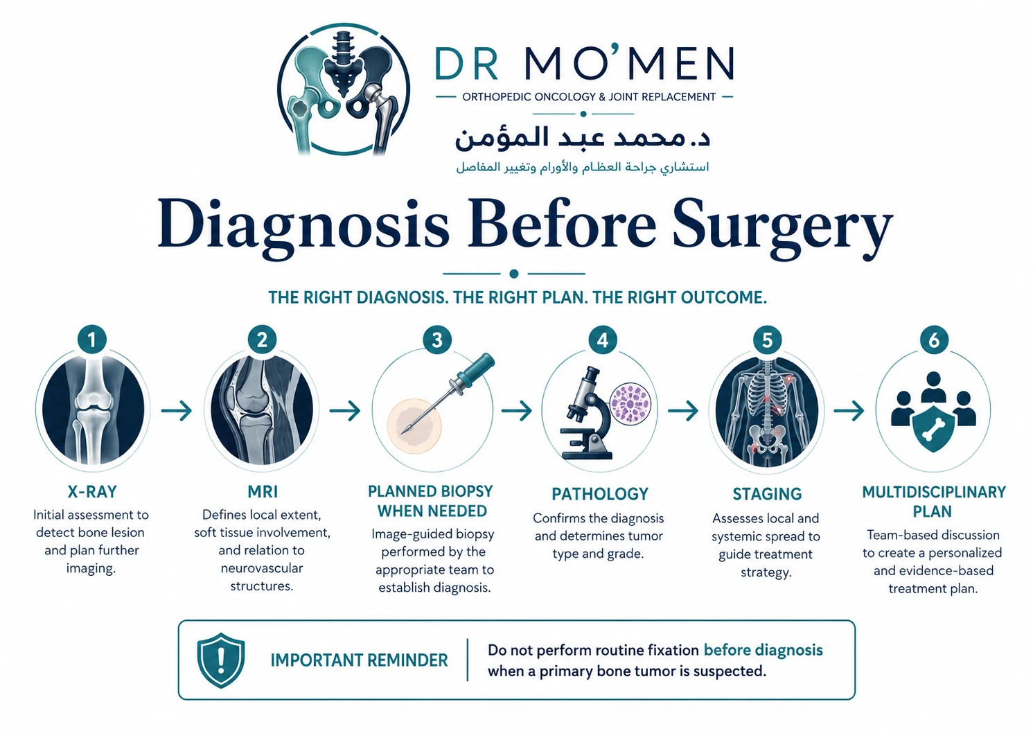
Diagnosis Comes Before Reconstruction
| Decision question | Why it matters | How it can change reconstruction |
|---|---|---|
| What is the diagnosis and biological behavior? | Benign, locally aggressive and malignant tumors require different margins and treatment pathways. | May range from surveillance or curettage to wide resection with oncological reconstruction. |
| How far does the tumor extend? | MRI defines intramedullary and soft-tissue extent and relationship to the joint and neurovascular structures. | Determines the length and compartments of resection and whether the natural joint can be preserved. |
| Has biopsy been planned correctly? | The biopsy tract must be compatible with later definitive surgery when malignancy is possible. | Poorly positioned biopsy can enlarge the tissue field that needs to be removed. |
| What bone and soft tissues will remain? | Durable reconstruction requires adequate fixation plus functional soft-tissue coverage and muscle/tendon reconstruction. | Influences joint preservation, allograft/prosthetic options, megaprosthesis design and rehabilitation expectations. |
| Can oncological margins and useful function both be achieved? | Keeping the limb is not the only goal; local control and expected function are central. | Determines whether limb salvage is appropriate and what reconstruction is reasonable for the individual patient. |
For proximal tibial tumors, reconstruction may require a proximal tibial megaprosthesis with extensor-mechanism repair.
A destructive distal femoral or proximal tibial lesion should not automatically be treated with a plate, intramedullary nail, joint replacement or curettage before the diagnosis is established. The same radiological region can contain tumors requiring completely different treatment.
Giant Cell Tumor
May sometimes be treated using joint-preserving extended curettage.
Osteosarcoma
Often requires systemic chemotherapy and oncological wide resection.
Chondrosarcoma
Surgery is particularly important for many conventional cases, with treatment depending on grade and subtype.
Ewing Sarcoma
Systemic chemotherapy is central, with local control using surgery, radiotherapy or selected combinations.
The treatment must therefore follow the pathology rather than simply the X-ray appearance.
Giant Cell Tumor Around the Knee
Giant cell tumor of bone frequently occurs near major joints. Around the knee it commonly affects the distal femur and proximal tibia. For appropriately selected tumors, one of the main treatment goals is preserving the patient’s native knee.
Extended curettage may involve thorough tumor removal, high-speed burr according to surgical technique, local adjuvant treatment depending on protocol, cement or bone-graft reconstruction, and fixation where mechanically necessary.
Related guide: Giant Cell Tumor of Bone: curettage, recurrence and treatment.
Can the Knee Be Preserved in Giant Cell Tumor?
In many selected cases, yes. The feasibility depends on remaining subchondral bone, articular-surface integrity, cortical destruction, soft-tissue extension, pathological fracture, recurrence and mechanical stability after curettage. Joint preservation should not be promised before complete imaging and surgical assessment.
Osteosarcoma Around the Knee
The distal femur and proximal tibia are important sites for osteosarcoma. Treatment commonly requires coordinated care involving chemotherapy, wide surgical resection, reconstruction and rehabilitation. The surgical plan depends heavily on MRI and response to treatment.
Related guide: Osteosarcoma: diagnosis, chemotherapy and limb salvage.
Chondrosarcoma Around the Knee
Chondrosarcoma may also affect the femur or tibia. The treatment strategy depends on grade, subtype, location, extent and joint involvement. For many conventional chondrosarcomas, appropriate surgical resection is central when feasible.
Related guide: Chondrosarcoma: diagnosis and treatment.
Ewing Sarcoma Around the Knee
Ewing sarcoma may involve long bones near the knee. The treatment differs from many other tumors because systemic chemotherapy is a central component. Local control may involve surgery, radiotherapy or selected combinations depending on anatomy and multidisciplinary assessment.
Related guide: Ewing Sarcoma: diagnosis and treatment.
When Is Limb-Salvage Surgery Possible?
Limb salvage means removing the tumor while preserving a useful limb. It may be possible when an adequate oncological resection can be achieved, critical nerves and vessels can be preserved or appropriately reconstructed, the remaining limb can be reconstructed, and expected postoperative function is acceptable.
Goal of limb salvage
Safe tumor treatment + durable reconstruction + useful function. The objective is not simply keeping the limb.
Related guide: limb salvage surgery for bone tumors.
Does Limb Salvage Always Preserve the Natural Knee?
No. There are two very different limb-salvage concepts.
Joint-Preserving Limb Salvage
The tumor is treated while the native knee remains. This may be possible in selected tumors depending on diagnosis, tumor extent, distance from the joint and remaining bone.
Limb Salvage With Joint Replacement
The limb is preserved, but the tumor-bearing bone and knee joint may need to be removed and reconstructed with a tumor prosthesis.
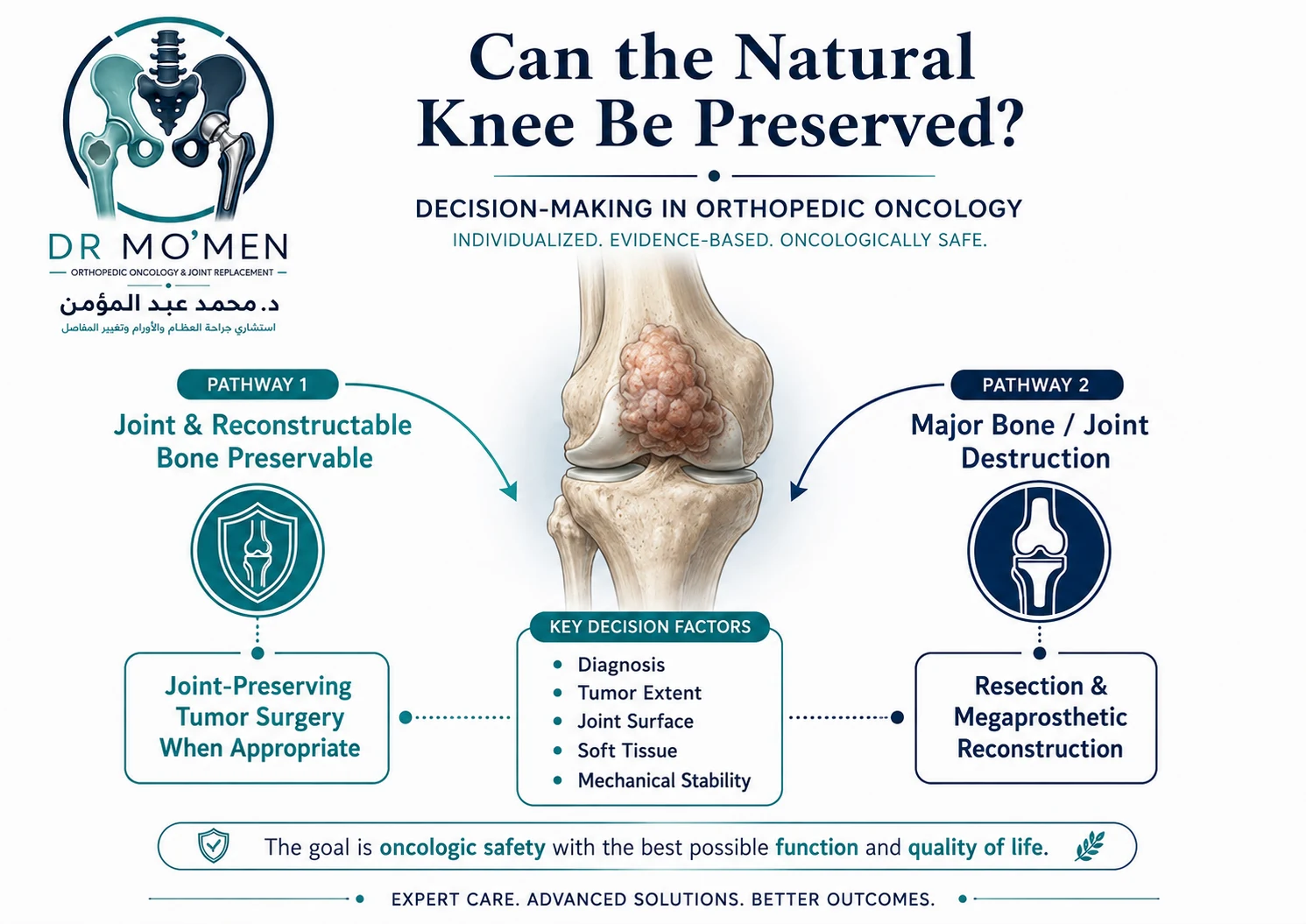
What Is a Distal Femoral Megaprosthesis?
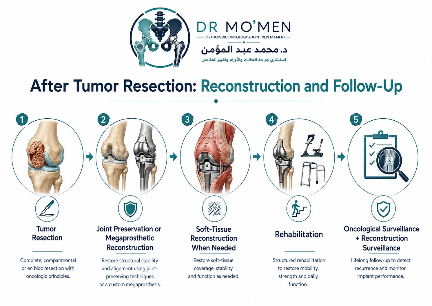
If a tumor requires removal of the distal femur and knee joint, the defect can sometimes be reconstructed using a modular distal femoral megaprosthesis. The implant replaces removed distal femoral bone, the knee articulation and the structural skeletal defect. The goal is to restore limb length, alignment, stability and walking function.
Related guide: megaprosthesis after bone tumor resection.
What Is Proximal Tibial Replacement?
If the proximal tibia must be removed, reconstruction may require a proximal tibial tumor prosthesis. This operation has additional challenges because the patellar tendon and extensor mechanism are closely related to the proximal tibia. Treatment may therefore require reconstruction of bone, knee joint, extensor mechanism and surrounding soft tissue.
Why Soft-Tissue Reconstruction Matters
After major tumor resection around the knee, good reconstruction is not only about the metal implant. The result also depends on muscles, tendons, skin coverage, vascularity, the extensor mechanism and wound healing. In selected complex cases, plastic or reconstructive surgical techniques may be required.
What If the Popliteal Vessels Are Close to the Tumor?
The major vessels behind the knee must be assessed carefully on imaging. A tumor may displace the vessels, contact the vessels, partially surround them or directly invade them. Vascular involvement does not automatically mean amputation, but it increases surgical complexity. Selected cases may require vascular surgical input.
What If a Major Nerve Is Involved?
The impact depends on which nerve is involved, degree of involvement, possibility of safe preservation and expected function after resection. Preserving a limb with severe loss of neurological function is not always the best functional result. This is why limb-salvage decisions must consider both oncological safety and expected function.
What If There Is a Pathological Fracture?
Bone tumors can weaken the femur or tibia enough to cause fracture. A pathological fracture does not automatically require amputation. However, it can affect local contamination, tumor extent, mechanical stability and reconstructive options. The lesion should be treated as a tumor first, not simply as an ordinary traumatic fracture.
Why Unplanned Fixation Can Be Harmful
Placing an intramedullary nail or plate through an undiagnosed primary bone tumor can contaminate additional regions of the bone or surrounding tissues. This may make later definitive resection more extensive. When a suspicious tumor is present, diagnosis and oncological planning should therefore precede routine fixation whenever clinically possible.
When Is Amputation Considered?
Modern orthopedic oncology allows limb salvage in many cases, but amputation remains an important option in selected situations. It may be considered when adequate tumor removal cannot otherwise be achieved, major neurovascular involvement prevents a functional reconstruction, severe infection compromises the limb, repeated local recurrence has destroyed reconstructive options, or limb salvage would produce poor function.
Amputation should not be regarded as automatic simply because a tumor is large.
What Determines the Reconstruction?
The reconstruction depends on the exact tumor diagnosis, amount of bone removed, joint preservation, remaining muscle, nerve function, blood vessels, patient age, expected activity, chemotherapy or radiotherapy, previous surgery and expected durability required. There is no single “best” reconstruction for every bone tumor around the knee.
Rehabilitation After Knee Tumor Surgery
Rehabilitation depends strongly on the operation.
After Curettage
- protected weight-bearing when required
- knee motion exercises
- strengthening
- fracture-risk precautions
After Megaprosthetic Reconstruction
- early mobilization when appropriate
- walking aids
- muscle strengthening
- gait training
- knee range-of-motion work
After proximal tibial reconstruction, rehabilitation may be more protective because the extensor mechanism may require healing.
When Can the Patient Walk?
There is no fixed timetable. Weight-bearing depends on type of reconstruction, implant fixation, bone quality, soft-tissue repair, pathological fracture, wound healing and the surgeon’s protocol. Internet timelines should not replace the individual postoperative plan.
What Complications Can Occur?
- infection
- wound complications
- stiffness
- weakness
- thrombosis
- fracture
- nerve injury
- vascular injury
- instability
- aseptic loosening
- mechanical implant failure
- soft-tissue failure
- local tumor recurrence
- need for revision surgery
The risks should be discussed in relation to the specific procedure rather than as one generic percentage.
What If Pain Develops Around a Tumor Prosthesis Later?
Possible causes include infection, implant loosening, mechanical problems, fracture, soft-tissue problems or tumor recurrence. New pain does not automatically mean that the tumor has returned. Evaluation may require examination, X-rays, blood tests where appropriate, additional imaging and infection assessment.
Follow-Up After Bone Tumor Treatment Around the Knee
Follow-up usually has at least two components.
Oncological Surveillance
- monitoring for local recurrence
- monitoring for metastatic disease where relevant
Reconstruction Surveillance
- implant and bone assessment
- soft-tissue assessment
- joint function
- mechanical complications
Patients treated with joint-preserving surgery also require monitoring of the reconstructed bone and adjacent knee.
When Should You Seek Specialist Orthopedic Oncology Assessment?
- imaging shows a destructive lesion around the knee
- biopsy has been recommended
- diagnosis is uncertain
- joint replacement has been proposed
- amputation has been proposed
- limb salvage is being considered
- pathological fracture has occurred
- previous surgery was performed before diagnosis
- the tumor has recurred
- major reconstruction has been recommended
International Patients: What Should You Send Before Travelling?
- X-rays
- MRI
- CT where available
- biopsy/pathology report
- pathology slides or blocks when appropriate
- staging investigations
- previous surgery reports
- previous chemotherapy records
- current treatment plan
For a patient who has not yet undergone biopsy, review before biopsy can be particularly valuable.
Send Your Imaging Before You Travel
A preliminary review may help assess whether the imaging is complete, whether biopsy planning needs review, whether the knee appears preservable, whether limb-salvage surgery should be assessed, whether a megaprosthesis may be necessary, whether additional reconstruction specialists might be needed, and whether travel to Cairo for specialist assessment is appropriate.
Remote review does not replace examination, pathology, staging or multidisciplinary treatment planning.
Request an Orthopedic Oncology Second Opinion →
Has Imaging Shown a Bone Tumor Around the Knee?
Before biopsy, fixation or major reconstruction, specialist review may help clarify what the lesion may represent, whether biopsy is required, how biopsy should be planned, whether the natural knee may be preserved, whether limb-salvage surgery is feasible, and whether a distal femoral or proximal tibial megaprosthesis may be necessary.
For appointments or review of X-rays and MRI: +20 102 1690693
About Dr. Mohammed Abdelmoemen Abuelhadid
Dr. Mohammed Abdelmoemen Abuelhadid is an Orthopedic Oncology & Joint Replacement Surgeon and Lecturer of Orthopedic Surgery at Ain Shams University. His clinical areas include orthopedic oncology, bone and soft-tissue tumors, limb-salvage surgery, tumor resection and reconstruction, megaprosthetic reconstruction, complex orthopedic reconstruction and second medical opinions for complex orthopedic cases.
For a bone tumor around the knee, the treatment objective is not simply removal of the lesion. It is accurate diagnosis, appropriate oncological control and the most suitable reconstruction for long-term function.
Frequently Asked Questions
Is every bone tumor around the knee cancer?
No. Several benign, locally aggressive and malignant tumors can occur around the knee. Diagnosis requires appropriate imaging and sometimes biopsy.
Can a bone tumor around the knee be removed without replacing the knee?
Sometimes. Joint-preserving treatment depends on the exact tumor, its extent and how much healthy joint-supporting bone remains.
Does a large tumor automatically require amputation?
No. Many large tumors can still be assessed for limb-salvage surgery, but oncological safety and expected limb function determine the final decision.
Why must biopsy be planned?
A poorly placed biopsy can contaminate tissue that would otherwise not need to be removed, making definitive surgery more complicated.
What is a distal femoral megaprosthesis?
It is a modular tumor implant used to replace the distal femur and knee after major oncological resection when the native joint cannot be preserved.
Can a pathological fracture still be treated with limb salvage?
Sometimes. The decision depends on diagnosis, fracture pattern, tumor extent, surrounding soft tissues and reconstructive possibilities.
Can I send MRI scans for review before travelling?
Yes. Imaging and available reports can be reviewed preliminarily before travel, although final decisions require direct specialist assessment.
Medical Disclaimer
Disclaimer: This article is for patient education and does not replace clinical examination, review of imaging, pathology, staging investigations, or individualized multidisciplinary medical advice.
How this article was reviewed
This patient-education article was reviewed for clinical accuracy, biopsy-planning safety, patient-facing clarity and consistency with the site’s orthopedic oncology content framework by Dr. Mohammed Abdelmoemen Abuelhadid. It does not replace direct review of imaging, pathology, staging or multidisciplinary treatment planning.

Author and medical reviewer
Dr. Mohammed Abdelmoemen Abuelhadid — Orthopedic Oncology & Joint Replacement Surgeon and Lecturer of Orthopedic Surgery at Ain Shams University.