
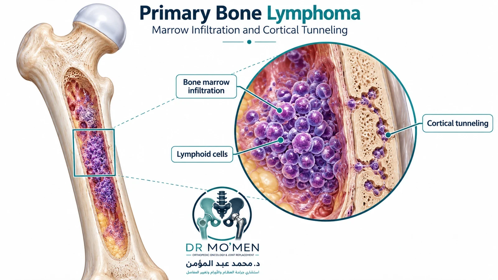
What is primary bone lymphoma?
Lymphoma is a cancer of immune-system lymphocytes. Primary bone lymphoma describes lymphoma presenting primarily within the skeleton rather than spread to bone from an established lymphoma elsewhere. Most primary bone lymphomas are B-cell lymphomas, particularly DLBCL, but other pathological subtypes occur and can require different treatment. Some definitions include a single bone lesion with regional lymph nodes, while others also discuss multifocal bone-only disease; classification must follow the hematopathology and staging team's assessment.
This is not an osteosarcoma, Ewing sarcoma or bone metastasis. Even when an X-ray suggests destructive bone disease, imaging cannot reliably determine the cell type. That distinction is why the team should complete the appropriate investigations before any unplanned curettage, major resection or internal fixation. For more detail, read when a bone tumor needs a planned biopsy.
Which symptoms should raise concern?
The most common complaint is localized, progressively persistent pain in a long bone, pelvis or spine. Pain may occur at rest or at night, but night pain alone cannot diagnose cancer. Other findings include swelling, tenderness, a limp or increasing pain during weight-bearing. A pathological fracture may occur after minimal trauma, and spinal involvement can sometimes cause nerve symptoms. Fever, drenching night sweats and unexplained weight loss may occur, but many patients do not have so-called B symptoms.
Seek emergency assessment for sudden inability to bear weight, a new deformity following minor injury, rapidly progressive limb weakness, numbness around the saddle area or new loss of bladder/bowel control. These may indicate fracture or spinal neural compression. Review bone tumor warning signs.
How is lymphoma of bone diagnosed?
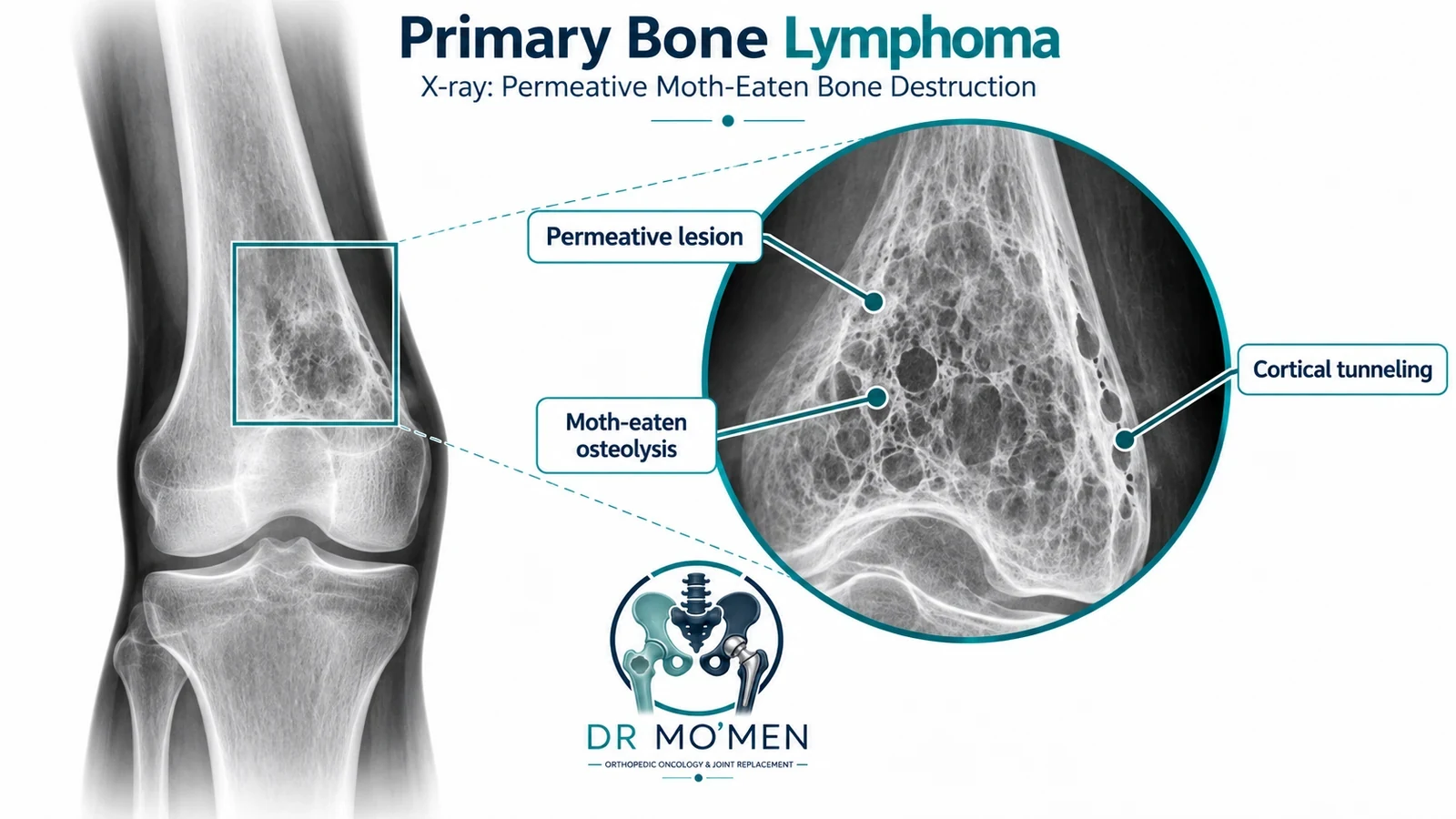
X-ray and CT: bone destruction and mechanical strength
Plain radiographs may show a permeative or moth-eaten pattern of bone loss, sometimes with cortical change or pathological fracture. Other patterns occur; no X-ray appearance is specific to PBL. CT can clarify the amount of remaining cortex, fracture morphology and an accessible biopsy target. A lesion that resembles a sarcoma or infection still requires careful investigation.
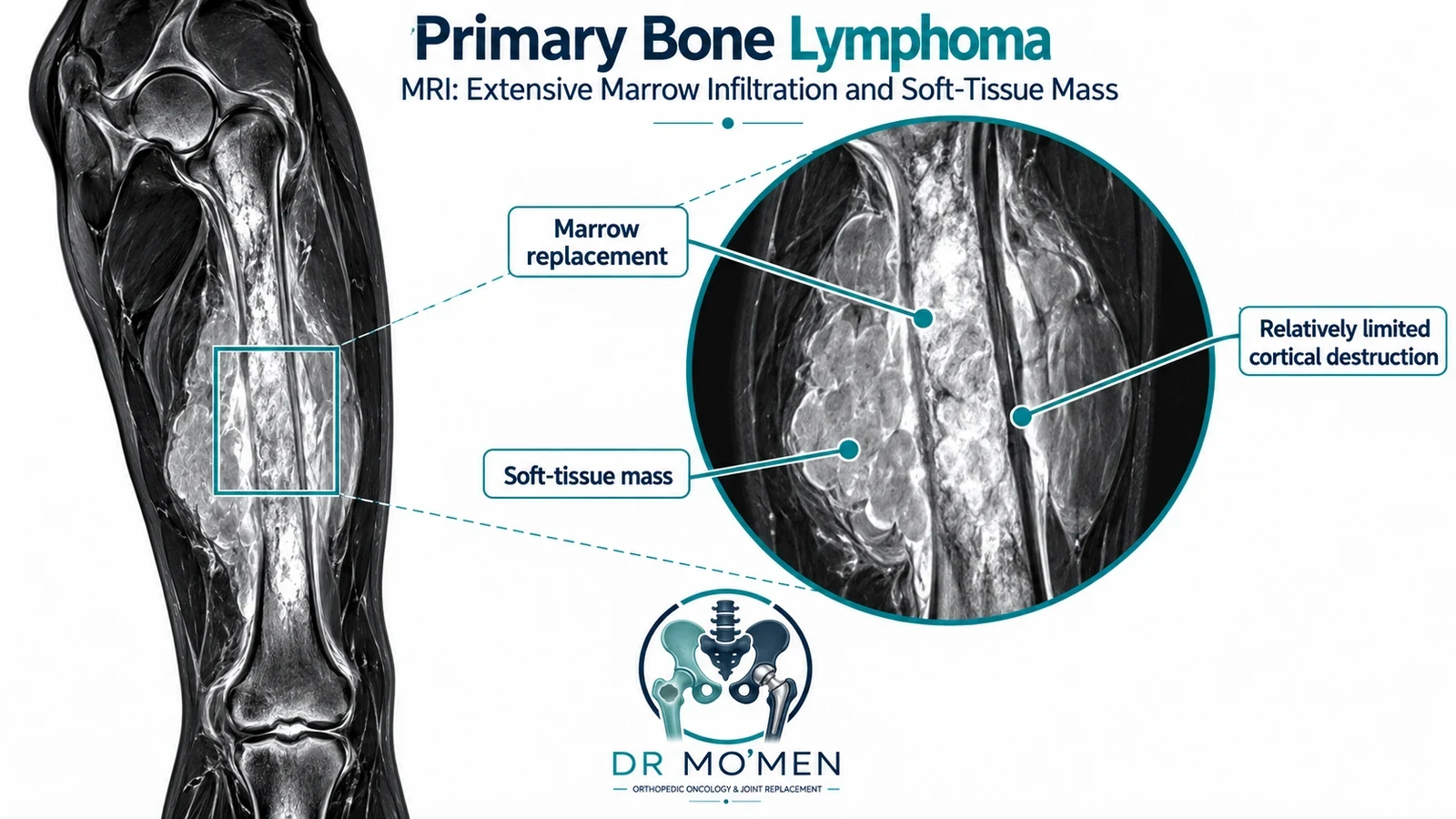
MRI: marrow and soft-tissue extent
MRI is useful for mapping intramedullary involvement, surrounding soft tissue, joints and neurovascular structures. Lymphoma can produce considerable marrow and extraosseous soft-tissue disease with surprisingly limited visible cortical destruction; this is a useful clue but not a diagnostic sign. MRI is also relevant when nerve or spinal canal involvement is suspected.
Staging with FDG PET/CT and blood tests
FDG PET/CT is routinely used in the staging and response assessment of FDG-avid DLBCL to establish whether disease is localized or present at other sites. The hematologist may order blood count, LDH, renal/liver profiles, viral screening and other risk-specific assessments. An iliac-crest bone marrow biopsy is not automatically required for every DLBCL patient when appropriate PET/CT staging is available; the hematology team decides whether its result would change management. The 2025 SEOM–GOTEL DLBCL guideline 2025 DLBCL clinical guideline discusses this PET/CT-based staging approach.
Planned tissue biopsy and pathology
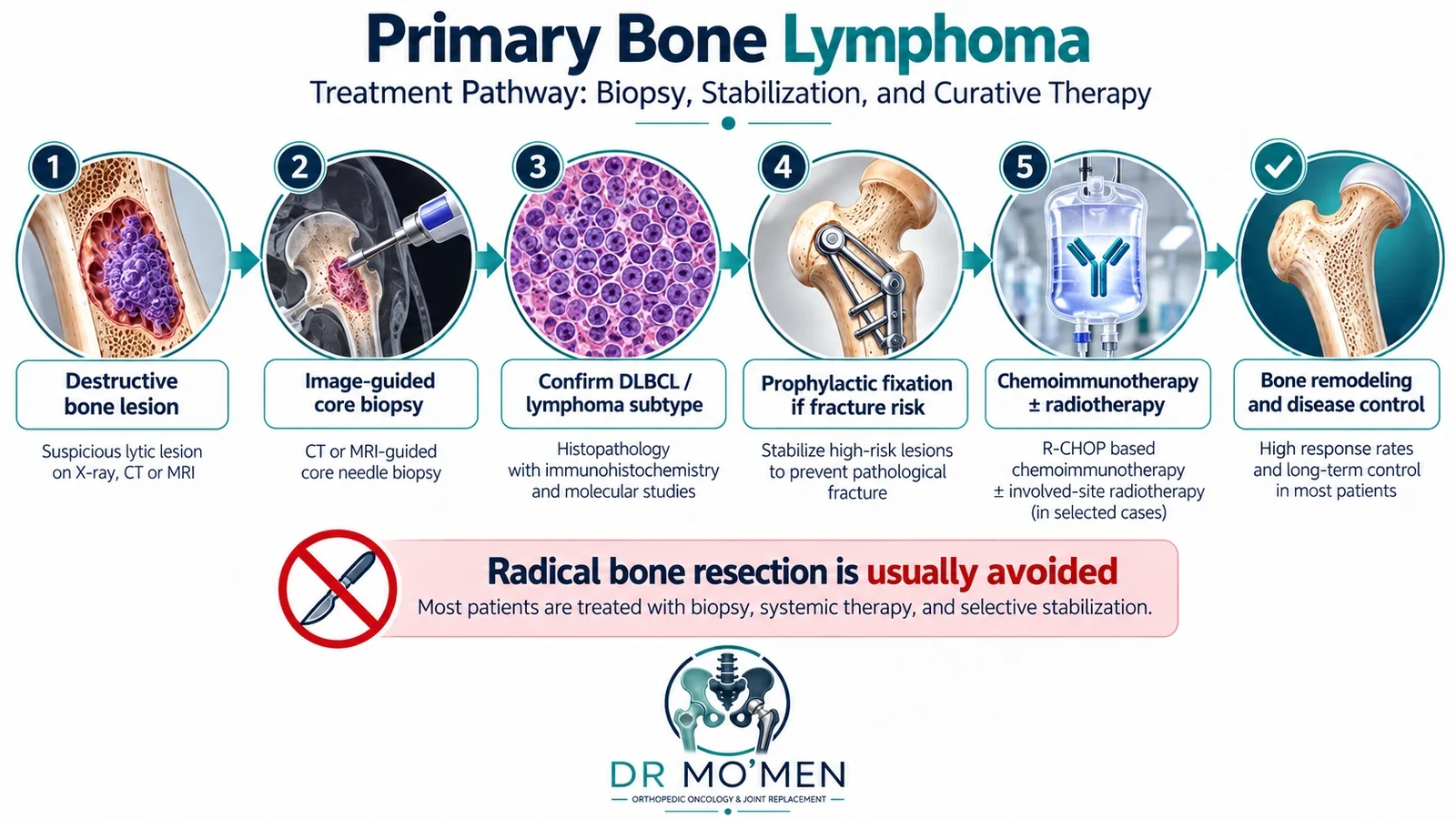
A well-planned image-guided core needle biopsy is commonly an appropriate first diagnostic procedure. The route should be coordinated with orthopedic oncology and interventional radiology when a primary bone sarcoma remains in the differential. Enough viable tissue is needed for morphology, immunohistochemistry, and—where necessary—flow cytometry and molecular studies. CD20, PAX5, CD79a, germinal-center markers, BCL2/MYC assessment and Ki-67 may be relevant depending on the case; no single stain confirms all lymphomas. If the core sample is insufficient, repeat core or incisional biopsy may be required. Fine-needle aspiration alone is often inadequate for initial lymphoma classification.
Never place an intramedullary nail into an undiagnosed aggressive solitary bone lesion before evaluating the possibility of a primary bone sarcoma: doing so can contaminate the medullary canal and alter later limb-salvage options. Read about pathological fracture assessment.
How does PBL differ from other bone lesions?
| Feature | Primary bone lymphoma | Osteosarcoma | Ewing sarcoma | Bone metastasis / myeloma |
|---|---|---|---|---|
| Origin | Lymphoid cells | Malignant osteoid-forming tumor | Ewing-family sarcoma | Other organ / plasma-cell malignancy |
| Imaging | Variable; permeative marrow lesion ± soft-tissue mass | Often aggressive; may show osteoid matrix | Often permeative ± soft-tissue component | Variable lytic, sclerotic or marrow lesions |
| Confirmation | Core biopsy + hematopathology | Planned biopsy + sarcoma pathology | Planned biopsy + molecular diagnosis | Clinical, laboratory, imaging and/or biopsy workup |
| Main treatment | Hematologist-led systemic lymphoma therapy ± selected RT | Chemotherapy and planned wide resection in conventional high-grade disease | Chemotherapy plus local control | Disease-specific systemic and structural treatment |
| Orthopedic operation | For diagnostic/structural/neurological indications | Often integral to local control | Selected local control | Selected fracture or instability indications |
Bone metastases from solid-organ carcinoma and multiple myeloma are distinct diagnoses; they share this comparison column only to illustrate differentials.
Imaging alone cannot reliably distinguish primary bone lymphoma from Ewing sarcoma or metastatic bone disease. Tissue diagnosis and staging are essential.
Why is radical bone resection usually avoided?
Unlike many conventional primary bone sarcomas, lymphoma is generally treated with systemic medication as the main anti-cancer treatment. Routine amputation, wide en bloc resection and replacement of the diseased bone are therefore not standard curative treatments for PBL. Such operations can impose substantial functional costs without being necessary to control the lymphoma. However, writing that any bone operation is “medically contraindicated” is too absolute: a rare patient may need resection or reconstruction for a severe structural problem or other exceptional indication.
What treatment is used instead?
For CD20-positive DLBCL of bone, rituximab-containing anthracycline-based chemoimmunotherapy, commonly an R-CHOP-based approach, has historically been central. Regimen and cycle count depend on stage, risk, disease biology, comorbidities, and current hematology protocols; not every PBL subtype receives R-CHOP. Radiation may be recommended for some bulky, residual, symptomatic or other selected sites. It is not obligatory after chemoimmunotherapy in every limited-stage case. A multicenter retrospective study of 112 patients with limited-stage primary bone DLBCL published in Haematologica in 2024 did not find an overall or relapse-free survival advantage from routinely adding consolidative radiotherapy, while selected partial responders may differ. This finding requires individualized interpretation, not abandonment of radiotherapy across all patients. See the 2024 multicenter PBL study 2024 PBL outcomes study and the 2025 ESMO lymphoma guideline 2025 ESMO lymphoma guideline.
Monitoring uses symptoms, clinical review and disease-appropriate response imaging—often PET/CT for DLBCL. Remission and survival are not the same measure, and population statistics should not be represented as an individual cure guarantee.
When does the orthopedic oncologist operate?
1. Fracture risk assessment and selective prophylactic fixation
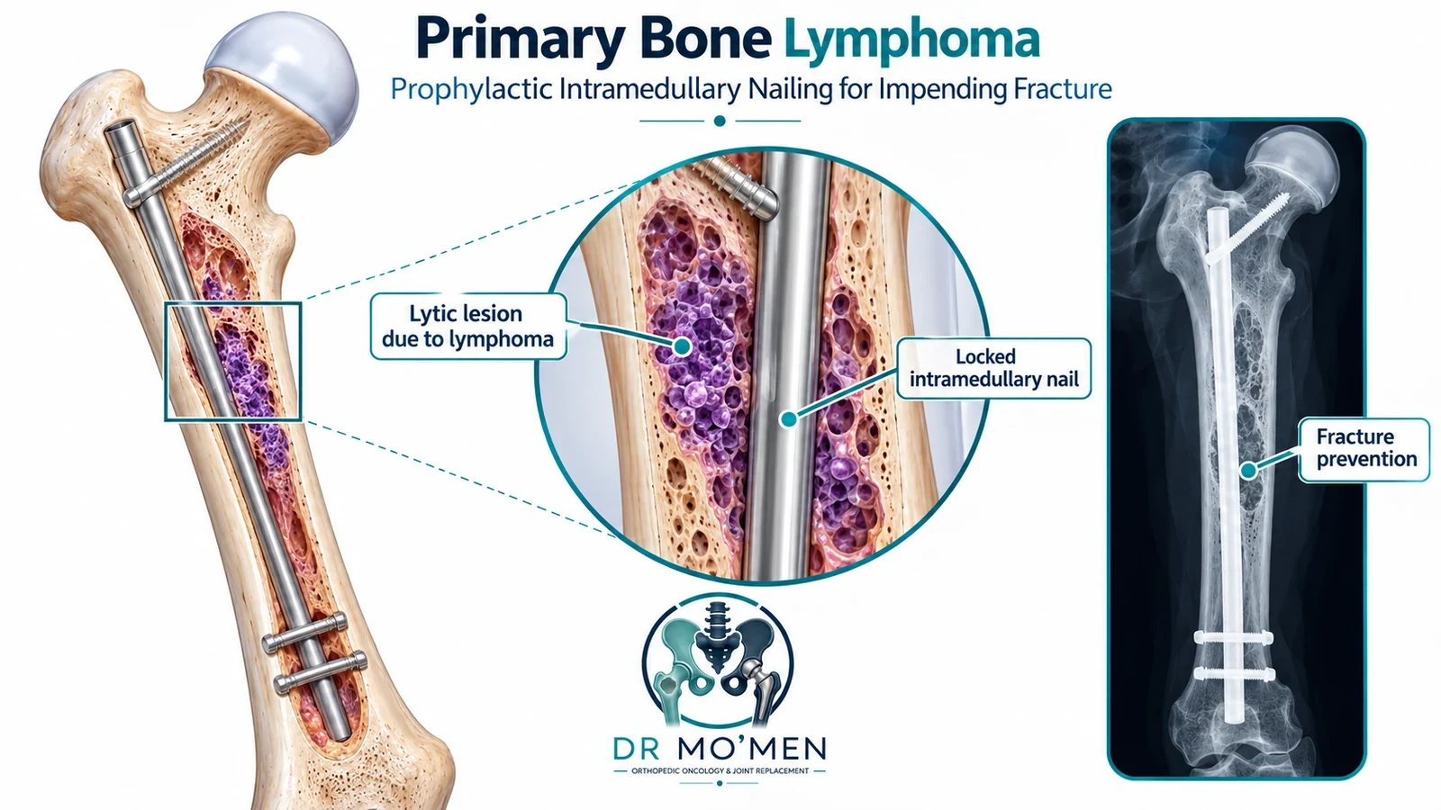
An orthopedic oncologist evaluates load-bearing pain, lesion location, cortical integrity, fracture status, expected response to lymphoma therapy, and whether stabilization would delay systemic treatment. Mirels was designed for metastatic long-bone lesions and is not a lymphoma-specific guarantee of fracture prediction. Some lesions respond medically and may be protected temporarily with restricted weight-bearing; others need plate fixation, intramedullary nailing or a more complex reconstruction. Stabilization is not automatic and should follow diagnosis and multidisciplinary planning.
2. Treatment of a pathological fracture
An established fracture may require stabilization to restore mobility, control mechanical pain and permit cancer therapy. The operative technique, timing, rehabilitation and weight-bearing plan are individualized; immediate unrestricted walking is not promised.
3. Spinal instability or neural compression
New neurological findings demand emergency assessment, spinal MRI when indicated and coordinated input from spine surgery, hematology and radiation oncology. Because lymphoma is often highly responsive to systemic treatment and radiotherapy, not every patient requires decompression surgery. Surgery is considered when mechanical instability, progressive neurological compromise or other patient-specific findings warrant it.
What happens to the bone after treatment?
Successful lymphoma treatment may allow partial or substantial remineralization and remodeling over time. Whether an affected bone recovers adequate strength depends on how much cortex was destroyed, whether a fracture occurred, the treatment delivered and subsequent healing. Persistent deformity or instability can remain; repeat imaging and functional review guide activity and rehabilitation.
When should you request a specialist opinion?
An orthopedic oncology review is appropriate when a destructive bone lesion lacks a confirmed diagnosis, biopsy has been suggested without adequate imaging, you are being offered major bone resection without hematopathology confirmation, weight-bearing pain raises concern for fracture, or reports disagree between sarcoma, infection, myeloma and lymphoma. A coordinated second opinion specialist second-opinion pathway may help assemble imaging and pathology for discussion with hematology before travel. Dr. Mohammed Abdelmoemen Abuelhadid evaluates the orthopedic aspects of complex bone lesions—biopsy planning, stability and reconstruction—in collaboration with the relevant oncology disciplines.
Appointments or imaging review: 01021690693.
Evidence and guideline references
- Rezazadeh A et al. Outcomes of limited stage primary bone diffuse large B-cell lymphoma in the rituximab era: a multicenter, retrospective study. Haematologica. 2024;109(5):1439–1444. https://doi.org/10.3324/haematol.2023.283210
- Gumà J et al. SEOM–GOTEL clinical guidelines on diffuse large B-cell lymphoma (update 2025). Clin Transl Oncol. 2025;27:4381–4392. https://doi.org/10.1007/s12094-025-04080-z
- ESMO. Lymphomas: ESMO Clinical Practice Guideline for diagnosis, treatment and follow-up. Ann Oncol. 2025;36(11):1263–1284. https://doi.org/10.1016/j.annonc.2025.07.014
- Primary Bone Lymphoma: A Review of the Literature (background review, 2023). https://pmc.ncbi.nlm.nih.gov/articles/PMC10047712/
Educational information only. Decisions require review of imaging, tissue diagnosis and hematology-led assessment. This draft awaits physician medical approval.