Key takeaways
- Oncological safety comes first. The tumor must be diagnosed, staged and removed with an appropriate oncological margin before reconstruction is selected.
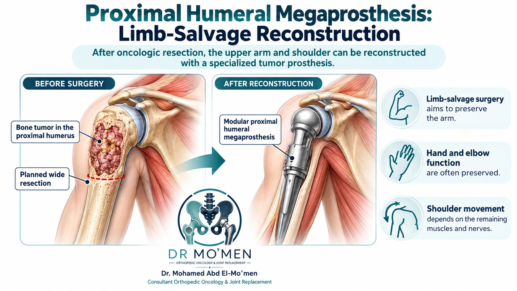
- Preserving a useful upper limb is the main functional goal. Hand, wrist and elbow function can often be preserved when the musculocutaneous, radial, median and ulnar nerves, blood supply and distal muscles can be safely maintained.
- Shoulder function is more variable. Active elevation depends heavily on the deltoid, axillary nerve, remaining cuff/capsule, soft-tissue reconstruction and implant design.
- Reverse and anatomical megaprostheses are not interchangeable. Reverse reconstruction generally requires adequate glenoid fixation plus a functioning, innervated deltoid; anatomical reconstruction may be used when reverse mechanics are unsuitable, but active elevation is often more limited.
- Biopsy planning is part of treatment. For many proximal humeral lesions, a longitudinal route through the anterior one-third of the deltoid is preferred so the tract can be removed with the definitive specimen, but the route must be individualized to the MRI and planned surgery.
- Not every proximal humerus tumor needs a megaprosthesis. Benign lesions, limited metastases and some fractures may be treated with observation, curettage, fixation, cement augmentation, radiotherapy, systemic treatment or another reconstruction depending on the diagnosis and mechanical problem.
What is a proximal humeral megaprosthesis?
A proximal humeral megaprosthesis is a large modular endoprosthesis that replaces the upper part of the humerus—and usually the shoulder articulation—after a substantial segment of bone has been removed. It is different from routine shoulder replacement for arthritis because tumor surgery may remove not only the humeral head but also a long segment of bone, the rotator-cuff insertions, capsule and portions of surrounding muscle.
The operation therefore has two separate objectives: first, oncological resection of the tumor; second, reconstruction of the resulting skeletal and soft-tissue defect. A technically successful implant cannot compensate for an inadequate tumor resection, and a wide resection alone is not enough if the remaining limb cannot be reconstructed safely and usefully.
For more detail, see megaprosthetic reconstruction after bone tumor resection.
Which tumors of the proximal humerus may require limb-salvage reconstruction?
The proximal humerus is a recognized site for primary bone sarcomas, including osteosarcoma, chondrosarcoma and Ewing sarcoma. It is also a common region for metastatic involvement of the humerus; the humerus overall is the second most commonly involved long bone after the femur in metastatic bone disease.
Primary malignant bone tumors
Osteosarcoma usually requires systemic chemotherapy together with planned surgery for localized high-grade extremity disease. When a proximal humeral tumor can be removed with an adequate margin while preserving a useful arm, limb-salvage reconstruction may be considered.
Chondrosarcoma is treated according to subtype and grade. Conventional chondrosarcoma is primarily a surgical disease, and wide resection is central when the lesion is malignant and resectable.
Ewing sarcoma requires systemic therapy and coordinated local control with surgery, radiotherapy or selected combinations. Reconstruction is chosen only after the local-control strategy has been defined.
Metastatic bone disease and myeloma
Metastatic lesions can weaken the proximal humerus and cause severe pain or an impending or completed pathological fracture. However, a megaprosthesis is not automatically required. Intramedullary nailing, plate-and-cement reconstruction, radiotherapy, systemic treatment or other strategies may be more appropriate depending on the location and extent of destruction, bone quality, tumor biology, expected survival and the mechanical stability required.
Endoprosthetic replacement becomes more relevant when the humeral head and metaphysis are extensively destroyed, when the joint surface cannot be preserved, or when previous fixation has failed and durable structural reconstruction is required.
For more detail, see pathological fracture fixation vs reconstruction in bone metastasis.
Aggressive benign and intermediate tumors
Most benign proximal humeral lesions do not require a megaprosthesis. Observation, curettage, grafting, cementation, fixation or another joint-preserving procedure may be more appropriate. Rarely, a locally aggressive lesion such as a giant cell tumor with extensive joint destruction may require segmental resection and reconstruction.
For more detail, see simple bone cyst of the proximal humerus.
What tests are needed before deciding on surgery?
The decision should not start with the implant. It starts with the diagnosis.
Typical assessment may include:
- plain X-rays of the humerus and shoulder;
- MRI to define marrow and soft-tissue extension and the relationship to the deltoid, rotator cuff, axillary nerve and neurovascular structures;
- CT when cortical detail or mineralized matrix needs clarification;
- appropriate staging imaging according to the suspected tumor type;
- pathology from a planned biopsy when tissue diagnosis is required;
- multidisciplinary review for malignant primary tumors and complex metastatic disease.
A pathology report should always be interpreted alongside the imaging. If pathology and imaging are biologically discordant, the case may require pathology review or repeat sampling rather than proceeding directly to definitive surgery.
Why is biopsy placement especially important in proximal humerus tumors?
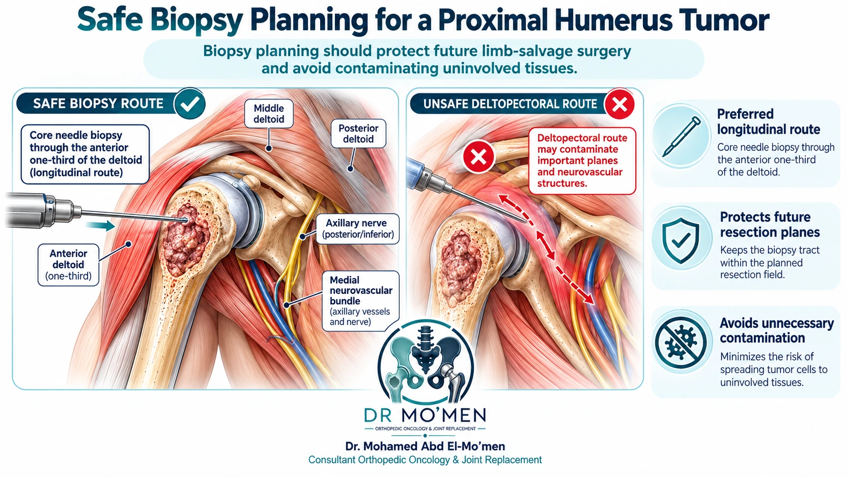
A biopsy can affect the final operation. Inappropriately placed biopsy tracts may contaminate tissue planes that would otherwise be preserved and can make later resection more extensive.
For many proximal humeral lesions, orthopedic oncology literature recommends a longitudinal biopsy route through the anterior one-third of the deltoid rather than the deltopectoral interval, because the tract can then be removed with the definitive specimen while limiting unnecessary contamination. This is not a universal rule for every lesion: the exact trajectory must be chosen after MRI review by the team responsible for definitive treatment.
The biopsy should avoid crossing uninvolved compartments, major neurovascular structures and tissue planes that would not otherwise be removed. The biopsy tract is generally planned so it can be excised en bloc at definitive surgery.
For more detail, see core needle vs open biopsy and why the route matters.
How does proximal humeral limb-salvage surgery work?
The procedure is individualized, but it usually has four coordinated components.
1. Oncological resection
The tumor-bearing segment is removed according to the planned margin, together with the biopsy tract and any involved soft tissue that must be sacrificed for tumor control. Margin strategy depends on diagnosis, grade, response to systemic treatment when applicable, and the relationship to critical structures.
2. Neurovascular preservation when oncologically safe
The surgeon identifies and protects structures needed for a useful distal limb whenever they are free of tumor. These may include the brachial artery and veins, the musculocutaneous nerve for elbow flexion, and the radial, median and ulnar nerves for forearm, wrist and hand function.
Preservation is never allowed to compromise tumor clearance. If a major nerve is directly involved, the functional consequences and reconstructive options must be discussed before surgery.
3. Skeletal reconstruction
A modular stem is fixed into the remaining humeral shaft. Cemented or uncemented fixation may be used according to implant design, bone quality, resection length and surgeon judgment. Modular segments restore the length of the resected bone and reconstruct the shoulder articulation.
4. Soft-tissue reconstruction
Soft-tissue reconstruction is crucial because the rotator cuff and deltoid attachments may be partly or completely removed with the specimen. Synthetic attachment tubes or mesh, tendon suturing, muscle transfers and capsular repair may be used to create a stable envelope around the implant. The exact technique depends on which structures remain after oncological resection.
Anatomical vs reverse proximal humeral megaprosthesis: what is the difference?
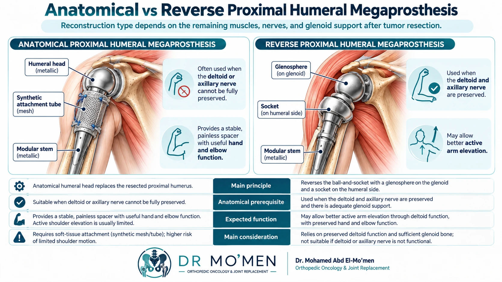
The implant is selected according to the anatomy that remains after safe resection—not simply according to patient preference.
| Reconstruction | Basic concept | Key prerequisites | Expected functional emphasis | Important limitations |
|---|---|---|---|---|
| Anatomical proximal humeral megaprosthesis | A humeral head component articulates with the native glenoid; soft tissues are reconstructed around the implant. | A reconstructable shoulder envelope; reverse mechanics are unsuitable or not possible. | Provides skeletal continuity and a stable platform for elbow, wrist and hand use. | Active shoulder elevation is often limited when cuff or deltoid attachments have been sacrificed; instability or migration can occur. |
| Reverse proximal humeral megaprosthesis | A glenosphere on the scapular side articulates with a socket on the humeral prosthesis, allowing the deltoid to drive elevation. | Adequate glenoid bone/fixation and a functioning, innervated deltoid with a usable axillary nerve. | Usually offers better active forward elevation than anatomical reconstruction in appropriately selected patients. | Results vary with resection extent and soft tissues; glenoid complications, instability and mechanical failure remain possible. |
| Allograft-prosthetic composite (APC) | Structural donor bone is combined with a prosthetic component to provide attachment sites for soft tissues. | Selected patients with healing potential, appropriate anatomy and access to graft resources. | May facilitate tendon and soft-tissue reattachment. | Graft nonunion, fracture, resorption and infection are additional risks. |
| Biological reconstruction | Autograft/allograft-based techniques reconstruct the defect without relying entirely on a large metallic segment. | Highly selected cases, particularly younger patients or special reconstructive goals. | Preserves biological bone stock in selected patients. | Longer healing, graft fracture/nonunion and donor-site morbidity may occur. |
Evidence synthesis shows that reverse endoprosthetic reconstruction tends to provide better postoperative range of motion and functional scores than anatomical endoprostheses when adequate abductor/deltoid function is preserved, but outcomes remain heterogeneous and no technique is best for every patient.
What function can patients expect after surgery?
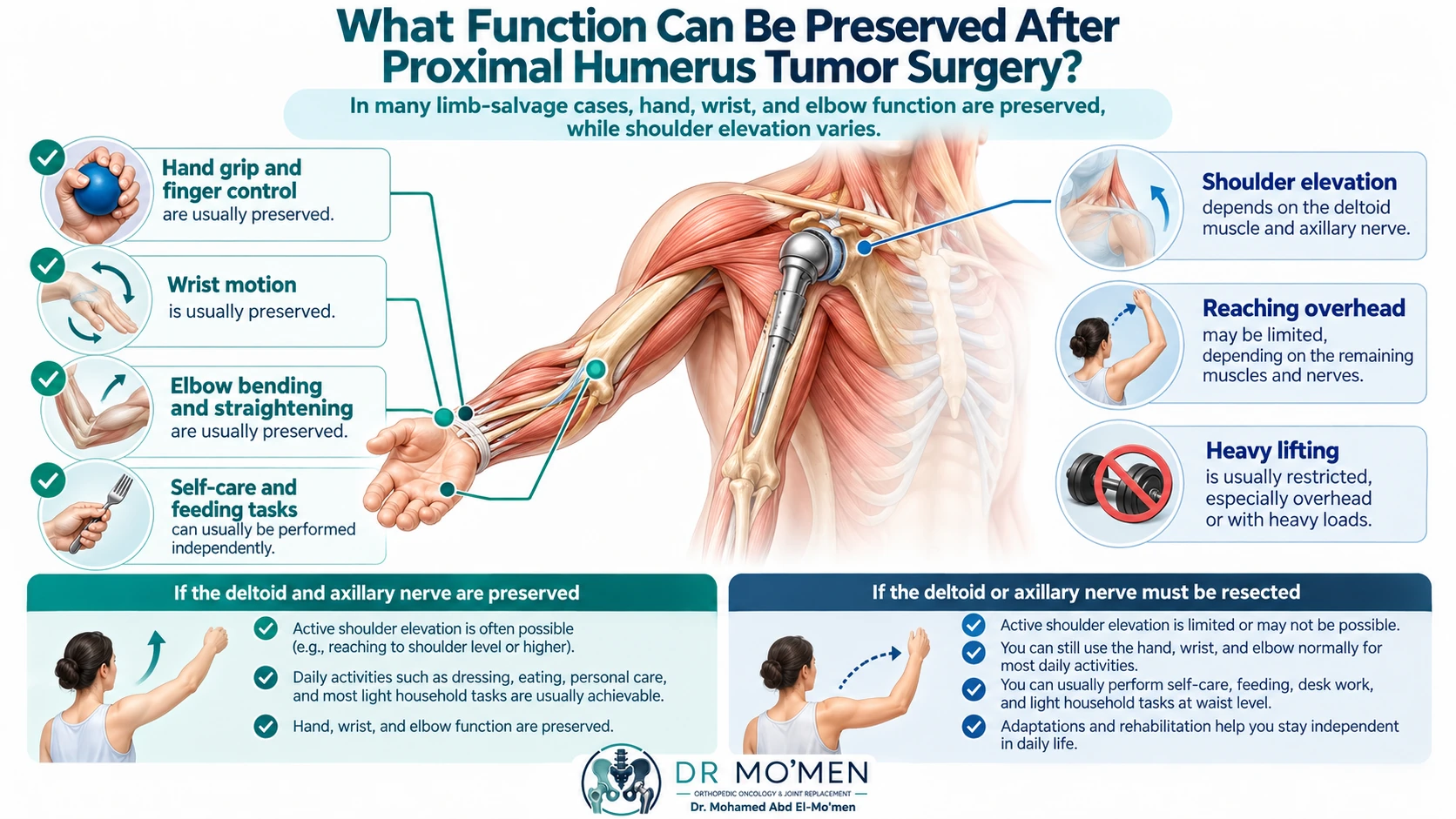
The most important preoperative counseling point is that shoulder elevation and hand function are different outcomes. A patient may have limited active shoulder elevation but still retain a useful hand, wrist and elbow.
| Function | When major distal nerves and muscles are preserved | What may limit it |
|---|---|---|
| Hand grip, typing, writing, phone use | Often well preserved | Direct nerve involvement, perioperative nerve injury, severe edema, systemic illness |
| Wrist and finger movement | Often well preserved | Radial/median/ulnar nerve compromise or distal muscle dysfunction |
| Elbow flexion and extension | Often useful or near baseline when musculocutaneous and radial function is preserved | Nerve involvement, muscle sacrifice, postoperative stiffness |
| Waist- and chest-level activities | Often achievable with rehabilitation | Shoulder instability, pain, muscle weakness, extent of resection |
| Active shoulder elevation | Highly variable | Deltoid and axillary nerve status, cuff/capsule loss, reconstruction type, soft-tissue healing |
| Heavy lifting and repetitive overhead loading | Usually restricted | Risk of instability, glenoid or stem stress, soft-tissue failure and implant wear |
There is no responsible way to promise a specific number of degrees of shoulder elevation before surgery without knowing the resection, nerve status, implant and soft-tissue reconstruction.
What is recovery and rehabilitation like?
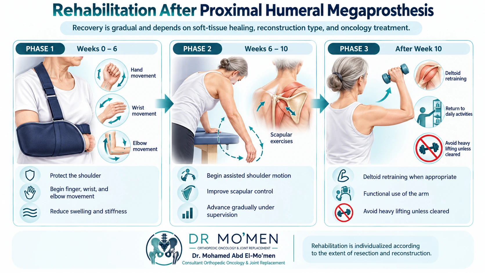
Rehabilitation after tumor reconstruction is individualized. Published protocols vary, and the treating team must adapt therapy to soft-tissue repair, implant design, wound healing, oncology treatment and neurological status.
A typical framework may look like this:
Early protection phase
The shoulder is protected in a sling or prescribed support while the soft-tissue reconstruction heals. Hand, wrist and elbow movement is usually encouraged early when safe to reduce distal stiffness and swelling.
Assisted-motion phase
Once the wound and repair are sufficiently stable, supervised pendulum, scapular and passive or assisted shoulder exercises may be introduced. Timing depends on the reconstruction and surgeon instructions.
Functional strengthening phase
In reverse reconstructions with a functioning deltoid, therapy later emphasizes deltoid retraining, scapular control and functional reaching. Resistance and lifting are advanced cautiously, and many patients remain on permanent limits for heavy or repetitive overhead loading.
A 2026 consensus-based rehabilitation publication specifically includes a protocol for proximal humerus resection reconstructed with a reverse shoulder megaprosthesis, reinforcing the need for procedure-specific rather than generic shoulder-replacement rehabilitation.
What complications need long-term monitoring?
Upper-limb tumor megaprostheses do not bear body weight during walking, but they have their own soft-tissue and mechanical risks.
Instability or soft-tissue failure
Loss of the rotator cuff, capsule and muscle attachments can lead to subluxation, migration or dislocation. Soft-tissue reconstruction and appropriate implant selection aim to reduce this risk.
Infection
Deep periprosthetic infection is a serious complication of oncological reconstruction. Risk varies according to the extent of surgery, wound condition, previous procedures, systemic treatment and patient factors. Persistent drainage, increasing redness, fever or new deep pain requires prompt assessment.
For more detail, see megaprosthesis infection: diagnosis and treatment.
Nerve dysfunction
Neurological deficits may result from tumor involvement, traction, dissection, postoperative swelling or necessary nerve resection. Recovery depends on the mechanism and severity; it should never be assumed to be temporary without assessment.
Aseptic loosening and mechanical failure
Stem loosening may occur, although the mechanical environment differs from weight-bearing lower-limb megaprostheses. Reverse constructs also introduce glenoid-side fixation and its own mechanical risks.
Local recurrence
Tumor surveillance remains separate from implant surveillance. Follow-up depends on the original diagnosis, grade, stage and oncology protocol.
When should a patient seek urgent specialist review after surgery?
Prompt assessment is warranted for:
- increasing redness, warmth, swelling or wound drainage;
- fever or chills with new shoulder pain;
- sudden deformity or a new sense of instability;
- sudden new weakness of wrist, fingers or elbow;
- a new or enlarging mass near the operative site;
- new unexplained pain after a previously stable recovery.
When is a specialist second opinion particularly useful?
A second opinion can be valuable before a biopsy, before a major proximal humeral resection, when the diagnosis is uncertain, when different surgeons have proposed different reconstructions, after previous surgery, or when amputation versus limb salvage is being considered.
Patients outside Egypt can send original imaging and reports before travel for a preliminary specialist review. The remote review does not replace physical examination when an in-person assessment is required.
Internal links:
- Limb-Salvage & Reconstructive Surgery Service
- Second opinion for bone tumors
- How to send MRI/CT/X-ray files
- Related recurrent humerus case — An educational case on reassessment and limb-preservation planning; the reconstruction type has not been verified.
Frequently asked questions
Will I still be able to use my hand and elbow after proximal humerus tumor surgery?
Often, yes—if the nerves, vessels and distal muscles responsible for elbow, wrist and hand function can be preserved. Function is not guaranteed, and it depends on tumor extent, nerve involvement, the operation and postoperative recovery.
Why can some patients raise their arm better than others after surgery?
Active shoulder elevation depends mainly on the amount of deltoid that remains functional, the axillary nerve, the remaining soft tissues and the reconstruction type. Reverse reconstruction can improve elevation in selected patients when the deltoid and axillary nerve remain functional.
How is a tumor megaprosthesis different from a standard shoulder replacement?
A standard shoulder replacement treats the joint surfaces. A tumor megaprosthesis may replace a long segment of the proximal humerus after oncological resection and therefore requires much more extensive soft-tissue reconstruction.
Does every proximal humeral metastasis need a megaprosthesis?
No. Fixation, cement augmentation, radiotherapy, systemic treatment or another strategy may be more appropriate. Megaprosthetic replacement is considered when destruction is extensive, the joint cannot be preserved, previous fixation has failed or durable segmental reconstruction is needed.
How long does a proximal humeral megaprosthesis last?
There is no single fixed lifespan. Many reconstructions remain functional for years, but longevity depends on implant design, soft-tissue stability, infection, instability, fixation, tumor recurrence, subsequent treatment and activity level. Long-term surveillance is therefore important.
Specialist assessment
Complex proximal humerus bone tumors require diagnosis-specific planning before biopsy or definitive surgery. For specialist review with Dr. Mohammed Abdelmoemen Abuelhadid, imaging and reports can be reviewed before an in-person visit when appropriate.
For booking or imaging review: 01021690693
Clinic: 50 El Hegaz Street, Heliopolis, Cairo. Saturday 3–5 PM, Monday 7–9 PM, Wednesday 5–7 PM.
Medical evidence and references
- Fiore M et al. Anatomical vs reverse megaprostheses after oncologic resection (2022).
- Su F, Garcia-Lopez E, Wustrack R, Lansdown DA. Proximal humeral tumor endoprosthetic reconstruction (2025).
- Strauss SJ et al. Bone sarcomas: ESMO clinical practice guidelines (2021).
- Denissen JJPM et al. Physical therapy protocols after tumor megaprosthesis reconstruction (2026).
- Functional outcomes after reverse shoulder megaprosthesis: systematic review (2023).
- Intramedullary nailing versus cemented plate for metastatic proximal humeral pathological fracture.
Medical disclaimer
This article is for patient education and does not replace clinical examination, review of original imaging, pathology, staging or multidisciplinary medical assessment.