
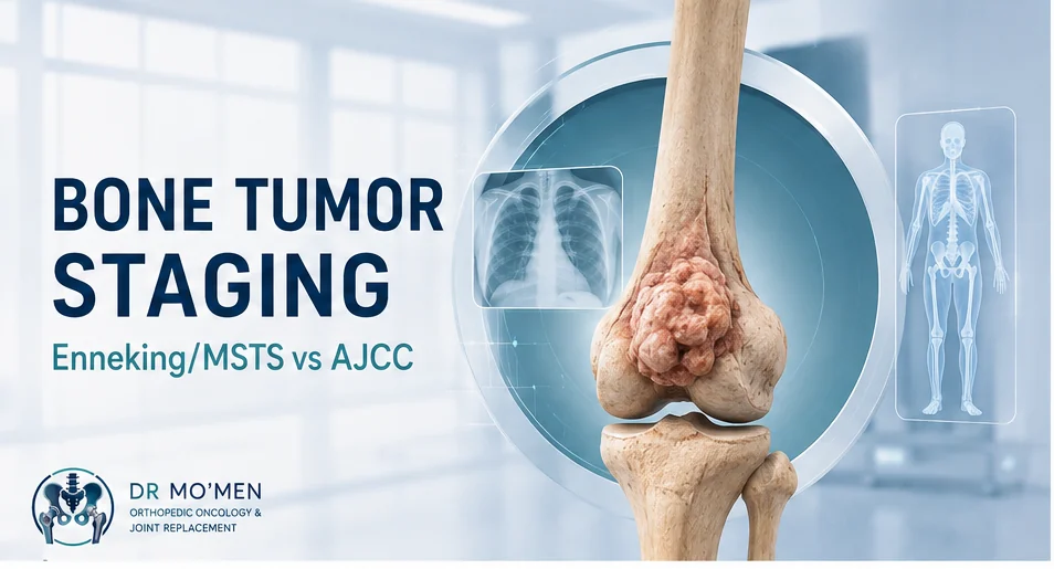
Being told that a bone tumor has a particular “stage” can be confusing. Patients may hear terms such as Stage IIB, high grade, extracompartmental, T2, or metastatic disease and assume they all mean the same thing. They do not.
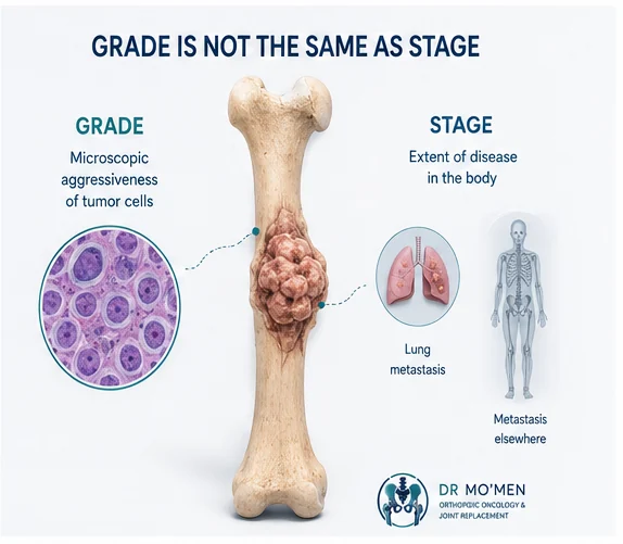
Tumor grade describes the biological appearance and aggressiveness of the tumor under the microscope, while tumor stage describes how extensive the disease is in the body.
For malignant bone tumors, two staging frameworks are commonly discussed: the Enneking/MSTS staging system and the AJCC TNM staging system.
Neither should be interpreted from an MRI report alone. Correct staging requires the diagnosis, pathology, local imaging and appropriate assessment for disease elsewhere in the body to be considered together.
If a primary bone tumor is suspected, biopsy should ideally be planned by, or in coordination with, the team responsible for definitive tumor surgery. The biopsy tract can affect later resection and reconstruction, so unplanned biopsy or excision should be avoided.
What Is Bone Tumor Staging?
Staging is a structured way of describing how far a malignant tumor has progressed at the time of assessment. Depending on the tumor and staging framework, this may include:
- tumor grade;
- size of the primary tumor;
- whether it remains within a particular anatomical compartment;
- whether it extends into surrounding soft tissues;
- whether separate tumor deposits are present within the same bone;
- regional lymph-node involvement;
- lung metastases;
- metastases to other bones or organs.
Staging helps the multidisciplinary team communicate precisely and plan treatment. It also allows outcomes from different patients and research studies to be compared more consistently.
Tumor Grade and Tumor Stage Are Not the Same
Tumor grade
The grade comes mainly from examination of tumor tissue by the pathologist.
- Low-grade tumors generally have less aggressive microscopic features.
- High-grade tumors generally show more aggressive biological features and often have greater potential for progression or metastasis.
However, grade alone does not describe where the tumor has spread.
Tumor stage
The stage combines information about the tumor and its extent. A patient may therefore have a low-grade but locally extensive tumor, a high-grade tumor that remains localized, or disease that has already metastasized. A pathology report by itself does not provide the complete staging picture.
The Enneking/MSTS Staging System
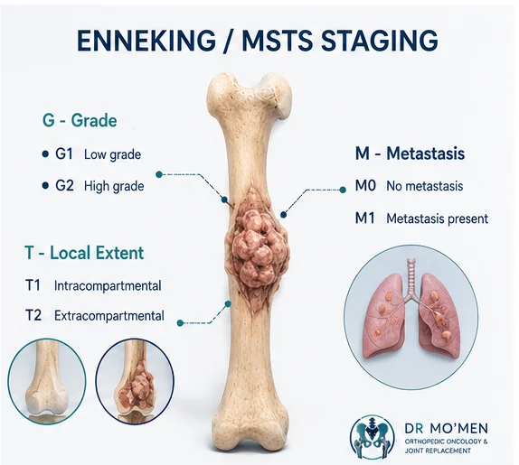
The Musculoskeletal Tumor Society system, often called the Enneking staging system, was developed specifically for musculoskeletal tumors. For malignant bone tumors it considers three major elements:
- G1 — Low grade
- Histologically low-grade malignant bone tumor.
- G2 — High grade
- Histologically high-grade malignant bone tumor.
- T1 — Intracompartmental
- The tumor remains within the anatomical boundaries of its compartment.
- T2 — Extracompartmental
- The tumor has extended beyond the anatomical compartment of origin.
- M0
- No metastasis identified.
- M1
- Metastatic disease is present.
1. Grade — G
- G1: low grade
- G2: high grade
2. Local extent — T
- T1: intracompartmental
- T2: extracompartmental
In practical terms, this describes whether the tumor remains within its anatomical compartment or has extended beyond it.
3. Metastasis — M
- M0: no metastasis identified
- M1: metastatic disease present
| Enneking/MSTS stage | Grade | Local extent | Metastasis |
|---|---|---|---|
| IA | Low grade | Intracompartmental | No |
| IB | Low grade | Extracompartmental | No |
| IIA | High grade | Intracompartmental | No |
| IIB | High grade | Extracompartmental | No |
| III | Any grade | Any local extent | Present |
What does Stage I mean?
Stage I refers to a low-grade localized malignant tumor.
What does Stage II mean?
Stage II refers to a high-grade localized malignant tumor.
What does Stage III mean?
Stage III indicates metastatic disease within the Enneking/MSTS malignant tumor framework.
What Does “Intracompartmental” or “Extracompartmental” Mean?
An intracompartmental tumor remains within the anatomical boundaries of its compartment. An extracompartmental tumor has extended beyond those boundaries.
For example, a malignant tumor beginning inside a long bone may break through the cortex and form a substantial soft-tissue component. This local extent can influence surgical margins, structures that need to be preserved or removed, feasibility and complexity of limb-salvage surgery, and reconstruction options.
Local extension alone does not automatically mean that the tumor has metastasized. A large tumor extending outside the bone can still be localized if staging investigations show no distant disease.
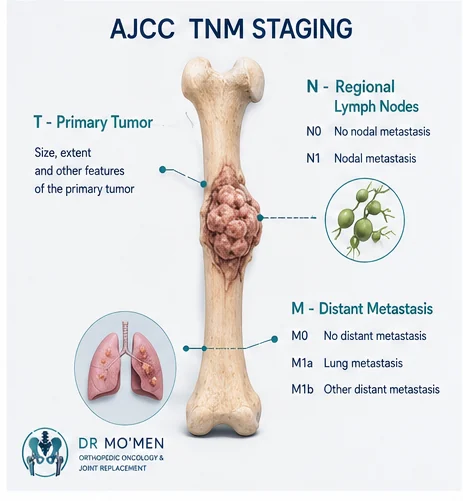
What Is AJCC TNM Staging for Bone Tumors?
The AJCC TNM system approaches staging differently.
T — Primary tumor
T describes characteristics of the primary tumor. For many primary bone cancers of the appendicular skeleton, the commonly presented framework distinguishes tumors according to size and the presence of discontinuous tumor deposits within the same bone.
- T1: tumor up to 8 cm
- T2: tumor larger than 8 cm
- T3: discontinuous tumors within the same primary bone site
This simplified framework should not be applied blindly to every skeletal site. AJCC staging has site-specific considerations, particularly for tumors involving areas such as the pelvis and spine.
N — Regional lymph nodes
- N0: no regional lymph-node metastasis
- N1: regional lymph-node metastasis
Regional lymph-node spread is relatively uncommon in many primary bone sarcomas, but when present it can significantly affect staging.
M — Distant metastasis
- M0: no distant metastasis
- M1a: lung metastasis
- M1b: metastasis to other distant sites
G — Grade
Histological grade is also incorporated into the stage grouping for applicable tumors.
AJCC Stages I to IV: A Patient-Friendly Overview
Stage I
Generally represents localized low-grade disease.
Stage II
Generally represents localized high-grade disease, with subgroups reflecting features such as tumor size.
Stage III
In the commonly described appendicular bone framework, Stage III can include high-grade tumors with discontinuous tumor deposits within the same bone. These are sometimes called skip lesions or skip metastases.
Stage IV
Indicates metastatic or certain advanced patterns of disease. In the commonly presented bone tumor framework, Stage IVA may describe lung-only distant metastasis, while Stage IVB includes certain other distant metastatic patterns or regional nodal disease.
The exact AJCC stage should be assigned using the appropriate rules for the tumor histology and anatomical site, rather than from a simplified online table alone.
Enneking/MSTS vs AJCC: What Is the Difference?
| Feature | Enneking/MSTS | AJCC TNM |
|---|---|---|
| Developed particularly for musculoskeletal tumors | Yes | General cancer staging framework adapted to specific cancers |
| Includes tumor grade | Yes | Yes, where applicable |
| Focuses on anatomical compartment | Yes | Not in the same way |
| Uses tumor size | Not as the principal T definition | Yes for several bone sites |
| Includes regional lymph nodes | Within metastatic assessment | Explicit N category |
| Distinguishes lung from other metastases | No | Yes in the commonly used bone framework |
| Common overall stages | I–III | I–IV |
| Particularly useful for surgical thinking | Yes | Useful for oncological staging and standardized reporting |
The two systems therefore describe overlapping but not identical aspects of the disease. One system is not simply a translation of the other.
Does Every Bone Sarcoma Use the Same Staging System?
No. This is an important limitation of simplified internet explanations.
For practical treatment purposes, osteosarcoma is commonly described as localized or metastatic, and formal prognostic stage groups are not the only framework used for treatment stratification. Similarly, Ewing sarcoma is commonly described clinically as localized, metastatic or recurrent.
The most useful staging language therefore depends on the exact pathology, the patient’s age, the anatomical site, the treatment protocol and the multidisciplinary team managing the case.
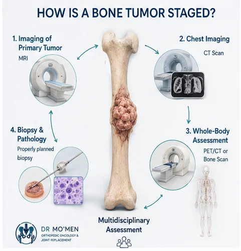
How Is a Bone Tumor Properly Staged?
Staging usually requires several pieces of information rather than one investigation.
1. Imaging of the primary tumor
MRI is particularly important for defining intramedullary tumor extent, soft-tissue extension, relationship to nerves and blood vessels, involvement near a joint and possible additional lesions within the involved bone. See the guide to bone tumors around the knee for location-specific context.
2. Chest imaging
The lungs are an important site of metastasis in several bone sarcomas. Chest CT is therefore commonly part of staging for tumors such as osteosarcoma and Ewing sarcoma.
3. Whole-body metastatic assessment when indicated
Depending on the suspected tumor and clinical situation, staging may involve PET/CT, PET/MRI in selected settings, bone scintigraphy or other targeted imaging. The appropriate study depends on the tumor type and treatment protocol; not every patient requires every test.
4. A Properly Planned Biopsy
Imaging should generally be completed before biopsy in a suspected primary malignant bone tumor. The biopsy must answer an essential question: What exactly is this tumor?
The way the biopsy is performed also matters. The biopsy tract may need to be removed during definitive tumor surgery. A poorly positioned biopsy can contaminate tissues that otherwise might not have needed removal and can complicate limb-salvage surgery. Read more about bone tumor biopsy planning.
Why Does Accurate Staging Matter?
Staging does more than place a number beside the diagnosis. It can affect decisions about surgery, systemic treatment, metastatic disease management and reconstruction.
Surgery
The team needs to determine whether the tumor can be removed with an appropriate oncological margin and what reconstruction would be required. For tumors around major joints this may involve biological reconstruction, joint-preserving surgery in selected cases, endoprosthetic reconstruction, megaprosthesis or other limb-salvage techniques.
Chemotherapy
The role of chemotherapy depends heavily on tumor histology. Chemotherapy is central to treatment of conventional high-grade osteosarcoma and Ewing sarcoma, whereas its role is very different in most conventional chondrosarcomas.
Treatment of metastatic disease
If metastases are present, treatment planning may need to address both the primary tumor and metastatic disease. The exact approach depends on tumor biology, metastatic site, number of lesions, resectability, systemic treatment options and the patient’s overall condition.
Does a Higher Stage Automatically Mean Amputation?
No. Stage and limb-salvage feasibility are related issues, but they are not the same question. The decision may depend on:
- tumor anatomy;
- neurovascular involvement;
- ability to achieve an adequate surgical margin;
- extent of soft-tissue involvement;
- pathological fracture in certain circumstances;
- response to systemic treatment when relevant;
- reconstruction possibilities;
- infection and wound considerations;
- overall oncological strategy.
A stage number alone cannot determine whether a limb can be preserved.
Does Stage IV Mean There Is No Treatment?
No. Stage IV indicates advanced or metastatic disease within the relevant staging framework. It does not mean that treatment is automatically impossible or that every Stage IV patient has the same situation.
- systemic therapy;
- surgery;
- radiation therapy;
- treatment of lung or other metastases;
- stabilization or reconstruction of threatened bones;
- symptom control;
- multidisciplinary oncological care.
The realistic goals and available options must be assessed individually.
Can MRI Alone Determine the Stage?
No. MRI provides crucial information about the local tumor, but complete staging generally requires information about the rest of the body and confirmation of the tumor diagnosis.
Local imaging + biopsy/pathology + chest imaging + appropriate metastatic assessment, depending on the suspected tumor.
A Practical Example
Consider a patient with a destructive tumor around the distal femur. MRI may show that the lesion has extended through the cortex, formed a soft-tissue mass and approached the knee joint. But this still does not tell us everything.
- What is the pathological diagnosis?
- Is it low or high grade?
- Is there another lesion within the same bone?
- Are the lungs clear?
- Is there disease elsewhere?
- Was the biopsy properly positioned?
- Can an adequate oncological margin be obtained?
- What reconstruction is possible after resection?
Only after these elements are brought together can staging and treatment planning become clinically meaningful.
Special Consideration: Pelvic and Spinal Bone Tumors
Patients should be particularly careful about trying to apply a general “8 cm rule” to every bone tumor. Bone staging schemas distinguish between the appendicular skeleton, spine and pelvis because the anatomical definitions of local tumor extent are not identical across these locations.
For example, pelvic tumors require consideration of which pelvic segments and surrounding structures are involved rather than simply measuring maximum tumor diameter. See Pelvic Bone Tumors: Diagnosis, Biopsy and Limb Salvage.
When Should You Seek an Orthopedic Oncology Assessment?
Specialist assessment is appropriate when imaging shows or suggests:
- an aggressive or destructive bone lesion;
- cortical destruction;
- a large soft-tissue component;
- a suspected primary malignant bone tumor;
- unexplained persistent or progressive bone pain associated with an abnormal lesion;
- a pathological fracture through a suspicious lesion;
- a previously treated bone tumor that may have returned;
- a biopsy being considered for an unexplained bone tumor.
A suspicious primary bone tumor should ideally be evaluated before an unplanned biopsy or surgery because the initial approach can affect later definitive treatment. See bone tumor warning signs.
The Role of Specialist Review in Complex Bone Tumor Cases
Accurate bone-tumor treatment planning requires more than identifying a lesion on MRI. Orthopedic oncology assessment integrates clinical examination, X-rays, MRI, staging investigations, biopsy planning, pathology, tumor location, relationship to major nerves and vessels, potential surgical margins, reconstruction options and multidisciplinary oncology input.
For patients outside Egypt, an initial orthopedic oncology second-opinion review of imaging, pathology reports and previous medical records may help determine what additional investigations are required before travel. The International Patients pathway explains how records can be reviewed before travel when appropriate.
Patients outside Egypt can send available X-rays, MRI/CT images, pathology reports and previous treatment records for an initial orthopedic oncology review before arranging travel. This may help identify what further assessment is needed, but it does not replace examination or guarantee that surgery is appropriate. International Patients pathway · Send staging scans and reports via WhatsApp.
Frequently Asked Questions
Is tumor grade the same as tumor stage?
No. Grade describes the microscopic biological characteristics of the tumor. Stage describes the extent of disease. Both may influence treatment planning.
Which is better: Enneking or AJCC?
Neither is universally better. They describe disease differently and their usefulness varies with tumor type, anatomical site and clinical purpose.
Can my MRI report tell me whether I have Stage II or Stage III cancer?
Usually not by itself. Complete staging requires the pathological diagnosis and appropriate assessment of local and distant disease.
Why is a chest CT commonly requested for bone sarcoma?
The lungs are an important metastatic site for several primary bone sarcomas, including osteosarcoma and Ewing sarcoma. Chest CT is therefore commonly included in staging investigations for these diseases.
Does metastatic bone sarcoma always mean surgery is impossible?
No. Surgical feasibility and the overall treatment strategy depend on the primary tumor, metastatic pattern, response to treatment, resectability and the patient’s individual circumstances.
Key Message
The stage of a bone tumor cannot be understood from a number alone. A meaningful assessment requires answering four fundamental questions:
- What is the exact tumor?
- How aggressive is it?
- How far has it extended locally?
- Has it spread elsewhere?
For suspected malignant bone tumors, these questions should ideally be answered through coordinated imaging, properly planned biopsy, pathology review and multidisciplinary specialist assessment before definitive surgery.
Appointments or imaging review
If you have imaging or pathology reports for a suspected bone tumor, specialist review can help clarify what additional staging or biopsy planning may be required.